规格 价格 库存
 50ul RMB¥ 1250 现货
 100ul RMB¥ 2300 现货
 200ul RMB¥ 3000 现货

货期: 当天发货

联系销售

产品描述

来源:
Mouse
应用:
WB 1:1000-1:10000, IHC 1:50-1:200
*The optimal dilutions should be determined by the end user.
*Tips:

WB: 适用于变性蛋白样本的免疫印迹检测. IHC: 适用于组织样本的石蜡(IHC-p)或冰冻(IHC-f)切片样本的免疫组化/荧光检测. IF/ICC: 适用于细胞样本的荧光检测. ELISA(peptide): 适用于抗原肽的ELISA检测.

反应:
Human, Mouse, Rat
克隆:
Monoclonal [AFfirm8015]
特异性:
p38 MAPK Antibody detects endogenous levels of p38 MAPK .
偶联:
Unconjugated. 130
纯化:
Affinity-chromatography.
保存:
Mouse IgG1 in phosphate buffered saline (without Mg2+ and Ca2+), pH 7.4, 150mM NaCl, 0.02% sodium azide and 50% glycerol. Store at -20 °C. Stable for 12 months from date of receipt.
别名:

展开/折叠

CSAID Binding Protein 1; CSAID binding protein; CSAID-binding protein; Csaids binding protein; CSBP 1; CSBP 2; CSBP; CSBP1; CSBP2; CSPB1; Cytokine suppressive anti-inflammatory drug-binding protein; EXIP; MAP kinase 14; MAP kinase MXI2; MAP kinase p38 alpha; MAPK 14; MAPK14; MAX interacting protein 2; MAX-interacting protein 2; Mitogen Activated Protein Kinase 14; Mitogen activated protein kinase p38 alpha; Mitogen-activated protein kinase 14; Mitogen-activated protein kinase p38 alpha; MK14_HUMAN; Mxi 2; MXI2; p38 ALPHA; p38; p38 MAP kinase; p38 MAPK; p38 mitogen activated protein kinase; p38ALPHA; p38alpha Exip; PRKM14; PRKM15; RK; SAPK2A;

抗原和靶标

免疫原:

A synthesized peptide derived from human p38 MAPK.

Uniprot:
基因/基因ID:
表达:
Q16539 MK14_HUMAN:

Brain, heart, placenta, pancreas and skeletal muscle. Expressed to a lesser extent in lung, liver and kidney.

序列:
MSQERPTFYRQELNKTIWEVPERYQNLSPVGSGAYGSVCAAFDTKTGLRVAVKKLSRPFQSIIHAKRTYRELRLLKHMKHENVIGLLDVFTPARSLEEFNDVYLVTHLMGADLNNIVKCQKLTDDHVQFLIYQILRGLKYIHSADIIHRDLKPSNLAVNEDCELKILDFGLARHTDDEMTGYVATRWYRAPEIMLNWMHYNQTVDIWSVGCIMAELLTGRTLFPGTDHIDQLKLILRLVGTPGAELLKKISSESARNYIQSLTQMPKMNFANVFIGANPLAVDLLEKMLVLDSDKRITAAQALAHAYFAQYHDPDDEPVADPYDQSFESRDLLIDEWKSLTYDEVISFVPPPLDQEEMES

研究背景

功能:

Serine/threonine kinase which acts as an essential component of the MAP kinase signal transduction pathway. MAPK14 is one of the four p38 MAPKs which play an important role in the cascades of cellular responses evoked by extracellular stimuli such as proinflammatory cytokines or physical stress leading to direct activation of transcription factors. Accordingly, p38 MAPKs phosphorylate a broad range of proteins and it has been estimated that they may have approximately 200 to 300 substrates each. Some of the targets are downstream kinases which are activated through phosphorylation and further phosphorylate additional targets. RPS6KA5/MSK1 and RPS6KA4/MSK2 can directly phosphorylate and activate transcription factors such as CREB1, ATF1, the NF-kappa-B isoform RELA/NFKB3, STAT1 and STAT3, but can also phosphorylate histone H3 and the nucleosomal protein HMGN1. RPS6KA5/MSK1 and RPS6KA4/MSK2 play important roles in the rapid induction of immediate-early genes in response to stress or mitogenic stimuli, either by inducing chromatin remodeling or by recruiting the transcription machinery. On the other hand, two other kinase targets, MAPKAPK2/MK2 and MAPKAPK3/MK3, participate in the control of gene expression mostly at the post-transcriptional level, by phosphorylating ZFP36 (tristetraprolin) and ELAVL1, and by regulating EEF2K, which is important for the elongation of mRNA during translation. MKNK1/MNK1 and MKNK2/MNK2, two other kinases activated by p38 MAPKs, regulate protein synthesis by phosphorylating the initiation factor EIF4E2. MAPK14 interacts also with casein kinase II, leading to its activation through autophosphorylation and further phosphorylation of TP53/p53. In the cytoplasm, the p38 MAPK pathway is an important regulator of protein turnover. For example, CFLAR is an inhibitor of TNF-induced apoptosis whose proteasome-mediated degradation is regulated by p38 MAPK phosphorylation. In a similar way, MAPK14 phosphorylates the ubiquitin ligase SIAH2, regulating its activity towards EGLN3. MAPK14 may also inhibit the lysosomal degradation pathway of autophagy by interfering with the intracellular trafficking of the transmembrane protein ATG9. Another function of MAPK14 is to regulate the endocytosis of membrane receptors by different mechanisms that impinge on the small GTPase RAB5A. In addition, clathrin-mediated EGFR internalization induced by inflammatory cytokines and UV irradiation depends on MAPK14-mediated phosphorylation of EGFR itself as well as of RAB5A effectors. Ectodomain shedding of transmembrane proteins is regulated by p38 MAPKs as well. In response to inflammatory stimuli, p38 MAPKs phosphorylate the membrane-associated metalloprotease ADAM17. Such phosphorylation is required for ADAM17-mediated ectodomain shedding of TGF-alpha family ligands, which results in the activation of EGFR signaling and cell proliferation. Another p38 MAPK substrate is FGFR1. FGFR1 can be translocated from the extracellular space into the cytosol and nucleus of target cells, and regulates processes such as rRNA synthesis and cell growth. FGFR1 translocation requires p38 MAPK activation. In the nucleus, many transcription factors are phosphorylated and activated by p38 MAPKs in response to different stimuli. Classical examples include ATF1, ATF2, ATF6, ELK1, PTPRH, DDIT3, TP53/p53 and MEF2C and MEF2A. The p38 MAPKs are emerging as important modulators of gene expression by regulating chromatin modifiers and remodelers. The promoters of several genes involved in the inflammatory response, such as IL6, IL8 and IL12B, display a p38 MAPK-dependent enrichment of histone H3 phosphorylation on 'Ser-10' (H3S10ph) in LPS-stimulated myeloid cells. This phosphorylation enhances the accessibility of the cryptic NF-kappa-B-binding sites marking promoters for increased NF-kappa-B recruitment. Phosphorylates CDC25B and CDC25C which is required for binding to 14-3-3 proteins and leads to initiation of a G2 delay after ultraviolet radiation. Phosphorylates TIAR following DNA damage, releasing TIAR from GADD45A mRNA and preventing mRNA degradation. The p38 MAPKs may also have kinase-independent roles, which are thought to be due to the binding to targets in the absence of phosphorylation. Protein O-Glc-N-acylation catalyzed by the OGT is regulated by MAPK14, and, although OGT does not seem to be phosphorylated by MAPK14, their interaction increases upon MAPK14 activation induced by glucose deprivation. This interaction may regulate OGT activity by recruiting it to specific targets such as neurofilament H, stimulating its O-Glc-N-acylation. Required in mid-fetal development for the growth of embryo-derived blood vessels in the labyrinth layer of the placenta. Also plays an essential role in developmental and stress-induced erythropoiesis, through regulation of EPO gene expression. Isoform MXI2 activation is stimulated by mitogens and oxidative stress and only poorly phosphorylates ELK1 and ATF2. Isoform EXIP may play a role in the early onset of apoptosis. Phosphorylates S100A9 at 'Thr-113'.

(Microbial infection) Activated by phosphorylation by M.tuberculosis EsxA in T-cells leading to inhibition of IFN-gamma production; phosphorylation is apparent within 15 minute and is inhibited by kinase-specific inhibitors SB203580 and siRNA.

翻译修饰:

Dually phosphorylated on Thr-180 and Tyr-182 by the MAP2Ks MAP2K3/MKK3, MAP2K4/MKK4 and MAP2K6/MKK6 in response to inflammatory citokines, environmental stress or growth factors, which activates the enzyme. Dual phosphorylation can also be mediated by TAB1-mediated autophosphorylation. TCR engagement in T-cells also leads to Tyr-323 phosphorylation by ZAP70. Dephosphorylated and inactivated by DUPS1, DUSP10 and DUSP16. PPM1D also mediates dephosphorylation and inactivation of MAPK14.

Acetylated at Lys-53 and Lys-152 by KAT2B and EP300. Acetylation at Lys-53 increases the affinity for ATP and enhances kinase activity. Lys-53 and Lys-152 are deacetylated by HDAC3.

Ubiquitinated. Ubiquitination leads to degradation by the proteasome pathway.

细胞定位:

Cytoplasm. Nucleus.

Extracellular region or secreted Cytosol Plasma membrane Cytoskeleton Lysosome Endosome Peroxisome ER Golgi apparatus Nucleus Mitochondrion Manual annotation Automatic computational assertionSubcellular location
组织特异性:

Brain, heart, placenta, pancreas and skeletal muscle. Expressed to a lesser extent in lung, liver and kidney.

亚基结构:

Component of a signaling complex containing at least AKAP13, PKN1, MAPK14, ZAK and MAP2K3. Within this complex, AKAP13 interacts directly with PKN1, which in turn recruits MAPK14, MAP2K3 and ZAK. Binds to a kinase interaction motif within the protein tyrosine phosphatase, PTPRR (By similarity). This interaction retains MAPK14 in the cytoplasm and prevents nuclear accumulation (By similarity). Interacts with SPAG9 and GADD45A (By similarity). Interacts with CDC25B, CDC25C, DUSP1, DUSP10, DUSP16, NP60, SUPT20H and TAB1. Interacts with casein kinase II subunits CSNK2A1 and CSNK2B. Interacts with PPM1D. Interacts with CDK5RAP3; recruits PPM1D to MAPK14 and may regulate its dephosphorylation.

蛋白家族:

The TXY motif contains the threonine and tyrosine residues whose phosphorylation activates the MAP kinases.

Belongs to the protein kinase superfamily. CMGC Ser/Thr protein kinase family. MAP kinase subfamily.

研究领域

· Cellular Processes > Cell growth and death > Cellular senescence.   (View pathway)

· Cellular Processes > Cellular community - eukaryotes > Signaling pathways regulating pluripotency of stem cells.   (View pathway)

· Environmental Information Processing > Signal transduction > MAPK signaling pathway.   (View pathway)

· Environmental Information Processing > Signal transduction > Rap1 signaling pathway.   (View pathway)

· Environmental Information Processing > Signal transduction > FoxO signaling pathway.   (View pathway)

· Environmental Information Processing > Signal transduction > Sphingolipid signaling pathway.   (View pathway)

· Environmental Information Processing > Signal transduction > TNF signaling pathway.   (View pathway)

· Human Diseases > Drug resistance: Antineoplastic > Endocrine resistance.

· Human Diseases > Neurodegenerative diseases > Amyotrophic lateral sclerosis (ALS).

· Human Diseases > Infectious diseases: Bacterial > Epithelial cell signaling in Helicobacter pylori infection.

· Human Diseases > Infectious diseases: Bacterial > Shigellosis.

· Human Diseases > Infectious diseases: Bacterial > Salmonella infection.

· Human Diseases > Infectious diseases: Bacterial > Pertussis.

· Human Diseases > Infectious diseases: Parasitic > Leishmaniasis.

· Human Diseases > Infectious diseases: Parasitic > Chagas disease (American trypanosomiasis).

· Human Diseases > Infectious diseases: Parasitic > Toxoplasmosis.

· Human Diseases > Infectious diseases: Bacterial > Tuberculosis.

· Human Diseases > Infectious diseases: Viral > Hepatitis C.

· Human Diseases > Infectious diseases: Viral > Influenza A.

· Human Diseases > Infectious diseases: Viral > Epstein-Barr virus infection.

· Human Diseases > Cancers: Overview > Proteoglycans in cancer.

· Organismal Systems > Circulatory system > Adrenergic signaling in cardiomyocytes.   (View pathway)

· Organismal Systems > Development > Osteoclast differentiation.   (View pathway)

· Organismal Systems > Immune system > Platelet activation.   (View pathway)

· Organismal Systems > Immune system > Toll-like receptor signaling pathway.   (View pathway)

· Organismal Systems > Immune system > NOD-like receptor signaling pathway.   (View pathway)

· Organismal Systems > Immune system > RIG-I-like receptor signaling pathway.   (View pathway)

· Organismal Systems > Immune system > IL-17 signaling pathway.   (View pathway)

· Organismal Systems > Immune system > Th1 and Th2 cell differentiation.   (View pathway)

· Organismal Systems > Immune system > Th17 cell differentiation.   (View pathway)

· Organismal Systems > Immune system > T cell receptor signaling pathway.   (View pathway)

· Organismal Systems > Immune system > Fc epsilon RI signaling pathway.   (View pathway)

· Organismal Systems > Immune system > Leukocyte transendothelial migration.   (View pathway)

· Organismal Systems > Nervous system > Neurotrophin signaling pathway.   (View pathway)

· Organismal Systems > Nervous system > Retrograde endocannabinoid signaling.   (View pathway)

· Organismal Systems > Nervous system > Dopaminergic synapse.

· Organismal Systems > Sensory system > Inflammatory mediator regulation of TRP channels.   (View pathway)

· Organismal Systems > Endocrine system > Progesterone-mediated oocyte maturation.

· Organismal Systems > Endocrine system > Prolactin signaling pathway.   (View pathway)

· Organismal Systems > Endocrine system > Relaxin signaling pathway.

文献引用

1). Ly6G+ Neutrophils and Interleukin-17 Are Essential in Protection against Rodent Malaria Caused by Plasmodium berghei ANKA. Research (Washington, D.C.), 2024 (PubMed: 39703777) [IF=11.0]

2). Cetuximab promotes RSL3-induced ferroptosis by suppressing the Nrf2/HO-1 signalling pathway in KRAS mutant colorectal cancer. Cell Death & Disease, 2021 (PubMed: 34775496) [IF=8.1]

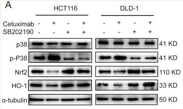
Application: WB    Species: Human    Sample: HCT116 and DLD-1 cells

Fig. 4 Cetuximab activates p38 MAPK and regulates the Nrf2/HO-1 axis. A Western blot analysis of p-p38, total p38, Nrf2 and HO-1 expression levels in HCT116 and DLD-1 cells incubated with cetuximab (100 μg/ml), SB202190 (1 μM) or cetuximab in combination with SB202190 for 24 h. B HCT116 and DLD-1 cells were treated with cetuximab (100 μg/ml) or RSL3 (1 μM) in the absence or presence of SB202190 (1 μM) for 24 h, and cell viability was assessed by the CCK-8 assay. C The protein levels of Nrf2 and HO-1 in HCT116 and DLD-1 cells or Nrf2 overexpressed HCT116 and DLD-1 cells treated with RSL3 (1 μM) combination with cetuximab (100 μg/ml) for 24 h. D HCT116 and DLD-1 cells with overexpression Nrf2 were treated with or without RSL3 (1 μM) combination with cetuximab (100 μg/ml) for 24 h. Cell viability was assessed by CCK-8 assays. E The protein level of HO-1 in HCT116 and DLD-1 cells or HO-1 overexpressed HCT116 and DLD-1 cells treated with RSL3 (1 μM) combination with cetuximab (100 μg/ml) for 24 h. F HCT116 and DLD-1 cells with overexpression HO-1 were treated with or without RSL3 (1 μM) combination with cetuximab (100 μg/ml) for 24 h. Cell viability was assessed by CCK-8 assays. G–H HCT116 and DLD-1 cells were treated with cetuximab (100 μg/ml) or RSL3 (1 μM) with or without t-BHQ (20 μM) or hemin (20 μM) for 24 h, and cell viability was assessed by the CCK-8 assay. **P < 0.01.

3). Protective effect of synbiotic combination of Lactobacillus plantarum SC-5 and olive oil extract tyrosol in a murine model of ulcerative colitis. Journal of translational medicine, 2024 (PubMed: 38528541) [IF=7.4]

4). DADLE promotes motor function recovery by inhibiting cytosolic phospholipase A2 mediated lysosomal membrane permeabilization after spinal cord injury. British journal of pharmacology, 2024 (PubMed: 37766498) [IF=6.8]

5). Candida utilis Ameliorates Dextran Sulfate Sodium-Induced Colitis in Mice via NF-κB/MAPK Suppression and Gut Microbiota Modulation. International journal of molecular sciences, 2025 (PubMed: 40076616) [IF=5.6]

6). Integrated Multi-Omics Analysis Reveals Mountain-Cultivated Ginseng Ameliorates Cold-Stimulated Steroid-Resistant Asthma by Regulating Interactions among Microbiota, Genes, and Metabolites. International journal of molecular sciences, 2024 (PubMed: 39201796) [IF=5.6]

Application: WB    Species: Mouse    Sample: lung tissues

Figure 9. Integrated analysis of potential mechanisms and validation of PI3K-Akt/MAPK signaling pathway in the combination of DEX and MCG for the treatment of CSRA. (A) Correlation network of DEGs and DEMs. (B) Heatmap of correlation between DEGs and flora. (C) Heatmap of correlation between DEMs and flora. (D) ROC curve analysis. (E) Flora–gene–metabolite–pathway network. (F) Representative western blotting images. (G) Densitometric quantification of protein expression. Reference: GAPDH. (ns, non-significant; * p < 0.05; ** p < 0.01; *** p < 0.001).

7). Protective effect of remdesivir against pulmonary fibrosis in mice. Frontiers in Pharmacology, 2021 (PubMed: 34512328) [IF=5.6]

Application: WB    Species: Mice    Sample: lung tissues

FIGURE 6 Remdesivir inhibits TGF-β1-induced activation of Smad and non-Smad signaling pathway in lung fibroblasts (A) Luciferase assays of CAGA-NIH3T3 cells. Cells were pretreated with Remdesivir (0–50 μM) for 30 min and then incubated with TGF-β1 (5 ng ml−1) for 24 h, then analyzed with luciferase assay. SB431542 is a TGF-β1/Smad pathway inhibitor and serves as a positive control (B) NIH-3T3 cells were co-treated with TGF-β1 (5 ng ml−1) and Remdesivir (12.5, 25, 50 μM) for 1 h. P-Smad3 and Smad3 were assessed using western blot. GAPDH was used as the internal control (C) PPF cells were co-treated with TGF-β1 (5 ng ml−1) and Remdesivir (12.5, 25, 50 μM) for 1 h. P-Smad3 and Smad3 were assessed using western blot. GAPDH was used as the internal control (D) NIH-3T3 cells were co-treated with TGF-β1 (5 ng ml−1) and Remdesivir (12.5, 25, 50 μM) for 1 h and the phosphorylation levels of P-38, JNK, ERK and Akt were analyzed by Western blot. β-tubulin was used as a loading control in grayscale analysis. Scale bar = 60 μm. Data was presented as the means ± SD, n = 3. * p < 0.05, ** p < 0.01, *** p < 0.001, **** p < 0.0001.

8). The probiotic Lactobacillus plantarum alleviates colitis by modulating gut microflora to activate PPARγ and inhibit MAPKs/NF-κB. European journal of nutrition, 2024 (PubMed: 39607600) [IF=5.0]

9). ROCK2 increases drug resistance in acute myeloid leukemia via metabolic reprogramming and MAPK/PI3K/AKT signaling. International immunopharmacology, 2024 (PubMed: 39126734) [IF=4.8]

10). Qingyihuaji Formula promotes apoptosis and autophagy through inhibition of MAPK/ERK and PI3K/Akt/mTOR signaling pathway on pancreatic cancer in vivo and in vitro. Journal of Ethnopharmacology, 2023 (PubMed: 36690307) [IF=4.8]

加载更多

限制条款

产品的规格、报价、验证数据请以官网为准,官网链接:www.affbiotech.com | www.affbiotech.cn(简体中文)| www.affbiotech.jp(日本語)

产品的数据信息为Affinity所有,未经授权不得收集Affinity官网数据或资料用于商业用途,对抄袭产品数据的行为我们将保留诉诸法律的权利。

产品相关数据会因产品批次、产品检测情况随时调整,如您已订购该产品,请以订购时随货说明书为准,否则请以官网内容为准,官网内容有改动时恕不另行通知。

Affinity保证所销售产品均经过严格质量检测。如您购买的商品在规定时间内出现问题需要售后时,请您在Affinity官方渠道提交售后申请。

产品仅供科学研究使用。不用于诊断和治疗。 

产品未经授权不得转售。

Affinity Biosciences将不会对在使用我们的产品时可能发生的专利侵权或其他侵权行为负责。Affinity Biosciences, Affinity Biosciences标志和所有其他商标所有权归Affinity Biosciences LTD.