Phospho-IKK alpha/ beta (Ser180/Ser181) Antibody - #AF3013
, using Phospho-IKK alpha/ beta (Ser180/Ser181) Antibody at 1/1000 dilution.
5ug/NC membrane strip.
Exposure for 2min with Affinity™ ECL Kit(#KF8003).
Bands result from membrane strip incubation.")

 on the expression of hepatic (A) XBP1, (B) phospho-IKK alpha/beta (Thr 183+Tyr 185), (C) phospho-JNK1/2/3 (Ser180/181) and (D) phospho-IRS1 (Ser 307).")
transforming growth factor (TGF)-b1, (b) phosphorylated (p)-Smad2/3, (c) Smad2/3, (d) Smad4, (e) Smad7, (f) Toll-like receptor 4 (TLR4), (g) myeloid differentiation factor 88 (MyD88), (h) p-inhibitor of nuclear factor-kappa B (NF-jB) kinase b (p-IKKb), (i) IKKb, NF-jB in (j) cytoplasm and in (k)nucleus.")
 The expression levels of p-p38, p-JNK, p-ERK1/2, p-IKK-a/b, p-p65, and p65 were analyzed by western blotting.")
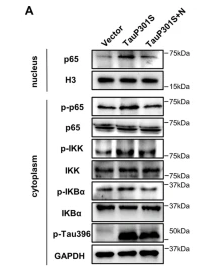
. Cells were administrated by dioscin (15 ng/mL)for 24 h, and then treated with MSU (100 μg/mL) for 12 h.d The protein expressions of TLR4,MyD88, p-IKKβ, IKKβ, p-p65 in cytoplasm, and NF-κB p65 innuclei were measured utilizing western blotting. GAPDH and Histone H3 were acted as internal references, respectively.")
 p-IκB and p-p65 protein levels in HUVECs were determined (n=5).")
 Relative protein levels of NF-κB p65 (cytoplasm), NF-κB p65 (nucleus), p-IKK Ser180/Ser181, IKK, p-IκB-α Ser32/36, and IκB-α in lung tissues of mice in different groups.")
 Relative mRNA levels of S1pr1 in lung tissues of mice in different groups. (b) Relative protein levels of S1PR1 in lung tissues of mice in different groups. (c) Relative protein levels of NF-κB p65 (cytoplasm), NF-κB p65 (nucleus), p-IKK Ser180/Ser181, IKK, p-IκB-α Ser32/36, and IκB-α in lung tissues of mice in different groups. (d) Representative images of dual immunofluorescence staining with S1PR1 and F4/80 in lung tissues of mice in different groups. (e) A luciferase reporter assay was used to measure the relative luciferase activities of S1PR1-3’-UTR wt and mut in RAW264.7 cells. Results were expressed as means ± SD, *P<0.05.")
 qRT-PCR analysis was performed to detect the relative mRNA expression of cathepsin K, TRAP, NFATc1, and MMP-9 in BMSCs. (b) Western blot analysis was performed to detect the relative protein expression of IκBα, p-IκBα, p65, p-p65, IKKα, p-IKKα, NFATc1, and cathepsin K in BMSCs (n = 3); results are represented as the mean ± SD. ▲P < 0.05, compared to the Con group; ▲▲P < 0.01, compared to the Con group; ∗P < 0.05, compared to the Ti group; ∗∗P < 0.01, compared to the Ti group. BMSCs: bone marrow stromal cells; Ti: titanium particles; Con: control.")
 to ACE2 detected via immunofluorescence. Single channel images of S-RBD, ACE2 and DAPI are shown. Moreover, a single overlapped image (the merge image) was merged and generated using the single channel images for S-RBD, ACE2, and DAPI. b: Effect of β-chitosan on S-RBD binding to ACE2 as determined using flow cytometry. Merge 1 was generated by merging the single channel images of S-RBD and ACE2. c: Western blot analysis of inflammation-related proteins. Cells were treated with S-RBD and/or β-chitosan as shown in Table 1. Relative IOD = IOD/β-actin. Scale bars, 50 μm (white).")
 and upregulated (c) KEGG pathway in overexpression zebrafish bmp8a 3T3-L1 cells. b, d After induction of adipogenic differentiation, the downregulated (b) and upregulated (d) KEGG pathway in overexpression mouse Bmp8a 3T3-L1 cells. e, f Immunoblot analysis and quantification of p-IKKα/β and p-p65 in Mock, LV-ZsGreen1, and LV-bmp8a 3T3-L1 cells (n = 3). g, h Immunoblot analysis and quantification of p-IKKα/β and p-p65 in Mock, LV-ZsGreen1, and LV-Bmp8a 3T3-L1cells. Protein expression levels were quantified by ImageJ software and normalized to total protein (n = 3). i Co-immunoprecipitation and immunoblot analysis of co-transfected with PPARγ and p65 (n = 3). j Schematic drawing of predicted PPRE site in Fabp4 promoter region. k Schematic drawing of WT and PPRE site mutation Luc-report plasmids. l, m Quantification of the activity of Fabp4-promoter (l) and Fabp4-promoter-ΔPPRE (m) luciferase reporters in mouse HEK293T cells transfected with Vector, pCMV-Pparγ, or co-transfected pCMV-Pparγ and pCMV-p65, respectively. Renilla luciferase was used as the internal control (n = 3). Data were from three independent experiments and were analyzed by One-way ANOVA and were presented as mean ± SD (ns not significant, **p")
 Representative bands of NF-κB, p-NF-κB, p-IKK-α, p-IKK-β, IκB, p-IκB, TNF and IL-6wereshown.")
 The serum inflammatory cytokines (i.e., TNF-α, IL-1β, IL-6, and IL-10) in serum were measured by ELISA. (E–J) Representative images of phosphorylation levels of P38, Erk1/2, JNK, NF-κB and IKK α/β were examined by western blot and the fold activation data analysis. Data are expressed as mean ± SD (at least n = 6/group), #p < 0.05 (vs. Sham group), *p < 0.05 (vs. CLP group).")

 Effects of TNFR2 knockdown on migratory and invasive capacities using Transwell assays in PANC-1 and ASPC-1 cells. (C) Western blotting determined that NF-κB signaling-related proteins were overexpressed in the PANC-1 and ASPC-1 cells. (D) Western blotting determined that NF-κB signaling-related proteins were downregulated after TNFR silencing in PANC-1 and ASPC-1 cells.")
 Effects of bilirubin on L-O2 cell morphology (100×). (B) CCK-8 assay of bilirubin-treated L-O2 cells. (C) mRNA expression of p65, IκB-α, and IKK-β in L-O2 cells stimulated by bilirubin. (D) Measurement of p65, p-p65, IKK-β, p-IKK-β, IκB-α, and p-IκB-α protein levels in L-O2 cells by Western blotting. (E) Statistical analysis of p-p65, p-IKK-β, and p-IκB-α protein levels in L-O2 cells (normalized to p65, IKK-β, or IκB-α). At least three replicate samples were used for each experiment. Statistical significance is shown as ** p < 0.01, *** p < 0.001, **** p < 0.0001.")
 Effects of bilirubin on L-O2 cell morphology (100×). (B) CCK-8 assay of bilirubin-treated L-O2 cells. (C) mRNA expression of p65, IκB-α, and IKK-β in L-O2 cells stimulated by bilirubin. (D) Measurement of p65, p-p65, IKK-β, p-IKK-β, IκB-α, and p-IκB-α protein levels in L-O2 cells by western blotting (normalized to p65, IKK-β, or IκB-α).")
 complex. (B) Representative images of immunofluorescence trial for p-NF-κB. The expression of NF-κB, p-NF-κB, and p-IKK α/β detected by western blot and the fold activation data analysis (C-E) (n = 3). In the statistical figure, the red line is the mean and the black horizontal line is the quartile. VC = vitamic C; JAK2 = Janus kinase 2; STAT3 = signal transducer and activator of transcription 3; NF-κB = nuclear factor kappa B; SIMI = sepsis-induced myocardial injury; 3D = three-dimensional.")
 of CD40 or STING. B Co-IP using anti-TRAF2 to pull down STING and immunoblot (IB) detection of CD40 and STING molecules. C Multiplex immunofluorescence demonstrated the interactions of CD40 and STING with TRAF2 (scale bar = 100 μm). D Impact of CD40 and STING stimulation in the NF-κB pathway. E Flow cytometry revealed the degrees of B cell differentiation under different stimuli (APC-CD19, PE-pre-BCR).")
. D Changes in protein ratios of iNOS and Arg1 (n = 6 independent experiments). E-H Western blot detects changes in protein levels of anti-inflammatory cytokines IL-4, IL-10, and TGF-β in mouse brain (n = 6 independent experiments). I-K Western blot detects changes in protein levels of anti-inflammatory cytokines IL-1β and IFN-γ in mouse substantia nigra (n = 6 independent experiments). L-R Western blot detected the protein levels of RANK, RANKL, OPG, p-NIK, NIK, p-IKK, IKK, p-NF-κB2, and NF-κB2 protein level changes (n = 6 independent experiments). iNOS is a marker for neurotoxic microglial, Arg1 (arginase-1) is a marker for neuroprotective microglial. Data are mean ± SEM.")

产品描述
*The optimal dilutions should be determined by the end user. For optimal experimental results, antibody reuse is not recommended.
*Tips:
WB: 适用于变性蛋白样本的免疫印迹检测. IHC: 适用于组织样本的石蜡(IHC-p)或冰冻(IHC-f)切片样本的免疫组化/荧光检测. IF/ICC: 适用于细胞样本的荧光检测. ELISA(peptide): 适用于抗原肽的ELISA检测.
引用格式: Affinity Biosciences Cat# AF3013, RRID:AB_2834452.
展开/折叠
chuk; CHUK1; Conserved Helix Loop Helix Ubiquitous Kinase; Conserved helix loop ubiquitous kinase; Conserved helix-loop-helix ubiquitous kinase; I Kappa B Kinase 1; I Kappa B Kinase Alpha; I-kappa-B kinase 1; I-kappa-B kinase alpha; IkappaB kinase; IkB kinase alpha subunit; IkBKA; IKK 1; IKK A; IKK a kinase; IKK-A; IKK-alpha; IKK1; IKKA; IKKA_HUMAN; Inhibitor Of Kappa Light Polypeptide Gene Enhancer In B Cells; Inhibitor Of Nuclear Factor Kappa B Kinase Alpha Subunit; Inhibitor of nuclear factor kappa-B kinase subunit alpha; NFKBIKA; Nuclear Factor Kappa B Inhibitor Kinase Alpha; Nuclear factor NF kappa B inhibitor kinase alpha; Nuclear factor NF-kappa-B inhibitor kinase alpha; Nuclear factor NFkappaB inhibitor kinase alpha; Nuclear Factor Of Kappa Light Chain Gene Enhancer In B Cells Inhibitor; TCF-16; TCF16; Transcription factor 16; I kappa B kinase 2; I kappa B kinase beta; I-kappa-B kinase 2; I-kappa-B-kinase beta; IkBKB; IKK beta; IKK-B; IKK-beta; IKK2; IKKB; IKKB_HUMAN; IMD15; Inhibitor of kappa light polypeptide gene enhancer in B cells, kinase beta; Inhibitor of nuclear factor kappa-B kinase subunit beta; NFKBIKB; Nuclear factor NF-kappa-B inhibitor kinase beta;
抗原和靶标
A synthesized peptide derived from human IKK- alpha/ beta around the phosphorylation site of Ser180/181.
研究领域
· Cellular Processes > Cell growth and death > Apoptosis. (View pathway)
· Environmental Information Processing > Signal transduction > MAPK signaling pathway. (View pathway)
· Environmental Information Processing > Signal transduction > Ras signaling pathway. (View pathway)
· Environmental Information Processing > Signal transduction > NF-kappa B signaling pathway. (View pathway)
· Environmental Information Processing > Signal transduction > FoxO signaling pathway. (View pathway)
· Environmental Information Processing > Signal transduction > mTOR signaling pathway. (View pathway)
· Environmental Information Processing > Signal transduction > PI3K-Akt signaling pathway. (View pathway)
· Environmental Information Processing > Signal transduction > TNF signaling pathway. (View pathway)
· Human Diseases > Drug resistance: Antineoplastic > Antifolate resistance.
· Human Diseases > Endocrine and metabolic diseases > Type II diabetes mellitus.
· Human Diseases > Endocrine and metabolic diseases > Insulin resistance.
· Human Diseases > Endocrine and metabolic diseases > Non-alcoholic fatty liver disease (NAFLD).
· Human Diseases > Infectious diseases: Bacterial > Epithelial cell signaling in Helicobacter pylori infection.
· Human Diseases > Infectious diseases: Bacterial > Shigellosis.
· Human Diseases > Infectious diseases: Parasitic > Chagas disease (American trypanosomiasis).
· Human Diseases > Infectious diseases: Parasitic > Toxoplasmosis.
· Human Diseases > Infectious diseases: Viral > Hepatitis C.
· Human Diseases > Infectious diseases: Viral > Hepatitis B.
· Human Diseases > Infectious diseases: Viral > Measles.
· Human Diseases > Infectious diseases: Viral > Influenza A.
· Human Diseases > Infectious diseases: Viral > Human papillomavirus infection.
· Human Diseases > Infectious diseases: Viral > HTLV-I infection.
· Human Diseases > Infectious diseases: Viral > Herpes simplex infection.
· Human Diseases > Infectious diseases: Viral > Epstein-Barr virus infection.
· Human Diseases > Cancers: Overview > Pathways in cancer. (View pathway)
· Human Diseases > Cancers: Overview > MicroRNAs in cancer.
· Human Diseases > Cancers: Specific types > Pancreatic cancer. (View pathway)
· Human Diseases > Cancers: Specific types > Prostate cancer. (View pathway)
· Human Diseases > Cancers: Specific types > Chronic myeloid leukemia. (View pathway)
· Human Diseases > Cancers: Specific types > Acute myeloid leukemia. (View pathway)
· Human Diseases > Cancers: Specific types > Small cell lung cancer. (View pathway)
· Organismal Systems > Immune system > Chemokine signaling pathway. (View pathway)
· Organismal Systems > Development > Osteoclast differentiation. (View pathway)
· Organismal Systems > Immune system > Toll-like receptor signaling pathway. (View pathway)
· Organismal Systems > Immune system > NOD-like receptor signaling pathway. (View pathway)
· Organismal Systems > Immune system > RIG-I-like receptor signaling pathway. (View pathway)
· Organismal Systems > Immune system > Cytosolic DNA-sensing pathway. (View pathway)
· Organismal Systems > Immune system > IL-17 signaling pathway. (View pathway)
· Organismal Systems > Immune system > Th1 and Th2 cell differentiation. (View pathway)
· Organismal Systems > Immune system > Th17 cell differentiation. (View pathway)
· Organismal Systems > Immune system > T cell receptor signaling pathway. (View pathway)
· Organismal Systems > Immune system > B cell receptor signaling pathway. (View pathway)
· Organismal Systems > Nervous system > Neurotrophin signaling pathway. (View pathway)
· Organismal Systems > Endocrine system > Insulin signaling pathway. (View pathway)
· Organismal Systems > Endocrine system > Adipocytokine signaling pathway.
文献引用

Application: WB Species: Mouse Sample: BMSCs
Application: WB Species: Mouse Sample: HT22 cells

Application: WB Species: Mouse Sample:
Application: WB Species: Mouse Sample: liver tissue
限制条款
产品的规格、报价、验证数据请以官网为准,官网链接:www.affbiotech.com | www.affbiotech.cn(简体中文)| www.affbiotech.jp(日本語)产品的数据信息为Affinity所有,未经授权不得收集Affinity官网数据或资料用于商业用途,对抄袭产品数据的行为我们将保留诉诸法律的权利。
产品相关数据会因产品批次、产品检测情况随时调整,如您已订购该产品,请以订购时随货说明书为准,否则请以官网内容为准,官网内容有改动时恕不另行通知。
Affinity保证所销售产品均经过严格质量检测。如您购买的商品在规定时间内出现问题需要售后时,请您在Affinity官方渠道提交售后申请。产品仅供科学研究使用。不用于诊断和治疗。
产品未经授权不得转售。
Affinity Biosciences将不会对在使用我们的产品时可能发生的专利侵权或其他侵权行为负责。Affinity Biosciences, Affinity Biosciences标志和所有其他商标所有权归Affinity Biosciences LTD.