产品: VEGFC 抗体
货号: DF7011
描述: Rabbit polyclonal antibody to VEGFC
应用: WB IHC
文献验证: WB
反应: Human, Mouse, Rat
预测: Pig, Bovine, Horse, Sheep, Rabbit, Dog, Chicken, Xenopus
蛋白号: P49767
RRID: AB_2838967

浏览相似产品>>

   规格 价格 库存
 50ul RMB¥ 1250 现货
 100ul RMB¥ 2300 现货
 200ul RMB¥ 3000 现货

货期: 当天发货

联系销售

产品描述

来源:
Rabbit
应用:
WB 1:500-1:2000, IHC 1:50-1:200
*The optimal dilutions should be determined by the end user.
*Tips:

WB: 适用于变性蛋白样本的免疫印迹检测. IHC: 适用于组织样本的石蜡(IHC-p)或冰冻(IHC-f)切片样本的免疫组化/荧光检测. IF/ICC: 适用于细胞样本的荧光检测. ELISA(peptide): 适用于抗原肽的ELISA检测.

反应:
Human, Mouse, Rat
克隆:
Polyclonal
特异性:
VEGFC Antibody detects endogenous levels of total VEGFC.
RRID:
AB_2838967
引用格式: Affinity Biosciences Cat# DF7011, RRID:AB_2838967.
偶联:
Unconjugated.
纯化:
The antiserum was purified by peptide affinity chromatography using SulfoLink™ Coupling Resin (Thermo Fisher Scientific).
保存:
Rabbit IgG in phosphate buffered saline , pH 7.4, 150mM NaCl, 0.02% sodium azide and 50% glycerol. Store at -20 °C. Stable for 12 months from date of receipt.
别名:

展开/折叠

Flt 4L; Flt4 ligand; FLT4 ligand DHM; Flt4-L; Flt4L; Vascular endothelial growth factor C; Vascular endothelial growth factor related protein; Vascular endothelial growth factor-related protein; VEGF C; VEGF-C; Vegfc; VEGFC_HUMAN; VRP;

抗原和靶标

免疫原:

A synthesized peptide derived from human VEGFC, corresponding to a region within the internal amino acids.

基因/基因ID:
描述:
The protein encoded by this gene is a member of the platelet-derived growth factor/vascular endothelial growth factor (PDGF/VEGF) family, is active in angiogenesis and endothelial cell growth, and can also affect the permeability of blood vessels. This secreted protein undergoes a complex proteolytic maturation, generating multiple processed forms which bind and activate VEGFR-3 receptors. Only the fully processed form can bind and activate VEGFR-2 receptors. This protein is structurally and functionally similar to vascular endothelial growth factor D.

研究领域

· Cellular Processes > Cellular community - eukaryotes > Focal adhesion.   (View pathway)

· Environmental Information Processing > Signal transduction > MAPK signaling pathway.   (View pathway)

· Environmental Information Processing > Signal transduction > Ras signaling pathway.   (View pathway)

· Environmental Information Processing > Signal transduction > Rap1 signaling pathway.   (View pathway)

· Environmental Information Processing > Signaling molecules and interaction > Cytokine-cytokine receptor interaction.   (View pathway)

· Environmental Information Processing > Signal transduction > PI3K-Akt signaling pathway.   (View pathway)

· Environmental Information Processing > Signal transduction > TNF signaling pathway.   (View pathway)

· Human Diseases > Cancers: Overview > Pathways in cancer.   (View pathway)

· Organismal Systems > Endocrine system > Relaxin signaling pathway.

文献引用

1). Neutrophil extracellular trap-mediated impairment of meningeal lymphatic drainage exacerbates secondary hydrocephalus after intraventricular hemorrhage. Theranostics, 2024 (PubMed: 38505607) [IF=12.4]

Application: WB    Species: Mouse    Sample:

Figure 7. NET degradation improves mLV damage. C57BL/6 mice were i.c.v. injected with or without DNase I after IVH. Twenty-four h post-IVH, the meninges were collected for observation. (A) Representative WB images showing the expression of CitH3, VEGFC, LYVE-1, PROX1, FOXC2, and VE-cadherin in the meninges in the Sham, IVH, and IVH+DNase I groups 24 h after IVH. GAPDH was used as an internal reference protein. (B-G) Quantification of CitH3, VEGFC, LYVE-1, PROX1, FOXC2, and VE-cadherin expression (n = 6, one-way ANOVA). (H, I) Relative mRNA levels of FOXC2 and VEGFC in the meninges in the various experimental groups (n = 6). (J, K) The concentrations of VEGFC and FOXC2 in the meninges were quantified by ELISA (n = 6, one-way ANOVA). (L) NO concentrations in the meninges were quantifid in the different groups 24 h after IVH (n = 6, one-way ANOVA). (M) A representative image showing the IF staining of the meninges with anti-LYVE-1 antibodies (red) in the COS. (N) The extent of anti-LYVE-1 antibody staining in the meninges of the COS was quantified and is presented as a percentage of the total area (n = 6, one-way ANOVA). (O) Images showing TUNEL and LYVE-1 staining in the TS of Sham-, IVH-, or DNase I-treated mice were selected as representative examples 24 h after IVH. (P) The number of TUNEL-positive LECs surrounding the TS in the meninges of mice (n = 6, one-way ANOVA). *P < 0.05, **p < 0.01. Data are means ± SEMs.

2). Black phosphorus quantum dot-modified ADSCs as a novel therapeutic for periodontitis bone loss coupling of osteogenesis and osteoimmunomodulation. Materials today. Bio, 2024 (PubMed: 38975241) [IF=8.7]

3). TRAF6 regulates the signaling pathway influencing colorectal cancer function through ubiquitination mechanisms. Cancer Science, 2022 (PubMed: 35179811) [IF=4.5]

Application: WB    Species: human    Sample: SW480 and HCT116 cells

FIGURE 3|Effect of TRAF6 expression on NF-κB, pNF-κB, and VEGF-C expression in SW480 and HCT116 cells. (A, B) Western blotting analysis of NF-κB, pNF-κB, and VEGF-C expression in SW480 and HCT116 cells transfected with pCDH and pCDH-TRAF6, respectively.GAPDH served as a loading control.

4). Early pregnancy human decidua gamma/delta T cells exhibit tissue resident and specific functional characteristics. Molecular Human Reproduction, 2022 (PubMed: 35758607) [IF=3.6]

Application: IF/ICC    Species: Human    Sample: γδ T+ Cells

Figure 5.Identification of growth-factor secretion in human γδ T+ Cells. (A) qRT-PCR results from eight selected genes using RNA-sequencing. (B) Immunofluorescence analysis of growth-factor expression in sorted γδ T cells from early decidua (n = 5). (C) Immunofluorescence analysis of growth-factor expression in sorted γδ T cells from term decidua (n = 5). (D) Immunofluorescence analysis of growth-factor expression in sorted γδ T cells from peripheral blood mononuclear cells (PBMCs) (n = 5). (E) Immunofluorescence analysis of growth-factor expression in sorted γδ T cells from recurrent spontaneous abortion (RSA) patients (n = 5; scale bar, 50 μm). (F) qRT-PCR results regarding growth factors in sorted γδ T cells from RSA patients (n = 5). P-value was calculated by unpaired Student’s t-test.

5). Compared with High-intensity Interval Exercise, Moderate Intensity Constant Load Exercise is more effective in curbing the Growth and Metastasis of Lung Cancer. Journal of Cancer, 2022 (PubMed: 35371324) [IF=3.3]

Application: WB    Species: mouse    Sample: lung cancer

Figure 4. | The effect of exercise on lung cancer tissue angiogenesis and lymphangiogenesis.E, F. The effect of exercise on the expression of VEGFA in lung cancer tissues (n=6). E, G. The effect of exercise on the expression of VEGFC in lung cancer tissues (n=6). Note: &, && respectively represent a significant difference compared with the Saline group (P<0.05),(P<0.01).

6). REC8 promotes tumor migration, invasion and angiogenesis by targeting the PKA pathway in hepatocellular carcinoma. CLINICAL AND EXPERIMENTAL MEDICINE, 2021 (PubMed: 33677646) [IF=3.2]

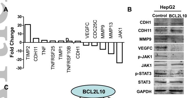
7). BCL2L10 inhibits growth and metastasis of hepatocellular carcinoma both in vitro and in vivo. MOLECULAR CARCINOGENESIS, 2017 (PubMed: 27770580) [IF=3.0]

Application: WB    Species: human    Sample: HepG2

Figure 6. Effect of BCL2L10 on its downstream gene expression profiles of human cancer pathway in HepG2 cells. (A) By human cancer pathway PCR array, ectopic expression of BCL2L10 up- or down-regulated several genes related to tumor proliferation, apoptosis, metastasis and angiogenesis. (B) Western blot was performed to confirm the downstream gene expression regulated by BCL2L10 in HepG2 cells. GAPDH was used as an internal control. (C) Schematic diagram of the molecular events for BCL2L10 function as a tumor suppressor through regulating cell cycle, proliferation, apoptosis metastasis and angiogenesis effectors.

8). The MIR181A2HG/miR-5680/VCAN-CD44 Axis Regulates Gastric Cancer Lymph Node Metastasis by Promoting M2 Macrophage Polarization. Cancer medicine, 2025 (PubMed: 39823128) [IF=2.9]

限制条款

产品的规格、报价、验证数据请以官网为准,官网链接:www.affbiotech.com | www.affbiotech.cn(简体中文)| www.affbiotech.jp(日本語)

产品的数据信息为Affinity所有,未经授权不得收集Affinity官网数据或资料用于商业用途,对抄袭产品数据的行为我们将保留诉诸法律的权利。

产品相关数据会因产品批次、产品检测情况随时调整,如您已订购该产品,请以订购时随货说明书为准,否则请以官网内容为准,官网内容有改动时恕不另行通知。

Affinity保证所销售产品均经过严格质量检测。如您购买的商品在规定时间内出现问题需要售后时,请您在Affinity官方渠道提交售后申请。

产品仅供科学研究使用。不用于诊断和治疗。 

产品未经授权不得转售。

Affinity Biosciences将不会对在使用我们的产品时可能发生的专利侵权或其他侵权行为负责。Affinity Biosciences, Affinity Biosciences标志和所有其他商标所有权归Affinity Biosciences LTD.