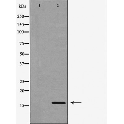
产品: Cystatin C 抗体
货号: DF6457
描述: Rabbit polyclonal antibody to Cystatin C
应用: WB IHC IF/ICC
文献验证: WB
反应: Human, Mouse, Rat, Monkey
蛋白号: P01034
RRID: AB_2838419

浏览相似产品>>

   规格 价格 库存
 50ul RMB¥ 1250 现货
 100ul RMB¥ 2300 现货
 200ul RMB¥ 3000 现货

货期: 当天发货

联系销售

产品描述

来源:
Rabbit
应用:
WB 1:500-1:2000, IHC 1:50-1:200, IF/ICC 1:100-1:500
*The optimal dilutions should be determined by the end user.
*Tips:

WB: 适用于变性蛋白样本的免疫印迹检测. IHC: 适用于组织样本的石蜡(IHC-p)或冰冻(IHC-f)切片样本的免疫组化/荧光检测. IF/ICC: 适用于细胞样本的荧光检测. ELISA(peptide): 适用于抗原肽的ELISA检测.

反应:
Human, Mouse, Rat, Monkey
克隆:
Polyclonal
特异性:
Cystatin C Antibody detects endogenous levels of total Cystatin C.
RRID:
AB_2838419
引用格式: Affinity Biosciences Cat# DF6457, RRID:AB_2838419.
偶联:
Unconjugated.
纯化:
The antiserum was purified by peptide affinity chromatography using SulfoLink™ Coupling Resin (Thermo Fisher Scientific).
保存:
Rabbit IgG in phosphate buffered saline , pH 7.4, 150mM NaCl, 0.02% sodium azide and 50% glycerol. Store at -20 °C. Stable for 12 months from date of receipt.
别名:

展开/折叠

AD 8; AD8; Amyloid angiopathy and cerebral hemorrhage; ARMD11; bA218C14.4 (cystatin C); bA218C14.4; Cst 3; Cst3; CST3 protein; Cystatin 3; Cystatin-3; Cystatin-C; Cystatin3; CystatinC; CYTC_HUMAN; Epididymis secretory protein Li 2; Gamma trace; Gamma-trace; HCCAA; HEL S 2; MGC117328; Neuroendocrine basic polypeptide; Post gamma globulin; Post-gamma-globulin;

抗原和靶标

免疫原:

A synthesized peptide derived from human Cystatin C, corresponding to a region within the internal amino acids.

基因/基因ID:
描述:
Cystatin C is a 14 kDa member of the Cystatin superfamily of cysteine protease inhibitors (1). Most cell types secrete Cystatin C (1). Cystatin C inhibits cathepsins, and thereby may function as a tumor suppressor by inhibiting cathepsin mediated tumor cell invasion (2). In addition, this tumor suppressor function can also be attributed to Cystatin C's ability to antagonize TGF-β1 signaling (2,3). Cystatin C may also modulate antigen presentation through its ability to inhibit cathepsins (4). Cystatin C is also a biomarker for kidney dysfunction (5).

研究领域

· Organismal Systems > Digestive system > Salivary secretion.

文献引用

1). Neutrophil extracellular traps drive intestinal microvascular endothelial ferroptosis by impairing Fundc1-dependent mitophagy. Redox biology, 2023 (PubMed: 37812880) [IF=10.7]

Application: WB    Species: Mouse    Sample:

Fig. 7. Effects of Fundc1 regulation on NET formation and endothelial mitochondrial quality control. A. Intestinal CitH3-DNA and MPO-DNA complexes were assayed using ELISAs. B–C. Protein expression levels of Ly6G and CitH3 were measured by Western blot analysis. D-E. Intestinal CD31-positive endothelial cells were isolated with CD31-coated magnetic beads. Mitochondrial (MitoTracker) and lysosomal (LysoTracker) immunofluorescence colocalization was performed to explore the mitophagy level. F-G. Endothelial mitophagy parameters (p62, LC3 II/I ratio and mito-LC3 II) and a mitochondrial protein (Tim23) were examined using western blotting. The relative grayscale values were measured using ImageJ. H–I. Cytc levels in the cytosol and isolated mitochondria were examined by western blotting. J-K. Quantification of cytoplasmic and mitochondrial ROS in the intestinal endothelium was conducted using DCFH‐DA and MitoSOX Red probes. L. Mitochondrial membrane potential changes in endothelial cells were detected by JC-1 staining. M-N. Key proteins of mitochondrial fusion (Mfn1 and Opa1) and fission (Mff and Drp1) were examined by a Western blot assay. O–P. Immunofluorescence confocal imaging of mitochondrial (Tom20, green) and Drp1 (red) colocalization was used to evaluate mitochondrial fission. AAV, adeno-associated virus; NC, negative control. Data are shown as the means ± SD, ns, not significant, *P < 0.05, **P < 0.01, ***P < 0.001.

2). Neutrophil extracellular traps aggravate intestinal epithelial necroptosis in ischaemia-reperfusion by regulating TLR4/RIPK3/FUNDC1-required mitophagy. Cell proliferation, 2024 (PubMed: 37691112) [IF=5.9]

Application: WB    Species: Mouse    Sample:

FIGURE 4 Increased NET levels inhibited mitophagy activation and aggravated Cytc leakage in the intestine. (A,B). Western blotting experiments to assess mitophagy flux (p62, LC3 II/I ratio and Mito-LC3 II), mitochondrial protein (TIM23) levels in human intestinal tissues. (C,D) The colocalization of LC3 and mitochondria (Tom20) in the intestine in WT and Pad4ΔPMN mice. Colocalization events of mitochondria and LC3 puncta are indicated in yellow. Scale bars = 100 μm. (E,F). Qualitative analysis of the number of mitophagosomes in intestinal epithelial cells via TEM. Scale bars = 5 μm. (G,H) Western blots were used to analyse the mitophagy parameters (p62, LC3 II/I ratio and Mito-LC3 II), mitochondrial biomarkers (Tim23) in animals. (I,J) The Cytc contents in the cytosol and in isolated mitochondria were examined by western blotting. Data are displayed as the mean values with SD. ns, no significance. *p 

3). Melatonin protects against sarcopenia in middle-aged mice. Histology and histopathology, 2024 (PubMed: 39385610) [IF=2.5]

Application: WB    Species: Mouse    Sample: GA tissues

Figure 4. The effects of MEL on the PGC-1α/TFAM pathway in GA tissues of middle-aged mice a-c. Western blot and qRT-PCR were applied to detect the effects of MEL on PGC-1α/TFAM pathwayrelated protein and mRNA levels, including cytochrome c oxidase subunit 4 (COX4), cystatin C (CYTC), nuclear respiratory factor 1 (NRF-1), mitochondrial transcription factor A (TFAM), p-P38, P38, and peroxisome proliferator-activated receptor γ coactivator-1α (PGC-1α), n=3. Data are manifested as mean ± SD * P

限制条款

产品的规格、报价、验证数据请以官网为准,官网链接:www.affbiotech.com | www.affbiotech.cn(简体中文)| www.affbiotech.jp(日本語)

产品的数据信息为Affinity所有,未经授权不得收集Affinity官网数据或资料用于商业用途,对抄袭产品数据的行为我们将保留诉诸法律的权利。

产品相关数据会因产品批次、产品检测情况随时调整,如您已订购该产品,请以订购时随货说明书为准,否则请以官网内容为准,官网内容有改动时恕不另行通知。

Affinity保证所销售产品均经过严格质量检测。如您购买的商品在规定时间内出现问题需要售后时,请您在Affinity官方渠道提交售后申请。

产品仅供科学研究使用。不用于诊断和治疗。 

产品未经授权不得转售。

Affinity Biosciences将不会对在使用我们的产品时可能发生的专利侵权或其他侵权行为负责。Affinity Biosciences, Affinity Biosciences标志和所有其他商标所有权归Affinity Biosciences LTD.