产品: Bim 抗体
货号: DF6093
描述: Rabbit polyclonal antibody to Bim
应用: WB
文献验证: WB
反应: Human, Mouse, Rat
预测: Pig, Horse, Sheep, Rabbit, Dog
蛋白号: O43521
RRID: AB_2838061

浏览相似产品>>

   规格 价格 库存
 50ul RMB¥ 1250 现货
 100ul RMB¥ 2300 现货
 200ul RMB¥ 3000 现货

货期: 当天发货

联系销售

产品描述

来源:
Rabbit
应用:
WB 1:500-1:2000
*The optimal dilutions should be determined by the end user.
*Tips:

WB: 适用于变性蛋白样本的免疫印迹检测. IHC: 适用于组织样本的石蜡(IHC-p)或冰冻(IHC-f)切片样本的免疫组化/荧光检测. IF/ICC: 适用于细胞样本的荧光检测. ELISA(peptide): 适用于抗原肽的ELISA检测.

反应:
Human, Mouse, Rat
克隆:
Polyclonal
特异性:
Bim Antibody detects endogenous levels of total Bim.
RRID:
AB_2838061
引用格式: Affinity Biosciences Cat# DF6093, RRID:AB_2838061.
偶联:
Unconjugated.
纯化:
The antiserum was purified by peptide affinity chromatography using SulfoLink™ Coupling Resin (Thermo Fisher Scientific).
保存:
Rabbit IgG in phosphate buffered saline , pH 7.4, 150mM NaCl, 0.02% sodium azide and 50% glycerol. Store at -20 °C. Stable for 12 months from date of receipt.
别名:

展开/折叠

BCL2 like 11; B2L11_HUMAN; BAM; Bcl 2 interacting protein Bim; Bcl 2 related ovarian death agonist; Bcl-2-like protein 11; BCL2 interacting mediator of cell death; BCL2 like 11 (apoptosis facilitator); BCL2 like protein 11; Bcl2-interacting mediator of cell death; Bcl2-L-11; Bcl2l11; BIM alpha6; BIM; BIM beta6; BIM beta7; BimEL; BimL; BOD;

抗原和靶标

免疫原:

A synthesized peptide derived from human Bim, corresponding to a region within the internal amino acids.

基因/基因ID:
描述:
Bim/Bod is a pro-apoptotic protein belonging to the BH3-only group of Bcl-2 family members including Bad, Bid, Bik, Hrk and Noxa that contain a BH3 domain but lack other conserved BH1 or BH2 domains (1,2). Bim induces apoptosis by binding to and antagonizing anti-apoptotic members of the Bcl-2 family. Interactions have been observed with Bcl-2, Bcl-xL, Mcl-1, Bcl-w, Bfl-1 and BHRF-1 (1,2). Bim functions in regulating apoptosis associated with thymocyte negative selection and following growth factor withdrawal, during which Bim expression is elevated (3-6). Three major isoforms of Bim are generated by alternative splicing: BimEL, BimL and BimS (1). The shortest form, BimS, is the most cytotoxic and is generally only transiently expressed during apoptosis. The BimEL and BimL isoforms may be sequestered to the dynein motor complex through an interaction with the dynein light chain and released from this complex during apoptosis (7). Apoptotic activity of these longer isoforms may be regulated by phosphorylation (8,9). Environmental stress triggers Bim phosphorylation by JNK and results in its dissociation from the dynein complex and increased apoptotic activity.

研究领域

· Cellular Processes > Cell growth and death > Apoptosis.   (View pathway)

· Cellular Processes > Cell growth and death > Apoptosis - multiple species.   (View pathway)

· Environmental Information Processing > Signal transduction > FoxO signaling pathway.   (View pathway)

· Environmental Information Processing > Signal transduction > PI3K-Akt signaling pathway.   (View pathway)

· Human Diseases > Drug resistance: Antineoplastic > EGFR tyrosine kinase inhibitor resistance.

· Human Diseases > Endocrine and metabolic diseases > Non-alcoholic fatty liver disease (NAFLD).

· Human Diseases > Cancers: Overview > Pathways in cancer.   (View pathway)

· Human Diseases > Cancers: Overview > MicroRNAs in cancer.

· Human Diseases > Cancers: Specific types > Colorectal cancer.   (View pathway)

文献引用

1). Overexpression of CD59 inhibits apoptosis of T-acute lymphoblastic leukemia via AKT/Notch1 signaling pathway. Cancer Cell International, 2019 (PubMed: 30636930) [IF=5.8]

Application: WB    Species: human    Sample: Jurkat cells

Fig.?4?|The activation of AKT, STAT5 and Notch1 signaling pathway are involve in the regulation of apoptosis by CD59 in Jurkat cells. The expression of CD59 (a), apoptosis-related proteins (c) and key proteins in signaling pathways (e) detected by western blot. Quantitative analysis of the protein expression of CD59 (b), apoptosis-related proteins (d) and key proteins in signaling pathways (f, g). All experiments were performed three times. *P?

2). Derlin1 functions as an oncogene in cervical cancer via AKT/mTOR signaling pathway. Biological Research, 2020 (PubMed: 30808417) [IF=4.3]

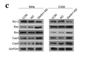
Application: WB    Species: human    Sample: CC cells

Fig.?4|Knockdown of Derlin1 induces the apoptosis of CC cells. a Apoptosis was detected by fow cytometry. b Quantitative results of apoptotic cell percentage. c The proteins expression of apoptosis-related proteins were detected by western blot. d Quantitative results of protein expression levels. N?=?3, *p?

3). Suppression of CHOP Reduces Neuronal Apoptosis and Rescues Cognitive Impairment Induced by Intermittent Hypoxia by Inhibiting Bax and Bak Activation. Neural Plasticity, 2021 (PubMed: 34471408) [IF=3.0]

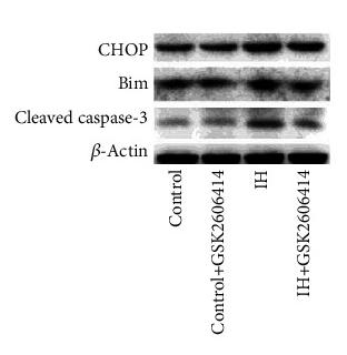
Application: WB    Species: Mouse    Sample: hippocampal tissues

Figure 3 GSK2606414 reduced the activation of caspase-3 via mitochondria-dependent apoptosis. (a) Representative western blots showed that GSK2606414 reduced the expression of CHOP, Bim, and cleaved caspase-3 in hippocampal tissue. The pooled data from three mice for each group are summarized in (b). (c) The western blot results showed that the expression of Bax and Bak in the mitochondria was obviously increased and that this effect was accompanied by a reduction in cytochrome c. These effects were prevented by the administration of GSK2606414. (d) Statistical data from three animals for each group are summarized. (e) Representative western blots showing that the cytoplasmic expression of Bax and Bak was obviously reduced and that of cytochrome C was significantly increased. GSK2606414 increased the expression of Bax and Bak and reduced cytochrome C. (f) Statistical data from three animals for each group are summarized. (g) The morphology of mitochondria in the hippocampal CA1 region in the (i) control group, (ii) GSK2606414 group, (iii) IH group, and (iv) IH + GSK2606414 group. In the control and GSK2606414 groups, intact mitochondria (indicated by arrowheads) with clear cristae were found. Much fewer cristae were found in the mitochondria from the IH group. GSK2606414 treatment preserved more intact mitochondria with discernable cristae. Scale bar: 0.25 μm. (h) The mitochondrial membrane potential (JC-1 fluorescence intensity ratio) was impaired by IH treatment and rescued by GSK2606414 treatment. ∗P < 0.05; ∗∗P < 0.01; ns: not significant.

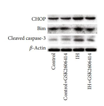
Application: WB    Species: mouse    Sample: hippocampal

Figure 3:| GSK2606414 reduced the activation of caspase-3 via mitochondria-dependent apoptosis. (a) Representative western blots showed that GSK2606414 reduced the expression of CHOP, Bim, and cleaved caspase-3 in hippocampal tissue.

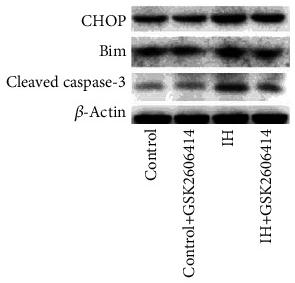
Application: WB    Species: Mice    Sample: hippocampal tissue

Figure 3 GSK2606414 reduced the activation of caspase-3 via mitochondria-dependent apoptosis. (a) Representative western blots showed that GSK2606414 reduced the expression of CHOP, Bim, and cleaved caspase-3 in hippocampal tissue. The pooled data from three mice for each group are summarized in (b). (c) The western blot results showed that the expression of Bax and Bak in the mitochondria was obviously increased and that this effect was accompanied by a reduction in cytochrome c. These effects were prevented by the administration of GSK2606414. (d) Statistical data from three animals for each group are summarized. (e) Representative western blots showing that the cytoplasmic expression of Bax and Bak was obviously reduced and that of cytochrome C was significantly increased. GSK2606414 increased the expression of Bax and Bak and reduced cytochrome C. (f) Statistical data from three animals for each group are summarized. (g) The morphology of mitochondria in the hippocampal CA1 region in the (i) control group, (ii) GSK2606414 group, (iii) IH group, and (iv) IH + GSK2606414 group. In the control and GSK2606414 groups, intact mitochondria (indicated by arrowheads) with clear cristae were found. Much fewer cristae were found in the mitochondria from the IH group. GSK2606414 treatment preserved more intact mitochondria with discernable cristae. Scale bar: 0.25 μm. (h) The mitochondrial membrane potential (JC-1 fluorescence intensity ratio) was impaired by IH treatment and rescued by GSK2606414 treatment. ∗P < 0.05; ∗∗P < 0.01; ns: not significant.

4). Quxie Capsule Inhibits Colon Tumor Growth Partially Through Foxo1-Mediated Apoptosis and Immune Modulation. INTEGRATIVE CANCER THERAPIES, 2019 (PubMed: 31030593) [IF=2.9]

Application: WB    Species: mouse    Sample: tumor

Figure 2.| Quxie capsule (QX) inhibited cell proliferation and induced apoptosis in tumor tissues. (A) Ki-67 staining of tumor sections obtained from mice treated with (a) vehicle control or (b) QX. Quantification of Ki-67-positive cells in the tumor sections (c). (B)TUNEL staining of tumor sections obtained from mice treated with (a) vehicle control or (b) QX. Quantification of TUNEL-positive cells in the tumor sections (c). (C) Western blotting of proapoptotic proteins Bim, FasL, and cleaved caspase-3 expression in tumor tissues of QX-treated mice or vehicle control-treated mice. Data are presented as mean ± SD. *P < .05, **P < .01 versus vehicle control.

限制条款

产品的规格、报价、验证数据请以官网为准,官网链接:www.affbiotech.com | www.affbiotech.cn(简体中文)| www.affbiotech.jp(日本語)

产品的数据信息为Affinity所有,未经授权不得收集Affinity官网数据或资料用于商业用途,对抄袭产品数据的行为我们将保留诉诸法律的权利。

产品相关数据会因产品批次、产品检测情况随时调整,如您已订购该产品,请以订购时随货说明书为准,否则请以官网内容为准,官网内容有改动时恕不另行通知。

Affinity保证所销售产品均经过严格质量检测。如您购买的商品在规定时间内出现问题需要售后时,请您在Affinity官方渠道提交售后申请。

产品仅供科学研究使用。不用于诊断和治疗。 

产品未经授权不得转售。

Affinity Biosciences将不会对在使用我们的产品时可能发生的专利侵权或其他侵权行为负责。Affinity Biosciences, Affinity Biosciences标志和所有其他商标所有权归Affinity Biosciences LTD.