产品: Cytokeratin 14 抗体
货号: AF5370
描述: Rabbit polyclonal antibody to Cytokeratin 14
应用: WB IHC IF/ICC
文献验证: IF/ICC
反应: Human, Mouse, Rat
预测: Rabbit, Dog
蛋白号: P02533
RRID: AB_2837855

浏览相似产品>>

   规格 价格 库存
 50ul RMB¥ 1250 现货
 100ul RMB¥ 2300 现货
 200ul RMB¥ 3000 现货

货期: 当天发货

联系销售

产品描述

来源:
Rabbit
应用:
WB 1:500-1:2000, IHC 1:50-1:200, IF/ICC 1:100-1:500
*The optimal dilutions should be determined by the end user.
*Tips:

WB: 适用于变性蛋白样本的免疫印迹检测. IHC: 适用于组织样本的石蜡(IHC-p)或冰冻(IHC-f)切片样本的免疫组化/荧光检测. IF/ICC: 适用于细胞样本的荧光检测. ELISA(peptide): 适用于抗原肽的ELISA检测.

反应:
Human, Mouse, Rat
克隆:
Polyclonal
特异性:
Cytokeratin 14 Antibody detects endogenous levels of total Cytokeratin 14.
RRID:
AB_2837855
引用格式: Affinity Biosciences Cat# AF5370, RRID:AB_2837855.
偶联:
Unconjugated.
纯化:
The antiserum was purified by peptide affinity chromatography using SulfoLink™ Coupling Resin (Thermo Fisher Scientific).
保存:
Rabbit IgG in phosphate buffered saline , pH 7.4, 150mM NaCl, 0.02% sodium azide and 50% glycerol. Store at -20 °C. Stable for 12 months from date of receipt.
别名:

展开/折叠

CK 14; CK-14; ck14; Cytokeratin 14; Cytokeratin-14; Cytokeratin14; Dowling Meara; EBS3; EBS4; Epidermolysis bullosa simplex; K14; K1C14_HUMAN; Keratin 14 (epidermolysis bullosa simplex, Dowling-Meara, Koebner); Keratin 14; Keratin; Keratin type I cytoskeletal 14; Keratin, type I cytoskeletal 14; Keratin-14; Keratin14; Koebner; Krt 14; Krt14; NFJ; OTTHUMP00000164624; type I cytoskeletal 14;

抗原和靶标

免疫原:

A synthesized peptide derived from human Cytokeratin 14, corresponding to a region within N-terminal amino acids.

基因/基因ID:
描述:
The nonhelical tail domain is involved in promoting KRT5-KRT14 filaments to self-organize into large bundles and enhances the mechanical properties involved in resilience of keratin intermediate filaments in vitro.

研究领域

· Organismal Systems > Endocrine system > Estrogen signaling pathway.   (View pathway)

文献引用

1). Small extracellular vesicles derived from hypoxic preconditioned dental pulp stem cells ameliorate inflammatory osteolysis by modulating macrophage polarization and osteoclastogenesis. Bioactive Materials, 2023 (PubMed: 36311048) [IF=18.9]

2). Three-dimensional bioprinted cell-adaptive hydrogel with anisotropic micropores for enhancing skin wound healing. International journal of biological macromolecules, 2024 (PubMed: 39343255) [IF=7.7]

3). Dental follicle stem cells rescue the regenerative capacity of inflamed rat dental pulp through a paracrine pathway. Stem Cell Research & Therapy, 2020 (PubMed: 32746910) [IF=7.5]

Application: IF/ICC    Species: mouse    Sample: rDFSCs

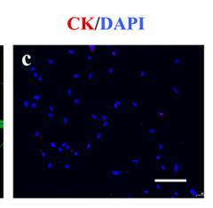
Fig. 1 Isolation and identification of rDFSCs and rDPCs. Representative images of rDFSCs (a) and rDPCs (f). rDFSCs and rDPCs were both positive for vimentin (b, g) and negative for cytokeratin (c, h). After osteogenic induction for 14 days, mineralized nodules were detected in rDFSCs (d) and rDPCs (i) by Alizarin Red staining. After adipogenic induction for 14 days, lipid droplets were detected in rDFSCs (e) and rDPCs (j) by Oil Red O staining. Scale bar 100 μm. Representative flow cytometric results showed the expression pattern of CD markers in P3 rDFSCs (k–q) and rDPCs (r–x)

4). Hypoxia‑induced mitophagy regulates proliferation, migration and odontoblastic differentiation of human dental pulp cells through FUN14 domain‑containing 1. International Journal of Molecular Medicine, 2022 (PubMed: 35362539) [IF=5.7]

Application: IF/ICC    Species: Human    Sample: HDPCs

Figure 2 Identification of HDPCs. (A) Representative images of HDPCs at passage 3. HDPCs are (B) positive for vimentin and (C) negative for cytokeratin. (D) Representative image of mineralized nodules in HDPCs after osteogenic induction for 28 days following Alizarin Red staining. (E) Representative image of chondroblasts in HDPCs after chondrogenic induction for 21 days following Alcian Blue staining. (F) Representative image of lipid droplets in HDPCs after adipogenic induction for 28 days following Oil Red O staining. Scale bars, 50 µm. (G) Representative flow cytometry histograms showing the expression profile of markers in the p3 HDPCs. HDPCs, human dental pulp cells; VIM, vimentin; CK, cytokeratin.

5). Autophagy inhibition facilitates wound closure partially dependent on the YAP/IL-33 signaling in a mouse model of skin wound healing. FASEB journal : official publication of the Federation of American Societies for Experimental Biology, 2021 (PubMed: 34547141) [IF=4.4]

6). Extended characterization of IL-33/ST2 as a predictor for wound age determination in skin wound tissue samples of humans and mice. International journal of legal medicine, 2023 (PubMed: 37246991) [IF=2.2]

限制条款

产品的规格、报价、验证数据请以官网为准,官网链接:www.affbiotech.com | www.affbiotech.cn(简体中文)| www.affbiotech.jp(日本語)

产品的数据信息为Affinity所有,未经授权不得收集Affinity官网数据或资料用于商业用途,对抄袭产品数据的行为我们将保留诉诸法律的权利。

产品相关数据会因产品批次、产品检测情况随时调整,如您已订购该产品,请以订购时随货说明书为准,否则请以官网内容为准,官网内容有改动时恕不另行通知。

Affinity保证所销售产品均经过严格质量检测。如您购买的商品在规定时间内出现问题需要售后时,请您在Affinity官方渠道提交售后申请。

产品仅供科学研究使用。不用于诊断和治疗。 

产品未经授权不得转售。

Affinity Biosciences将不会对在使用我们的产品时可能发生的专利侵权或其他侵权行为负责。Affinity Biosciences, Affinity Biosciences标志和所有其他商标所有权归Affinity Biosciences LTD.