


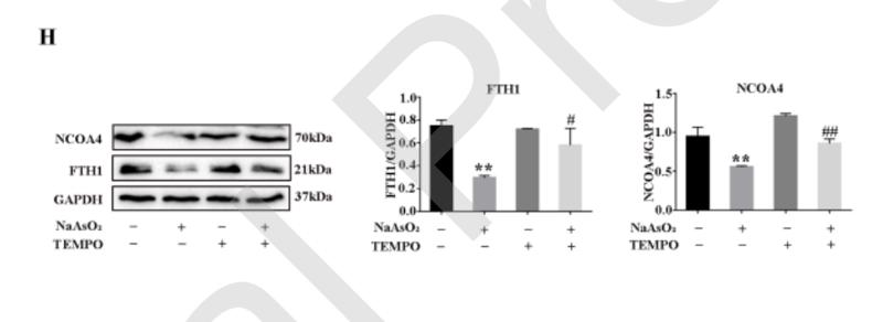
. (A) The Fe2þ content was measured in
the brain. (B) Western blot analysis for ferroptosis-related protein expression, such as that of TFR1, NCOA4 and FTH1. (C) Western blot analysis for Nrf2, nuclear Nrf2 and GPX4. Data
are expressed as the mean ± SD. &P < 0.05 and &&P < 0.01 vs. SAMR1 group; *P < 0.05 and **P < 0.01 vs. SAMP8 group.")
 (n = 3, scale bar: 25 μm). The expression quantity of TFR was assessed by immunofluorescence staining of HOS cells after the indicated treatment (b, e) (n = 3, scale bar: 50 μm). The expression of iron metabolism-related proteins in UA- and/or Cis-treated HOS cells was determined by western blotting (c) (n = 3). The data are presented as the mean ± SD. ∗p < 0.05 vs. the control group.")
. Macroscopic view of xenograft tumours at the endpoint of the experiment (b) (n = 3). The average tumour volume and weight and body weight of the different groups (c–e) (n = 3). H&E staining and immunofluorescence staining of autophagy-, ferroptosis-, and iron metabolism-related proteins in xenograft tissue sections (f) (n = 3, scale bar: 50 μm). The data are presented as the mean ± SD. ∗p < 0.05 vs. the control group.")
 Confocal images of ferritin (red) and LAMP2 (green) were shown in LPS-treated MLE-12 cells, while lysosomes were stained with LAMP2 (green). (B) Quantification measurement of lysosome fluorescence intensity. (C) Quantification measurement of ferritin fluorescence intensity. (D–G) Data showed the expression contents of LC3II/LC3I (E), NCOA4 (F), and FTH1 (G). Data are expressed as mean ± SD. n = 3; *p < 0.05, **p < 0.01, ***p < 0.001. NS: no significance, P>0.05.")
 CCK8 assay result of B-3 cells when treated with UVB+DMSO, UVB+MT, UVB+Z-VAD-FMK and UVB+Z-VAD-FMK+MT compared to UVB+DMSO group. (b-c) Total iron concentration and MDA level in HLE B-3 cells when treated with DMSO, MT, UVB+DMSO and UVB+MT compared to UVB+DMSO group. Three independent experiments. (d) The relative mRNA levels of SIRT6 and Nrf2 in HLE B-3 cells treated with increasing energy of UVB compared to control group. Three independent experiments. (e-f) The western blot analysis and relative protein levels of SIRT6 and p-Nrf2(S40) in HLE B-3 cells treated with increasing energy of UVB compared to control group. Three independent experiments. (g-h) The western blot analysis and relative protein levels of SIRT6, p-Nrf2(S40), GPX4, FTH1 and NCOA4 in B-3 cells treated with DMSO, MT, UVB+DMSO and UVB+MT compared to DMSO group. Three independent experiments. (i) IF staining and relative fluorescence intensity of SIRT6 in B-3 cells when treated to DMSO, UVB+DMSO and UVB+MT compared to DMSO group. Scale bar, 40 µm. Three independent experiments.")
 Western blot analysis of ferroptosis-related protein expression levels in HG-induced 661W cells. GAPDH was used as a control. (B) Expression of GPX4 and SLC7A11 proteins was significantly downregulated in HG-stimulated 661W cells after 12, 18, and 24 h. HG induced obvious upregulation in the expression of ACSL4, FTH1, and NCOA4 in 661W cells compared with the Ctrl group. (C) Immunofluorescence staining of localization of ferroptosis-related proteins (red) and nuclear (blue) in HG-induced 661W cells after 18 h. Data are shown as mean ± SEM, n = 3 per group for Western blotting. p = not significant [ns], * p < 0.05, ** p < 0.01, *** p < 0.001 versus Ctrl group. Scale bar: 50 μm.")
 Western blot analysis of ferroptosis-related protein expression levels in HG-induced 661W cells. GAPDH was used as a control. (B) Expression of GPX4 and SLC7A11 proteins was significantly downregulated in HG-stimulated 661W cells after 12, 18, and 24 h. HG induced obvious upregulation in the expression of ACSL4, FTH1, and NCOA4 in 661W cells compared with the Ctrl group. (C) Immunofluorescence staining of localization of ferroptosis-related proteins (red) and nuclear (blue) in HG-induced 661W cells after 18 h. Data are shown as mean ± SEM, n = 3 per group for Western blotting. p = not significant [ns], * p < 0.05, ** p < 0.01, *** p < 0.001 versus Ctrl group. Scale bar: 50 μm.")
; the dark brown deposits shown in the image represent calcium deposits. We compared the level of NCOA4 protein relative to that of GAPDH in the two groups of kidneys (b). Images of immunohistochemical staining were used to compare the NCOA4-positive staining in the renal tubules of the two groups (c); the higher the proportion of positive staining, the higher the protein level. Images in (a) and (c) were all taken under a 200× field of view under an automatic microscope. The levels of NCOA4 proteins relative to the GAPDH in the NC and Ox group (d). Data are presented as the means ± SEM from three independent experiments. ∗P < 0.05, ∗∗P < 0.01, ∗∗∗P < 0.001, and ∗∗∗∗P < 0.0001 versus the NC group.")
 suppresses ferroptosis by regulating iron metabolism and reducing lipid peroxidation. (a) Analysis of high-fat diet (HFD)-induced iron deposition in the aortic root of mice. The aortic root sections were stained with Prussian Blue and the iron content was quantified, n = 10. (b) Immunofluorescence analysis of GPX4 in aortic root sections from apoE−/− mice, and mean fluorescence intensity was quantified, n = 10. (c–e) Serum MDA, SOD and CAT levels of apoE−/− mice were measured by Elisa assay kits, n = 10. (f) Quantification of expression of genes encoding for lipid peroxide clearance (Gpx4 and Slc7A11), peroxide generation (Acsl4 and Lpcat3) and iron metabolism (Fth), with qRT-PCR, in peritoneal macrophages from apoE−/− mice, n = 6. (g) Quantification of NCOA4, SLC7A11, FtH and GPX4 protein expression via western blotting in peritoneal macrophages from apoE−/− mice, n = 6. Data shown are means ± SEM (n = 6). *P < 0.05, significantly different from control (Ctrl).")

 Representative immunofluorescence staining images of neurons (NeuN, green) and autophagy markers (LC3B, red) after MCAO2h/R24h in rats. (B) P62 and LC3B were probed by WB (n = 3 independent experiments). (C) Typical images of NeuN (green) and NCOA4 (red) stained by immunofluorescence. (D) NCOA4, GPX4, Ferritin, COX2 and FPN1 indexes of ferritinophagy and ferroptosis in rats were detected by WB. Mice were sacrificed in 24 h after reperfusion to evaluate the effects of MCAO/R and HSYA on ferritinophagy/ferroptosis (n = 3 independent experiments). (E) The level of ferrous ions in rat brain homogenate was detected by an iron assay kit (n = 5 independent experiments). (F) The level of ferritin in rat brain homogenate (n = 5 independent experiments). Results are expressed as the mean ± SD. ** p < 0.01, *** p < 0.001 vs. the MCAO/R group. A one-way analysis of variance (ANOVA) was employed to analyze data from three groups, while pairwise comparisons were conducted using the t-test.")
 Representative western blot results of placental tissues. (B) Representative western blot results showing HTR8/SVeno and TEV-1 expression. Data shown in the bar chart are presented as mean ± SD. Statistical analysis between two groups in (A) was performed by the student t-test, inter-group statistical analysis in (B) was performed by one-way ANOVA. ** in (A) represent p 0.05, and ** represent p")
 ELISA and enzyme assay show the effect of PCB2 on inflammatory and oxidative stress responses in the whole brain of CPZ mice ( ± SD, n = 3). *p")
 The mRNA and protein level of NCOA4. (C) The expression level of NCOA4 in liver tissues of rats in each group was determined by immunohistochemical staining. (scale bar = 100 μm). (D) The protein level of LC3B in liver tissue. (E) The protein level of p62 in liver tissue. (F) The expression level of FTH1 protein in liver tissues of rats in each group was determined by immunofluorescence staining, and the colocalization of FTH1 (red) and lysosome (green) was observed. (scale bar = 20 μm). n = 6. All data are expressed as mean ± S.D. of three replicates. ns, p ≥ 0.05; *, p")
产品描述
*The optimal dilutions should be determined by the end user. For optimal experimental results, antibody reuse is not recommended.
*Tips:
WB: 适用于变性蛋白样本的免疫印迹检测. IHC: 适用于组织样本的石蜡(IHC-p)或冰冻(IHC-f)切片样本的免疫组化/荧光检测. IF/ICC: 适用于细胞样本的荧光检测. ELISA(peptide): 适用于抗原肽的ELISA检测.
引用格式: Affinity Biosciences Cat# DF4255, RRID:AB_2836606.
展开/折叠
70 kDa androgen receptor activator; 70 kDa androgen receptor coactivator; 70 kDa AR activator; 70 kDa AR-activator; Androgen receptor coactivator 70 kD; Androgen receptor coactivator 70 kDa protein; Androgen receptor-associated protein of 70 kDa; ARA 70; ARA70; DKFZp762E1112; ELE 1; ELE1; ELE1/ret TK; NCOA 4; NCoA-4; NCOA4; NCOA4_HUMAN; Nuclear receptor coactivator 4; Papillary thyroid carcinoma 3; PTC 3; PTC3; RET activating gene ELE1; Ret activating protein ELE1; Ret-activating protein ELE1; RFG;
抗原和靶标
A synthesized peptide derived from human NCOA4, corresponding to a region within C-terminal amino acids.
研究领域
· Cellular Processes > Cell growth and death > Ferroptosis. (View pathway)
· Human Diseases > Cancers: Overview > Pathways in cancer. (View pathway)
· Human Diseases > Cancers: Specific types > Thyroid cancer. (View pathway)
文献引用
Application: WB Species: Rat Sample: MIN6 cells
Application: WB Species: Mouse Sample: GC-1 spg cells

Application: IF/ICC Species: Mouse Sample: GC-2 cells

Application: WB Species: Mouse Sample: GC-2 cells

Application: WB Species: Mouse Sample: GC-2 cells

Application: WB Species: Mouse Sample: GC-2 cells
限制条款
产品的规格、报价、验证数据请以官网为准,官网链接:www.affbiotech.com | www.affbiotech.cn(简体中文)| www.affbiotech.jp(日本語)产品的数据信息为Affinity所有,未经授权不得收集Affinity官网数据或资料用于商业用途,对抄袭产品数据的行为我们将保留诉诸法律的权利。
产品相关数据会因产品批次、产品检测情况随时调整,如您已订购该产品,请以订购时随货说明书为准,否则请以官网内容为准,官网内容有改动时恕不另行通知。
Affinity保证所销售产品均经过严格质量检测。如您购买的商品在规定时间内出现问题需要售后时,请您在Affinity官方渠道提交售后申请。产品仅供科学研究使用。不用于诊断和治疗。
产品未经授权不得转售。
Affinity Biosciences将不会对在使用我们的产品时可能发生的专利侵权或其他侵权行为负责。Affinity Biosciences, Affinity Biosciences标志和所有其他商标所有权归Affinity Biosciences LTD.