产品: CLCN7 抗体
货号: DF3932
描述: Rabbit polyclonal antibody to CLCN7
应用: WB IHC IF/ICC
文献验证: WB
反应: Human, Mouse, Rat
预测: Bovine, Rabbit
分子量: 89 KD; 89kD(Calculated).
蛋白号: P51798
RRID: AB_2836285

浏览相似产品>>

   规格 价格 库存
 50ul RMB¥ 1250 现货
 100ul RMB¥ 2300 现货
 200ul RMB¥ 3000 现货

货期: 当天发货

联系销售

产品描述

来源:
Rabbit
应用:
WB 1:500-1:1000, IF/ICC 1:100-1:500, IHC 1:50-1:200
*The optimal dilutions should be determined by the end user.
*Tips:

WB: 适用于变性蛋白样本的免疫印迹检测. IHC: 适用于组织样本的石蜡(IHC-p)或冰冻(IHC-f)切片样本的免疫组化/荧光检测. IF/ICC: 适用于细胞样本的荧光检测. ELISA(peptide): 适用于抗原肽的ELISA检测.

反应:
Human, Mouse, Rat
预测:
Bovine(82%), Rabbit(%)
克隆:
Polyclonal
特异性:
CLCN7 Antibody detects endogenous levels of total CLCN7.
RRID:
AB_2836285
引用格式: Affinity Biosciences Cat# DF3932, RRID:AB_2836285.
偶联:
Unconjugated.
纯化:
The antiserum was purified by peptide affinity chromatography using SulfoLink™ Coupling Resin (Thermo Fisher Scientific).
保存:
Rabbit IgG in phosphate buffered saline , pH 7.4, 150mM NaCl, 0.02% sodium azide and 50% glycerol. Store at -20 °C. Stable for 12 months from date of receipt.
别名:

展开/折叠

Chloride channel protein 7; CLC 7; ClC-7; ClC7; CLCN7; CLCN7_HUMAN; FLJ26686; FLJ39644; FLJ46423; H(+)/Cl(-) exchange transporter 7; OPTA2; OPTB4;

抗原和靶标

免疫原:
Uniprot:
基因/基因ID:
表达:
P51798 CLCN7_HUMAN:

Brain, testis, muscle and kidney.

序列:
MANVSKKVSWSGRDRDDEEAAPLLRRTARPGGGTPLLNGAGPGAARQSPRSALFRVGHMSSVELDDELLDPDMDPPHPFPKEIPHNEKLLSLKYESLDYDNSENQLFLEEERRINHTAFRTVEIKRWVICALIGILTGLVACFIDIVVENLAGLKYRVIKGNIDKFTEKGGLSFSLLLWATLNAAFVLVGSVIVAFIEPVAAGSGIPQIKCFLNGVKIPHVVRLKTLVIKVSGVILSVVGGLAVGKEGPMIHSGSVIAAGISQGRSTSLKRDFKIFEYFRRDTEKRDFVSAGAAAGVSAAFGAPVGGVLFSLEEGASFWNQFLTWRIFFASMISTFTLNFVLSIYHGNMWDLSSPGLINFGRFDSEKMAYTIHEIPVFIAMGVVGGVLGAVFNALNYWLTMFRIRYIHRPCLQVIEAVLVAAVTATVAFVLIYSSRDCQPLQGGSMSYPLQLFCADGEYNSMAAAFFNTPEKSVVSLFHDPPGSYNPLTLGLFTLVYFFLACWTYGLTVSAGVFIPSLLIGAAWGRLFGISLSYLTGAAIWADPGKYALMGAAAQLGGIVRMTLSLTVIMMEATSNVTYGFPIMLVLMTAKIVGDVFIEGLYDMHIQLQSVPFLHWEAPVTSHSLTAREVMSTPVTCLRRREKVGVIVDVLSDTASNHNGFPVVEHADDTQPARLQGLILRSQLIVLLKHKVFVERSNLGLVQRRLRLKDFRDAYPRFPPIQSIHVSQDERECTMDLSEFMNPSPYTVPQEASLPRVFKLFRALGLRHLVVVDNRNQVVGLVTRKDLARYRLGKRGLEELSLAQT

种属预测

种属预测:

score>80的预测可信度较高,可尝试用于WB检测。*预测模型主要基于免疫原序列比对,结果仅作参考,不作为质保凭据。

Species
Results
Score
Rabbit
100
Bovine
82
Pig
0
Horse
0
Sheep
0
Dog
0
Xenopus
0
Zebrafish
0
Chicken
0
Model Confidence:
High(score>80) Medium(80>score>50) Low(score<50) No confidence

研究背景

功能:

Slowly voltage-gated channel mediating the exchange of chloride ions against protons. Functions as antiporter and contributes to the acidification of the lysosome lumen.

细胞定位:

Lysosome membrane>Multi-pass membrane protein.

Extracellular region or secreted Cytosol Plasma membrane Cytoskeleton Lysosome Endosome Peroxisome ER Golgi apparatus Nucleus Mitochondrion Manual annotation Automatic computational assertionSubcellular location
组织特异性:

Brain, testis, muscle and kidney.

亚基结构:

Chloride channel 7 are heteromers of alpha (CLCN7) and beta (OSTM1) subunits.

蛋白家族:

Belongs to the chloride channel (TC 2.A.49) family. ClC-7/CLCN7 subfamily.

文献引用

1). Donor MSCs release apoptotic bodies to improve myocardial infarction via autophagy regulation in recipient cells. Autophagy, 2020 (PubMed: 31959090) [IF=14.6]

Application: WB    Species: human    Sample: human umbilical vein endothelial cells (HUVECs)

Figure S3. ABs upregulated the expression of lysosome-related proteins. The expression levels of SCPEP1, MCOLN1, CTSD and CLCN7 were detected by western blots in HUVECs treated with ABs (20 µg/ml). Quantitative analysis of the western blots was performed by ImageJ software. The results were representative of the data generated in at least 3 independent experiments and presented as mean ± SEM. ns, not significant; *P<0.05; **P<0.01 by one-way ANOVA.

2). Apoptotic vesicles activate autophagy in recipient cells to induce angiogenesis and dental pulp regeneration. Molecular Therapy, 2022 (PubMed: 35538661) [IF=12.1]

Application: WB    Species: Human    Sample: endothelial cells

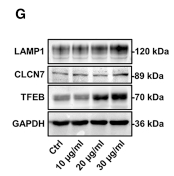
Figure 6 TUFM derived from hDPSC-apoVs activate EC autophagy and promote EC angiogenesis via the TFEB-induced autophagy-lysosome pathway (A) Western blotting analysis shows the presence of TUFM in apoVs derived from hDPSCs, si-NC-treated hDPSCs, and si-TUFM-treated hDPSCs. (B) Western blot analysis shows autophagy-associated gene (ATG7, Beclin-1, and LC3) expressions in ECs after incubation with apoVs or si-TUFM-apoVs. The angiogenic capacity of ECs, including tube formation (C), migration (D), and proliferation (E) were detected after treatment with apoVs or si-TUFM-apoVs. Scale bar, 100 μm. (F) Representative confocal microscopy images show the number of lysosomes in ECs after treatment with apoVs or Torin 1. Scale bar, 50 μm. (G) Western blot analysis shows lysosome-associated gene (LAMP1 and CLCN7) and TFEB expressions in ECs after incubation with apoVs at concentrations of 10, 20, or 30 μg/mL. (H) Expression of TFEB in cytosolic (Cyt.) and nuclear (Nuc.) fractions was detected by western blots after treatment with apoVs or Torin 1. (I) Representative confocal microscopy images show the locations of TFEB in ECs after treatment with apoVs or Torin1. Scale bar, 25 μm. (J) Western blot analysis shows autophagy-associated gene (ATG7, Beclin-1, and LC3) expressions in ECs after decreased expression of TFEB. (K) Western blot analysis shows autophagy-associated gene (ATG7, Beclin-1, and LC3) expressions in ECs after increased expression of TFEB. After transfection with si-TFEB, the angiogenic capacity of ECs, including tube formation (L), migration (M), and proliferation (N) were detected following coculture with apoVs (20 μg/mL). Scale bar, 100 μm; n = 3-5 per group. Data are presented as mean ± SD. Statistical analyses were performed by Student’s t test (two-tailed) for two-group comparisons and one-way ANOVA with Tukey’s post hoc test or Welch’s ANOVA with Games-Howell post hoc test for multiple-group comparisons.

3). CRISPR/Cas9-Mediated Gene Correction in Osteopetrosis Patient-Derived iPSCs. Frontiers in Bioscience-Landmark, 2023 (PubMed: 37395026) [IF=3.3]

Application: WB    Species: Mouse    Sample:

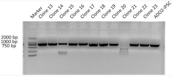
Fig. 2. Representative images showing the PCR assay for CLCN7 detection in different GC-ADO2-iPSC clones and ADO2-iPSCs. Clone 13-clone23 represent GC-ADO2-iPSC clone 13- GC-ADO2-iPSC clone23.

限制条款

产品的规格、报价、验证数据请以官网为准,官网链接:www.affbiotech.com | www.affbiotech.cn(简体中文)| www.affbiotech.jp(日本語)

产品的数据信息为Affinity所有,未经授权不得收集Affinity官网数据或资料用于商业用途,对抄袭产品数据的行为我们将保留诉诸法律的权利。

产品相关数据会因产品批次、产品检测情况随时调整,如您已订购该产品,请以订购时随货说明书为准,否则请以官网内容为准,官网内容有改动时恕不另行通知。

Affinity保证所销售产品均经过严格质量检测。如您购买的商品在规定时间内出现问题需要售后时,请您在Affinity官方渠道提交售后申请。

产品仅供科学研究使用。不用于诊断和治疗。 

产品未经授权不得转售。

Affinity Biosciences将不会对在使用我们的产品时可能发生的专利侵权或其他侵权行为负责。Affinity Biosciences, Affinity Biosciences标志和所有其他商标所有权归Affinity Biosciences LTD.