产品: RPL34 抗体
货号: DF3708
描述: Rabbit polyclonal antibody to RPL34
应用: WB IHC IF/ICC
反应: Human, Mouse, Rat, Monkey
预测: Pig, Zebrafish, Bovine, Horse, Sheep, Chicken, Xenopus
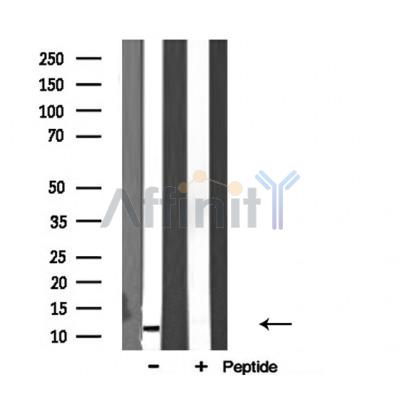
分子量: 13 KD; 13kD(Calculated).
蛋白号: P49207
RRID: AB_2836072

浏览相似产品>>

   规格 价格 库存
 50ul RMB¥ 1250 现货
 100ul RMB¥ 2300 现货
 200ul RMB¥ 3000 现货

货期: 当天发货

联系销售

产品描述

来源:
Rabbit
应用:
WB 1:500-1:1000, IF/ICC 1:100-1:500, IHC 1:50-1:200
*The optimal dilutions should be determined by the end user.
*Tips:

WB: 适用于变性蛋白样本的免疫印迹检测. IHC: 适用于组织样本的石蜡(IHC-p)或冰冻(IHC-f)切片样本的免疫组化/荧光检测. IF/ICC: 适用于细胞样本的荧光检测. ELISA(peptide): 适用于抗原肽的ELISA检测.

反应:
Human,Mouse,Rat,Monkey
预测:
Pig(100%), Zebrafish(100%), Bovine(100%), Horse(100%), Sheep(100%), Chicken(100%), Xenopus(100%)
克隆:
Polyclonal
特异性:
RPL34 Antibody detects endogenous levels of total RPL34.
RRID:
AB_2836072
引用格式: Affinity Biosciences Cat# DF3708, RRID:AB_2836072.
偶联:
Unconjugated.
纯化:
The antiserum was purified by peptide affinity chromatography using SulfoLink™ Coupling Resin (Thermo Fisher Scientific).
保存:
Rabbit IgG in phosphate buffered saline , pH 7.4, 150mM NaCl, 0.02% sodium azide and 50% glycerol. Store at -20 °C. Stable for 12 months from date of receipt.
别名:

展开/折叠

60S ribosomal protein L34; L34; MGC111005; Ribosomal protein L34;

抗原和靶标

免疫原:

A synthesized peptide derived from human RPL34, corresponding to a region within C-terminal amino acids.

Uniprot:
基因/基因ID:
序列:
MVQRLTYRRRLSYNTASNKTRLSRTPGNRIVYLYTKKVGKAPKSACGVCPGRLRGVRAVRPKVLMRLSKTKKHVSRAYGGSMCAKCVRDRIKRAFLIEEQKIVVKVLKAQAQSQKAK

种属预测

种属预测:

score>80的预测可信度较高,可尝试用于WB检测。*预测模型主要基于免疫原序列比对,结果仅作参考,不作为质保凭据。

Species
Results
Score
Pig
100
Horse
100
Bovine
100
Sheep
100
Xenopus
100
Zebrafish
100
Chicken
100
Dog
0
Rabbit
0
Model Confidence:
High(score>80) Medium(80>score>50) Low(score<50) No confidence

翻译修饰 - P49207 作为底物

Site PTM Type Enzyme
R10 Methylation
S12 Phosphorylation
Y13 Phosphorylation
T15 Phosphorylation
S17 Phosphorylation
K19 Ubiquitination
R29 Methylation
Y32 Phosphorylation
K36 Acetylation
K36 Ubiquitination
K37 Acetylation
K43 Ubiquitination
K62 Ubiquitination
T70 Phosphorylation
R76 Methylation
K85 Ubiquitination
R88 Methylation
K101 Ubiquitination
K105 Ubiquitination
K108 Ubiquitination
K115 Acetylation
K115 Ubiquitination

研究背景

功能:

Component of the large ribosomal subunit.

细胞定位:

Cytoplasm>Cytosol. Cytoplasm. Endoplasmic reticulum.
Note: Detected on cytosolic polysomes (PubMed:25957688). Detected in ribosomes that are associated with the rough endoplasmic reticulum (By similarity).

Extracellular region or secreted Cytosol Plasma membrane Cytoskeleton Lysosome Endosome Peroxisome ER Golgi apparatus Nucleus Mitochondrion Manual annotation Automatic computational assertionSubcellular location
亚基结构:

Component of the large ribosomal subunit.

蛋白家族:

Belongs to the eukaryotic ribosomal protein eL34 family.

研究领域

· Genetic Information Processing > Translation > Ribosome.

文献引用

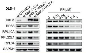
1). Dual Inhibition of DKC1 and MEK1/2 Synergistically Restrains the Growth of Colorectal Cancer Cells. Advanced Science (PubMed: 34026451) [IF=15.1]

Application: WB    Species: Human    Sample: DLD‐1 and HCT116 cells

Figure 3 Proteomic analysis reveals that DKC1 elicits ribosomal protein expression. A) Volcano plot of differentially expressed proteins obtained from proteomic analysis of triplicate samples of DKC1 knockdown DLD‐1 cells and control cells. A total of 114 downregulated proteins and 58 upregulated proteins were included. B,C) GO enrichment analysis for the cellular component category (B) and KEGG enrichment analysis (C) of differentially expressed proteins. D) Comparison of the mRNA abundance of 28 ribosomal proteins measured by qRT‐PCR and their protein levels measured by proteomic analysis of DKC1 knockdown DLD‐1 cells and control cells (All proteins with P < 0.05). E) qRT‐PCR assays evaluating the mRNA levels of the indicated genes in control and DKC1 knockdown DLD‐1 cells with or without enforced expression of wild‐type DKC1 or the DKC1 mutant (D125A) from three independent experiments (mean ± SD). F) Immunoblots assessing the abundance of the indicated ribosomal proteins in control and DKC1 knockdown DLD‐1 and HCT116 cells with or without enforced expression of wild‐type DKC1 or D125A and PF‐treated DLD‐1 and HCT116 cells (48 h). E.V: empty vector. *P < 0.05, **P < 0.01, ***P < 0.001, ****P < 0.0001, ns: no significance ((D) two‐sided Student's t‐test, (E) one‐way ANOVA with Bonferroni correction).

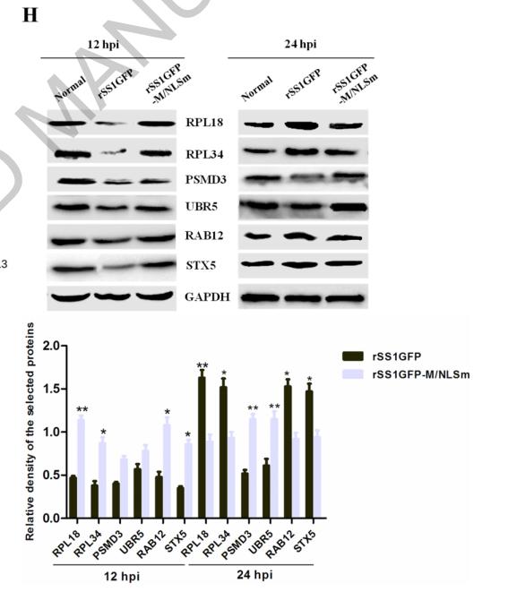
2). TMT-based quantitative proteomics analysis reveals the attenuated replication mechanism of Newcastle disease virus caused by nuclear localization signal mutation in viral matrix protein. Virulence (PubMed: 32420802) [IF=5.2]

Application: WB    Species: Mouse    Sample: BSR-T7/5 cells

Figure 8. rSS1GFP infection affects the expression of cellular translation, posttranslational modification and trafficking-associated proteins. (A) The heatmap of representative 20 DEPs related to “Translation, ribosomal structure and biogenesis” during rSS1GFP and rSS1GFP-M/NLSm infection at 12 and 24 h. (B) The protein-protein interactions of the DEPs related to “Translation, ribosomal structure and biogenesis” are analyzed by the STRING software. A red line indicates the presence of fusion evidence; a blue line indicates co-occurrence evidence; a light blue line indicates database evidence; a purple line indicates experimental evidence; a green line indicates neighborhood evidence; a black line indicates co-expression evidence. (C) The heatmap of representative 20 DEPs related to “Posttranslational modification, protein turnover, chaperones” during rSS1GFP and rSS1GFP-M/NLSm infection at 12 and 24 h. (D) The protein-protein interactions of the DEPs related to “Posttranslational modification, protein turnover, chaperones” are analyzed by the STRING software. (E) The heatmap of representative 20 DEPs related to “Intracellular trafficking, secretion, and vesicular transport” during rSS1GFP and rSS1GFP-M/NLSm infection at 12 and 24 h. (F) The protein-protein interactions of the DEPs related to “Intracellular trafficking, secretion, and vesicular transport” are analyzed by the STRING software. (G) The mRNA expression levels of six selected DEP genes in BSR-T7/5 cells infected with rSS1GFP and rSS1GFP-M/NLSm were verified by qRT-PCR. (H) The protein expression levels of six DEPs in BSR-T7/5 cells infected with rSS1GFP and rSS1GFP-M/NLSm were examined by Western blotting. The relative expression levels of six DEPs were compared with the control GAPDH expression.

限制条款

产品的规格、报价、验证数据请以官网为准,官网链接:www.affbiotech.com | www.affbiotech.cn(简体中文)| www.affbiotech.jp(日本語)

产品的数据信息为Affinity所有,未经授权不得收集Affinity官网数据或资料用于商业用途,对抄袭产品数据的行为我们将保留诉诸法律的权利。

产品相关数据会因产品批次、产品检测情况随时调整,如您已订购该产品,请以订购时随货说明书为准,否则请以官网内容为准,官网内容有改动时恕不另行通知。

Affinity保证所销售产品均经过严格质量检测。如您购买的商品在规定时间内出现问题需要售后时,请您在Affinity官方渠道提交售后申请。

产品仅供科学研究使用。不用于诊断和治疗。 

产品未经授权不得转售。

Affinity Biosciences将不会对在使用我们的产品时可能发生的专利侵权或其他侵权行为负责。Affinity Biosciences, Affinity Biosciences标志和所有其他商标所有权归Affinity Biosciences LTD.