


 and mouse anti-beta tubulin Ab(T0023 1:200) for 1 hour at 37°C. An AlexaFluor594 conjugated goat anti-rabbit IgG(H+L) Ab(Red) and an AlexaFluor488 conjugated goat anti-mouse IgG(H+L) Ab(Green) were used as the secondary antibody.
The nuclear counter stain is DAPI(blue).")
: the relative expression of TGF-β1 (B), p-Smad2/3 (C) and BAMBI (D) were analyzed in the liver of TLR4wild/ TLR4mut mice infected with or without C. sinensis-infected by Western blotting. The blots of each group were run under the same experimental conditions and the images were from the same gel. The data were obtained from 3~5 mice of three-independent experiment. The values were expressed as mean±SEM.")
 of
immunolabeling for Fn and nuclear staining with DAPI. Scale bar, 50 ~μm.")
 and pCX43 (C) showed a significant decrease but TGF-β1 (D), Smad3 (E), and pSmad3 (F) showed a significant increase in the bladder detrusor after SCI. Those changes were more significant in transection than in hemisection of sacral spine cord. CX45 was not changed among three groups.")
 + TGF-β1 (0 ng/mL), corilagin (0 μM) + TGF-β1 (5
ng/mL) and corilagin (25 μM) + TGF-β1 (5 ng/mL) for 12 h. Smad2/3 is shown by green fluorescence and nuclei were stained with DAPI, which emits blue
fluorescence. Scale bars = 50 μm. D. Protein levels of Smad7 examined by western blot assay after HSFs were treated with corilagin for 3 days. GAPDH served as
control. n = 3. E. Protein levels of MMP2, MMP9, MMP13 and TIMP1 in HSFs after treatment with corilagin for 3 days. GAPDH served as control. n = 3. Data are
show as mean ± SD. *p < 0.05, **p < 0.01, ***p < 0.001. (For interpretation of the references to color in this figure legend, the reader is referred to the web version
of this article.)")
, corilagin (0 μM) + TGF-β1 (5
ng/mL) and corilagin (25 μM) + TGF-β1 (5 ng/mL) for 48 h. α-SMA is shown by green fluorescence and nuclei were stained with DAPI, which emits blue fluorescence.
Scale bars = 50 μm. B. qRT-PCR results of α-SMA mRNA levels in HSFs after incubation with TGF-β1 and corilagin for 3 days. n = 3. C. α-SMA protein levels detected
using western blot assay after treatment with TGF-β1 and corilagin for 3 days, and quantification results normalized to GAPDH. n = 3. Data are mean ± SD. *p <
0.05, ***p < 0.001. ns, no significance. (For interpretation of the references to color in this figure legend, the reader is referred to the web version of this article.)")

 Expression of proliferation- and migration-associated genes (PCNA, MMP9 and TIMP-1) were evaluated using western blotting in HEY cells. (C and D) Western blotting of proteins involved in integrin-β1-FAK signaling pathway in the KRT7-overexpressing HEY cells. (E) Expression of MMPs after knockdown of KRT7 in OVCAR433 cells. (F and G) Expression of the TGF-β signaling pathway-related proteins was evaluated by western blotting in KRT7-overexpressing HEY cells and KRT7-knockdown OVCAR433 cells. All experiments were performed at least three times. Results are presented as the mean ± standard deviation. **P<0.01. FAK, focal adhesion kinase; PCNA, proliferating cell nuclear antigen; FN, fibronectin; TIMP-1, TIMP metallopeptidase inhibitor 1; p-, phosphorylated; MMP, matrix metalloproteinase; KRT7, keratin 7; sh, short hairpin RNA; NC, negative control.")
, and western blot assay disclosed that the expressions of exosome markers CD9, CD63, CD81, TGF-β1 Flotillin-1 and EEF2 were elevated in Exosome group than those in NC group (sample from serum-free medium) (B).")
. Different letters marked above the bars are significantly different by an ANOVA multiple test (p < 0.05).")

, IL-6 (b) and IL-10 (c). Three samples per group were examined, and representative images are shown (Scale bar represents 50 µm).")
. Scale bar, 100 μm. b The expression of profibrotic proteins TGF-β1, fibronectin, collagen I, and collagen III in the kidney of unilateral urethral obstruction (UUO) mice, and quantitative analysis")
 Te expression levels of the renal fbrosis-associated genes TGF-β1, FN, Smad3, Col1a1, Col1a2, and α-SMA were quantitated in each of the groups. All groups were compared with OC. *stands for p<0.05, **stands for p<0.01, and ***stands for p<0.001. (b) Te western blot analysis of TGF-β1, Smad3, a-SMA, FN, Col1a1 and Col1a2.")

 Representative western blots and quantitative analysis of (B) TGF‑β1/β‑actin, (C) p‑Smad2/3/Smad2/3 and (D) p‑ERK1/2/ERK1/2 levels in rats.")
 Immunofluorescence was conducted to evaluate the expression of α-SMA (red). Nucleus was stained with DAPI
(blue). Scale bar: 50 μm. (b) Relative mRNA expression of Col1a1 and α-SMA. (c, d) Expressions of p-MEK1/2, MEK1/2, p-ERK1/2, ERK1/
2 were detected by western blot. GAPDH was conducted as a loading control. One-way ANOVA, **p < 0.01, ***p < 0.001,
****p < 0.0001.")


 reduces peritoneal
fibrosis (PF) relative protein and gene expression
in mice tested by western blotting and real-time
qPCR, respectively. The results of western blotting (A, B) and real-time qPCR (C) demonstrate
that, after intraperitoneal administration of
4.25% peritoneal dialysis solution, the levels of
α-SMA, Col-I, TGF-β1, SGLT-2 and p-Smad3 increase, and E-cadherin decreases in the submesothelial compact zone on day 28. Treatment
with Emp significantly reduces the levels of
α-SMA, Col-I, TGF-β1, and p-Smad3, and increases the levels of E-cadherin compared with
the effect of the peritoneal dialysis solution alone.
*P < 0.05, **P < 0.01, ***P < 0.001 versus PF
mice with peritoneal dialysis solution injected. N
= 6 per group.")
 TGF-β1 mRNA and protein levels were increased in HT22 cells in the presence of hemin. (C and D) HT22 cells were stimulated with different doses (0, 2, 5, and 10 ng/mL) of recombinant TGF-β1, and then assayed for SERPINE1 expression using qRT-PCR assay and Western blot. (E and F) HT22 cells were transfected with TGF-β1 siRNA, and then assayed for SERPINE1 expression using qRT-PCR assay and Western blot. **p <0.01 and ***p <0.001.")

 Western blot analysis for α-smooth muscle actin (α-SMA), collagen I, transforming growth factor (TGF)-β1 and connective tissue growth factor (CTGF). The relative expression of (B) α-SMA, (C) collagen I, (D) TGF-β1 and (E) CTGF. Data were shown as mean value ± SD (n=6). ***P<0.001, versus Control group. &&P<0.01, &&&P<0.001, versus NC agomir group.")
.
(A) The mRNA expression of TGF-β1. (B) The mRNA expression of SMAD2. (C) Representative images of the western blotting analysis for the quantification of TGF-β1, p-SMAD2, and SMAD2 in the heart tissues. (D) The protein expression of TGF-β1 in the heart tissues. (E) The protein expression of p-SMAD2/SMAD2 in the heart tissues. The data are expressed as the means ± SDs. ∗, P < 0.05 and ∗∗, P < 0.01 compared with the Sham group. #, P < 0.05 and ##, P < 0.01 compared with the AMI group. △, P < 0.05 and △△, P < 0.01 compared with the AMI group.")
. D, F, G. The expression levels of MMP2 (D, F) and MMP9 (D, G) in lung cancer tissues (n=6). D, H. The expression level of TGF-β1 in lung cancer tissues (n=6). D, I. The expression level of collagen type I in lung cancer tissues(n=6). Note: &, &&&& respectively represent a significant difference compared with the Saline group (P<0.05), (P<0.0001).")
. (A–F) Representative immunohistochemical staining of Collagen I, Fibronectin, PAI-1, TGF-β, IL-1β and IL-18 in the 4 groups (scale bar = 20 μm). (G–L) Quantitative analysis of the Collagen I, Fibronectin, PAI-1, TGF-β, IL-1β, and IL-18 protein expression shown in panels (A–F). *p < 0.05 versus the control group; #p < 0.05 versus the Ad-SC-shRNA group.")
 TGF-β1 protein levels. (B) α-SMA protein levels. Data are presented as mean ± SEM (n > 3 for each group). #P < 0.05 vs. CON, ∗P < 0.05 vs. DKD.")
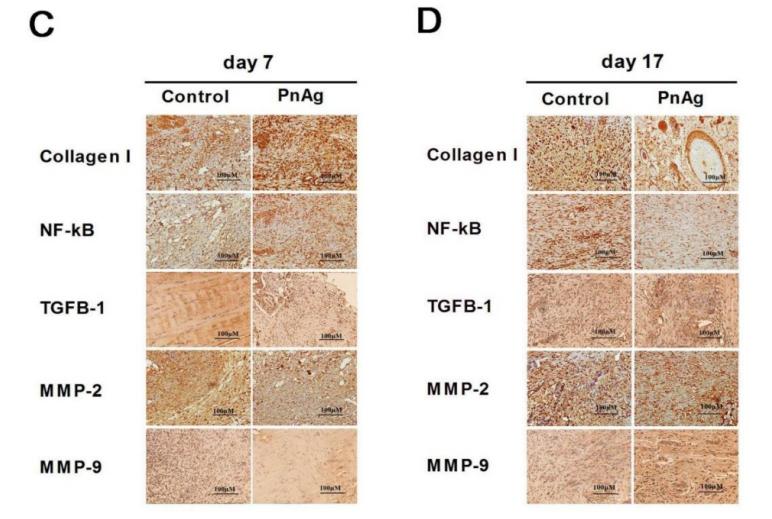
, α-SMA, fibronectin, and collagen I expression in tracheal
scar tissues from normal, saline, and nintedanib (10 mg, 20 mg) treated rat groups. (A) TGF-β1 was detected in both the epithelial and submucosal layers (200); (B) α-SMA was detected in fibroblasts in submucosal layers (200); (C,D) fibronectin and collagen I were expressed in the
intercellular substance and submucosal cells in the stenotic tissues (200). Average optical density (AOD) date of four antibodies are presented as the means SEM. *P < .05, **P < .01, ***P < .001, ****P < .0001 compared with saline-treated group; ns = not significant. [Color
figure can be viewed in the online issue, which is available at www.laryngoscope.com.]")
 Relative mRNA level of IL10, Arg-1, and TGF-β1in the colon was measured through qPCR. Expression of IL-10, Arg-1, and TGF-β1 in colon tissue lysates were determined by Western blot from DSS-challenged mice. n = 3,.means ± SEM.*p < 0.05, **p < 0.01, ***p < 0.001.")
 CX3CR1 (red, stained with Alexa Fluor 594) and TGFβ1 (green, stained with Alexa Fluor 488) double labeling. TGFβ1 colocalized with CX3CR1 at 3 and 7 dpi, but not pre-injury or at 14 dpi. The asterisks indicate the lesion epicenter. Colocalization of the proteins is shown in yellow. Nuclear staining (DAPI) is shown in blue, and white arrowheads indicate the colocalization observed with a 40× objective. Scale bars: 200 μm in the left three columns; 20 μm in the right column. (B) Percentage of TGFβ1+CX3CR1+ cells relative to the total number of CX3CR1+ cells in the injured spinal cord. The data are presented as the mean ± SEM (n = 3 independent experiments). ****P < 0.0001 (one-way analysis of variance followed by Tukey's post hoc test). CX3CR1: C-X3-C motif chemokine receptor 1; DAPI: 4′,6-diamidino-2-phenylindole, dihydrochloride; dpi: days post-injury; ND: not determined; TGFβ1: transforming growth factor-β1.")
 Western blotting shows the expression of M1 (iNOS) or M2 (Arg1) polarization markers and TGFβ1 in BV-2 cells after polarization induction. (B) Quantitative analysis of iNOS expression shown in (A). β-Actin was used as the loading control. iNOS was expressed at significantly higher levels in M1-type microglia than in M0- or M2-type microglia. (C) Quantitative analysis of Arg1 protein expression shown in (A). Arg1 was expressed at significantly higher levels in M2-type microglia than in M0- or M1-type microglia. (D) Quantitative analysis of TGFβ1 protein expression shown in (A). TGFβ1 was expressed at significantly higher levels in M1-type microglia than in M0- or M2-type microglia. The data are presented as the mean ± SEM (n = 4) and were analyzed by one-way analysis of variance, followed by Tukey's post hoc test. ***P < 0.001, ****P < 0.0001. (E) Representative immunocytochemistry images of TGFβ1 (green, stained with Alexa Fluor 488) in BV-2 cells after polarization induction. The TGFβ1 fluorescence intensity was strongest in M1-type microglia, suggesting that these cells expressed the highest levels of TGFβ1. DAPI (blue) was used to stain the nuclei. At least three independent replicates were performed for each experiment. Scale bar: 50 μm. Arg1: Arginine 1; DAPI: 4′,6-diamidino-2-phenylindole, dihydrochloride; iNOS: inducible nitric oxide synthase; TGFβ1: transforming growth factor-β1.")

 Representative results of Western blot analysis of α-SMA,
Collagen I and TGF-β1 in rats from all five groups. (B) Expression of α-SMA in all five groups with β-actin as the loading control is presented
as a bar graph. (C) Expression of Collagen I in all five groups with β-actin as the loading control is presented as a bar graph. (D) Expression
of TGF-β1 in all five groups with β-actin as the loading control is presented as a bar graph. (E) Representative images showing smooth
muscle and collagen staining in all five groups. Smooth muscle and collagen in the corpus cavernosum are stained red and blue, respectively.
Original magnification, 50× and 200×. (F) Representative results of immunohistochemistry for TGF-β1 in all five groups (50× and 200×). The
arrows indicate TGF-β1 expression. Data are expressed as the mean ± standard deviation. &P < .05 compared with the Con group. #
P < .05
compared with the DMED group. *
P < .05 compared with the DMED + CCSMC-EXO group. $
P < .05 compared with the DMED + BMSCEXO group. CCSMC-EXOs: exosomes derived from corpus cavernosum smooth muscle cells; BMSC-EXOs: exosomes derived from bone
marrow stem cells; ADSC-EXOs: exosomes derived from adipose-derived stem cells; DMED: diabetes mellitus-induced erectile dysfunction")
 Representative images showing the distribution of PAI-1 in cardiac tissues after immunofluorescence staining. Scar bar = 50 μm. (b, c) Relative mRNA and protein expression of urokinase plasminogen activator (uPA) and tissue-type plasminogen activator (tPA) in the cardiac tissues. (d) Western blot analysis for TGF-β1R, TGF-β1, p-Smad2/3, and Smad2/3. Relative protein expression of (e) PAI-1, (f) TGF-β1R, (g) TGF-β1, (h) p-Smad2/3, and (i) Smad2/3 in the cardiac tissues. Values are expressed as mean ± SD (n = 6). +++p < 0.001vs. sham group. ∗p < 0.05, ∗∗p < 0.01, ∗∗∗p < 0.001vs. MI/R group.")
 The representative band of Western blot. (b)–(h) The quantitative result of Western blot. Data were expressed as mean ± SD. #P < 0.05/##p < 0.01 vs. control; ∗p < 0.05/∗∗p < 0.01 vs. the BLM group. n = 3.")
 mHSCs were co-cultured with the CMs from RAW264.7 cells with LPS/IFNγ and PHI treatment for 12 h. (b-g) The expression of MMP2, TIMP1, TGF-β, α-SMA, COL1 and NF-κB mRNA in mHSCs after co-culture with macrophage-derived CMs for 24 h was detected by RT-qPCR (n = 3). (h) The expression of TGF-β, α-SMA, COL1, P65 and P-P65 proteins in mHSCs after co-culture with macrophage-derived CMs for 24 h was detected by western blotting. (i) The relative quantification of TGF-β, α-SMA, COL1 and P-P65/P65 protein expression in western blotting results was analyzed by ImageJ software (n = 3). (j) The expression of cytoplasm and nucleus NF-κB P65 proteins in mHSCs after co-culture with macrophage-derived CMs for 24 h was detected by western blotting. (k, l) The relative quantification of cytoplasm and nucleus NF-κB P65 protein expression in western blotting results was analyzed by ImageJ software (n = 3). (m) Immunofluorescence staining of α-SMA in mHSCs after co-culture with CMs from RAW264.7 cells with different treatments for 24 h (Scale bar=100 µm). Results are presented as mean ± SD. ###P")

. Western blotting analysis of NF-κB, TGF-β, PXR, IL-6, α-SMA in livers, and the datas were normalized to that of GAPDH (A–H), Western blotting analysis of Bax, and PXR in HepG2 cells, and the datas were normalized to that of GAPDH (H,I). Values are the mean ± standard deviation.")
. NC, normal control group; BLM, bleomycin-induced systemic sclerosis model group; PESV-L, low-dose PESV intervention group; PESV-M, medium-dose PESV intervention group; PESV-H, high-dose PESV intervention group; DXM, dexamethasone intervention group.")
 Immunohistochemical detection of arginase-1, (b) Statistical expressions of arginase-1 by an immunohistochemical assay (p")
 Expression levels of TGFb1, SMAD2/3, and quantification. (d‒g) Expression levels of TGFb2, TGFb3, pSMAD2/3 (Thr8), and quantification. (h‒k) Expression levels of Akt and pAkt and quantification. n ¼ 3 biological replicates. Data are represented as mean SD; one-way ANOVA with Fisher’s posthoc test. *P < 0.05 versus control and #P < 0.05 versus FB-Exo. Akt, protein kinase B; ESC-Exo, epidermal stem cellederived exosome; FB, fibroblast; FB-Exo, fibroblast-derived exosome; pAkt, phosphorylated protein kinase B; pSMAD, phosphorylated SMAD.")
. (A) Masson’s trichrome staining of uterine horns (×400). (B) Representative images of IHC for the fibrosis associated marker COL I (×400). (C) Representative images of IHC for the pro-fibrogenic mediator TGF-β 1 (×400). Scale bar=500 µm. (D) Ratio of the area with fibrosis to the total endometrial area in each group. (E) Quantification of COL I-positive expression. (F) Quantification of TGF-β1-positive expression. All data are expressed as the mean ± SEM (*P")
. As compared to the normal group, the smad3 and TGF-β1 proteins elevated in all the groups excepting the high dose of CA-treated group(P")
. As compared to the normal group, the smad3 and TGF-β1 proteins elevated in all the groups excepting the high dose of CA-treated group(P")
 and (B) Transmission electron microscopy of the enriched extracellular vesicles (EVs) isolated from the sera of E. multilocularis-infected (A) and uninfected mice (B). (C) The diameter distribution of the purified EVs. (D) Western blotting analysis of mammalian EV-marker CD63 in the purified EVs. (E) Heatmap of differentially abundance proteins between the serum exosomes from E. multilocularis-infected and uninfected mice. (F) Western blotting analysis the abundance of TGF-β1, GPATC8, LRP1, and AMY1 during E. multilocularis infection. https://doi.org/10.1371/journal.pntd.0010814.g001")
 of RIPK1, RIPK3, MLKL (necroptosis core mediators), NRGPI oncogenes (SERPINE1, GPX3, GRP, FCN1, CYTL1, CNTN1, PLCL1, and APOD), markers of WNT signaling pathway (WNT2B, WNT9A), TGF-β signaling pathway (TGFB1, TGFB3), and macrophage (CD63, CD206, CD163) in the clinical samples of stomach adenocarcinoma.")
 and high-dose aspirin (300 mg/kg + CCl4). ns, not significant. *P")
 macrophages. (A) Different morphologic appearances of THP-1 cells that were untreated (THP-1), treated with PMA (M0) or treated with PMA and subsequently with IL4/IL13 (M2). THP-1 cells grew in suspension, while M0 and M2 were larger with increased adherence and more granules in the cytoplasm. Scale bar: 50 μm. (B) Representative flow cytometric analysis of untreated THP-1, M0 and M2 cells stained with allophycocyanin-conjugated monoclonal antibody anti-CD206. The MFI is shown on the x-axis, and the relative cell number on the y-axis. (C) Representative immunofluorescence images showing increased expression of CD206 in M2 cells compared to M0 macrophages. Scale bars: 10 μm. (D) Quantification of the results in (C). **P")
 Representative images of Periodic Acid Schiff staining (PAS, ×400, bar: 20 μm). (B) Representative images of Masson's trichrome staining (Masson, ×400, bar: 20 μm). (C) Statistics of mesangial area. (D) Statistics of mesangial density. (E) Statistical result of Masson. (F) Representative WB images of TGF-β1, Smad2/3, and p-Smad2/3. (G) Statistical results of TGF-β1. (H) Statistical results of p-Smad2/3 to Smad2/3. Data are expressed as mean ± SD, n = 4. **P < 0.01 versus Control; #P < 0.05, ##P < 0.01 versus Model.")
. All experiments were repeated three times.")
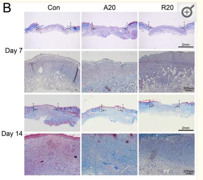
 Representative micrographs showing collagen deposition in obstructed kidneys. Mice were pretreated with Apcin (5 mg/kg per day) by intraperitoneal injection for 1 day. UUO surgery was then performed, and Apcin was administered continuously for the next 7 days until mice were sacrificed on day 7. Paraffin sections were subjected to Masson's trichrome staining for collagen deposition. Scale bar: 50 μm. (B) Graphic presentation showing the fibrotic area in kidney tissues among groups as indicated (n = 8 in vehicle Sham group; n = 7 in Apcin Sham group; n = 7 in vehicle UUO group; n = 7 in Apcin UUO group). The quantification of fibrotic area was performed by Image-Pro Plus. (C) Representative micrographs of Sirius red staining in mice after UUO. Scale bar: 50 μm. (D and E) Representative micrographs and quantitative data for FN1 immune staining in kidney tissues (n = 4). Scale bar: 50 μm. (F and G) Representative micrographs and quantitative data showing α-SMA expression in kidney tissues among groups as indicated (n = 4). Scale bar: 50 μm. Positive signal quantification was performed using Image-Pro Plus. (H and I) The mRNA level of Collagen I and Collagen III were evaluated by qRT-PCR (n = 8 in vehicle Sham group; n = 7 in Apcin Sham group; n = 7 in vehicle UUO group; n = 7 in Apcin UUO group). (J and K) Representative Western blot and quantitative data showing renal expression of Collagen I, Collagen III, TGF-β1 and CDC20 (n = 3 in sham group; n = 4 in UUO group). The values represent means ± SEM. P values were shown in figure.")
 Immunohistochemical staining of COL1 (400×, scale bar = 40 µm). (B) The mean density of COL1 staining. (C) The mRNA expression of COL1. (D) COL1, α-SMA and GAPDH were detected by WB. (E and F) Relative expressions of COL1 and α-SMA. (G) Samd2/3, p-Samd2/3, TGF-β and GAPDH were detected by WB. (H and I) Relative expressions of p-Samd2/3/Samd2/3 and TGF-β. (J) Relative expression of TGF-β mRNA. Data are expressed as mean ± SD, q-PCR n = 3, others n = 4. **P < 0.01 as compared with Control group. #P < 0.05, ##P < 0.01 as compared with HFD + STZ group.")
. #p")
, P < 0.05). The effect of ASP on the TGF-β/Smad4 pathway was further evaluated. TGF-β and Smad4 protein expressions were both dose-dependently decreased by ASP treatment, as shown by western blotting (Figure 2(b), P < 0.05).")
产品描述
*The optimal dilutions should be determined by the end user.
*Tips:
WB: 适用于变性蛋白样本的免疫印迹检测. IHC: 适用于组织样本的石蜡(IHC-p)或冰冻(IHC-f)切片样本的免疫组化/荧光检测. IF/ICC: 适用于细胞样本的荧光检测. ELISA(peptide): 适用于抗原肽的ELISA检测.
引用格式: Affinity Biosciences Cat# AF1027, RRID:AB_2835389.
展开/折叠
Cartilage-inducing factor; CED; Differentiation inhibiting factor; DPD1; LAP; Latency-associated peptide; Prepro transforming growth factor beta 1; TGF beta 1; TGF beta; TGF beta 1 protein; TGF-beta 1 protein; TGF-beta-1; TGF-beta-5; TGF-beta1; TGFB; Tgfb-1; tgfb1; TGFB1_HUMAN; TGFbeta; TGFbeta1; Transforming Growth Factor b1; Transforming Growth Factor beta 1; Transforming growth factor beta 1a; transforming growth factor beta-1; transforming growth factor, beta 1; Transforming Growth Factor-ß1;
抗原和靶标
A synthesized peptide derived from human TGF beta1, corresponding to a region within C-terminal amino acids.
研究领域
· Cellular Processes > Cell growth and death > Cell cycle. (View pathway)
· Cellular Processes > Cell growth and death > Cellular senescence. (View pathway)
· Environmental Information Processing > Signal transduction > MAPK signaling pathway. (View pathway)
· Environmental Information Processing > Signaling molecules and interaction > Cytokine-cytokine receptor interaction. (View pathway)
· Environmental Information Processing > Signal transduction > FoxO signaling pathway. (View pathway)
· Environmental Information Processing > Signal transduction > TGF-beta signaling pathway. (View pathway)
· Environmental Information Processing > Signal transduction > Hippo signaling pathway. (View pathway)
· Human Diseases > Endocrine and metabolic diseases > Non-alcoholic fatty liver disease (NAFLD).
· Human Diseases > Infectious diseases: Parasitic > Leishmaniasis.
· Human Diseases > Infectious diseases: Parasitic > Chagas disease (American trypanosomiasis).
· Human Diseases > Infectious diseases: Parasitic > Malaria.
· Human Diseases > Infectious diseases: Parasitic > Toxoplasmosis.
· Human Diseases > Infectious diseases: Parasitic > Amoebiasis.
· Human Diseases > Infectious diseases: Bacterial > Tuberculosis.
· Human Diseases > Infectious diseases: Viral > Hepatitis B.
· Human Diseases > Infectious diseases: Viral > HTLV-I infection.
· Human Diseases > Cancers: Overview > Pathways in cancer. (View pathway)
· Human Diseases > Cancers: Overview > Proteoglycans in cancer.
· Human Diseases > Cancers: Specific types > Colorectal cancer. (View pathway)
· Human Diseases > Cancers: Specific types > Renal cell carcinoma. (View pathway)
· Human Diseases > Cancers: Specific types > Pancreatic cancer. (View pathway)
· Human Diseases > Cancers: Specific types > Chronic myeloid leukemia. (View pathway)
· Human Diseases > Cancers: Specific types > Hepatocellular carcinoma. (View pathway)
· Human Diseases > Cancers: Specific types > Gastric cancer. (View pathway)
· Human Diseases > Immune diseases > Inflammatory bowel disease (IBD).
· Human Diseases > Immune diseases > Rheumatoid arthritis.
· Human Diseases > Cardiovascular diseases > Hypertrophic cardiomyopathy (HCM).
· Human Diseases > Cardiovascular diseases > Dilated cardiomyopathy (DCM).
· Organismal Systems > Development > Osteoclast differentiation. (View pathway)
· Organismal Systems > Immune system > Th17 cell differentiation. (View pathway)
· Organismal Systems > Immune system > Intestinal immune network for IgA production. (View pathway)
· Organismal Systems > Endocrine system > Relaxin signaling pathway.
文献引用

Application: WB Species: human Sample:
Application: IHC Species: rat Sample:
Application: IHC Species: Mice Sample: skin

Application: WB Species: Human Sample: HK2 cells
限制条款
产品的规格、报价、验证数据请以官网为准,官网链接:www.affbiotech.com | www.affbiotech.cn(简体中文)| www.affbiotech.jp(日本語)产品的数据信息为Affinity所有,未经授权不得收集Affinity官网数据或资料用于商业用途,对抄袭产品数据的行为我们将保留诉诸法律的权利。
产品相关数据会因产品批次、产品检测情况随时调整,如您已订购该产品,请以订购时随货说明书为准,否则请以官网内容为准,官网内容有改动时恕不另行通知。
Affinity保证所销售产品均经过严格质量检测。如您购买的商品在规定时间内出现问题需要售后时,请您在Affinity官方渠道提交售后申请。产品仅供科学研究使用。不用于诊断和治疗。
产品未经授权不得转售。
Affinity Biosciences将不会对在使用我们的产品时可能发生的专利侵权或其他侵权行为负责。Affinity Biosciences, Affinity Biosciences标志和所有其他商标所有权归Affinity Biosciences LTD.