, using Caspase 8 Antibody. The lane on the left was treated with blocking peptide.")


 and mouse anti-beta tubulin Ab(T0023) for 1 hour at 37°C. An AlexaFluor594 conjugated goat anti-rabbit IgG(H+L) Ab(Red) and an AlexaFluor488 conjugated goat anti-mouse IgG(H+L) Ab(Green) were used as the secondary antibody.
The nuclear counter stain is DAPI(blue).")
 and mouse anti-beta tubulin Ab(T0023) for 1 hour at 37°C. An AlexaFluor594 conjugated goat anti-rabbit IgG(H+L) Ab(Red) and an AlexaFluor488 conjugated goat anti-mouse IgG(H+L) Ab(Green) were used as the secondary antibody.
The nuclear counter stain is DAPI(blue).")
. The arrows show the positive expression of caspase-8. (A) Normal group; (B) model group; (C) positive control group (CTX 20 mg kg−1 ); (D) low concentration of the PPP-40 group (50 mg kg−1 ); (E) medium concentration of the PPP-40 group (150 mg kg−1 ); (F) high concentration of the PPP-40 group (300 mg kg−1 ); (G) quantity analysis (mean ± SD, n = 6). *p < 0.05 and **p < 0.01 compared with the normal group; #p < 0.05 and ##p < 0.01 compared with the model group.")
 BCL2L10 induced apoptosis in HepG2 and Huh7 cells, as determined by flow cytometry analysis following Annexin V and PI staining. The upper panel represents FACS images of HCC cells transfected with empty vector or BCL2L10, while the lower panel shows the quantitative analyses of early apoptotic and late apoptotic cells. The experiment was repeated three times in triplicate. Data are mean ± SD. (B) Protein expression of the active forms of apoptosis related genes caspase 3, caspase 8, and caspase 9 was evaluated by Western blot.")
 Representative proteins were investigated using western blot analysis. The cells of two group were treated for twenty-four hours. In Fig. B and C, NC/0: primary cells, N30/30: thirty generations of bovine mammary epithelial cells treated by IFN-γ (10 ng/ml). RNAseq: transcriptomics results. qRT-PCR: fluorescence quantitative PCR results. The data represent the means ± SEM of 3 independent experiments. Each bar represents the mean of three independent experiments. One-way ANOVA; **P < 0.01.")
 caspase 8, caspase 9, and caspase 3 were determined by immunoblotting in liver tissue. Mean ± S.E.M (n = 3), *Control vs NASH; #NASH vs NAR. *P < 0.05, **P < 0.01, ***P < 0.001; #P < 0.05, ##P < 0.01, ###P < 0.001.")
 WB analysis of the antiapoptotic and caspase proteins in HepG2 cells treated with C20/C22 at 2 μM for 0, 6, 12, and 24 h. The reported values correspond to the mean ± SD for three independent experiments. (D) Immunofluorescence assays employed to determine the expression of caspase3/7 in cells with indicated treatments. Scale bars, 10 µm. (E,F) Apoptotic cells detected by flow cytometry after indicated treatments (n = 3). The p-value was analyzed by one-way ANOVA followed by Tukey’s test using GraphPad Prism version 8.00. ** p < 0.01, *** p < 0.001, and **** p < 0.0001 vs. 0 h/control group.")
 Western blot of the Caspase-8, FADD, Caspase-3, and cytochrome C proteins. (B) Relative levels of Caspase-8. (C) Relative levels of FADD. (D) Relative levels of Caspase-3. (E) Relative levels of cytochrome C.")
 Expression of caspase-3, caspase-8, Bid, Bax and Bcl-2 were analyzed by western blot. (B) Statistical analysis. Expression of proteins level were quantified and standardized against GAPDH. Values are expressed as the mean ± SD of three independent experiments. *p < 0.05, **p < 0.01, ***p < 0.001, compared with 0 μM group.")
 Representative images of cell apoptosis in COV362 cells after silencing METTL3 or elevating METTL3 were analyzed by Tunel staining. Scale bar =50 µm. (B) FAS, FADD, p-FADD, Caspase-8, Caspase-3, Bcl-2, and Bax protein expressions were determined in COV362 cells with METTL3 knock-down or overexpression through western blotting. ▴P < 0.05, ▴▴P < 0.01 vs. si-NC group; ∗P < 0.05, ∗∗P < 0.01 vs. PcDNA3.1 group.")
 ROS level in RAW264.7 cells. Scale bars: 250 μm. (B–D) Representative immunoblot and relative quantification of Txnip and Trx-1 in RAW264.7 cells. (E,F) Representative immunofluorescence images of NLRP3-positive, NEK7-positive in RAW264.7 cells. Scale bars: 25 μm. (G–L) Representative immunoblot and relative quantification of NLRP3, Caspase-1, NEK7, Caspase-8, and IL18 in RAW264.7 cells. (M–O) Representative immunoblot and relative quantification of Cleaved Caspase-1 p10 and Cleaved Caspase-8 in RAW264.7 cells. (P–R) Representative immunoblot and relative quantification of GSDMD and GSDMD N-terminal in RAW264.7 cells. (S,T) Representative immunoblot and relative quantification of NLRP3 in RAW264.7 cells after MCC950 intervention. (U) Representative immunofluorescence images of NLRP3-positive in RAW264.7 cells after MCC950 intervention. Scale bars: 50 μm. Data are presented as mean ± SEM (n = 3). * p < 0.05 and ** p < 0.01 vs. the Con group. # p < 0.05, ## p < 0.01, ### p < 0.001, and #### p < 0.0001 vs. the LPS group. && p < 0.01 vs. the 7MF group.")
 Western blot analysis with caspase 8, RIPK1, p-RIPK1, NLRP3, IL-1β, NF-kB P65 and p-NF-kB P65 extracted from BMSCs treated with lentivirus infection and were induced by osteogenic medium for 1 and 7 days individually. (C) ARS staining and (D) Western blot analysis for caspase 8 associated proteins extracted from BMSCs treated with lentivirus infection and caspase 8 inhibitor Z-IETD-FMK for 7 days. All results are presented with three replicates. *p ≤ 0.05, ***p ≤ 0.001.")
 Immunofluorescence images of ASC (red), caspase 8 (green), and RIPK3 (rose red) co-localization with nuclear DAPI staining (blue). (B–D) Average fluorescence intensity of ASC, caspase 8, and RIPK3. (Mean ± SEM, n = 3, * p < 0.05 and ** p < 0.01 vs. model group). Dashed box shows the zoomed area.")
 and si-FGA (b) on the apoptosis of cells. The effects of overexpression of FGA (c) and si-FGA (d) on the mRNA levels of genes related to cell apoptosis. The effects of overexpression of FGA and si-FGA on the protein levels of genes related to cell apoptosis (e). CoIP detected that FGA binds to apoptosis-related proteins (f). CoIP reversed validation of apoptosis-related gene protein binding (g). Caspase3 in the nucleus and cytoplasm at the mRNA (h) and protein level (i). In all panels, the statistical significance of differences between means were assessed using Student’s t-test (* P")
, n = 3 (A/C/D/E). *P")
 ELISA assay was utilized to measure the expression levels of inflammatory cytokines after MK5-AS1 overexpression and knockdown (n=6); (e) the changes in apoptosis-related protein levels were measured using WB after MK5-AS1 overexpression and knockdown (n=3); (f) FCM experiment was adopted to detect cell apoptosis after MK5-AS1 overexpression and knockdown(n=3). Data were represented as mean ± SD. ns, not significant. vs. Control group, ***p < 0.001. vs. cells transfected with si-NC, #p < 0.05, ##p < 0.01, ###p < 0.001. vs. cells transfected with OV-NC, ▴p < 0.05, ▴▴p < 0.01, ▴▴▴p < 0.001. Statistical analysis was performed using the one-way ANOVA.")

产品描述
*The optimal dilutions should be determined by the end user. For optimal experimental results, antibody reuse is not recommended.
*Tips:
WB: 适用于变性蛋白样本的免疫印迹检测. IHC: 适用于组织样本的石蜡(IHC-p)或冰冻(IHC-f)切片样本的免疫组化/荧光检测. IF/ICC: 适用于细胞样本的荧光检测. ELISA(peptide): 适用于抗原肽的ELISA检测.
引用格式: Affinity Biosciences Cat# AF6442, RRID:AB_2835266.
展开/折叠
ALPS2B; Amyotrophic lateral sclerosis 2 chromosomal region candidate gene 12 protein; Apoptotic cysteine protease; Apoptotic protease Mch-5; Apoptotic protease Mch5; CAP4; CASP-8; CASP8; CASP8_HUMAN; Caspase 8; Caspase 8 apoptosis related cysteine peptidase; Caspase-8 subunit p10; CED 3; FADD Like ICE; FADD-homologous ICE/CED-3-like protease; FADD-like ICE; FLICE; FLJ17672; ICE-like apoptotic protease 5; MACH alpha 1/2/3 protein; MACH; MACH beta 1/2/3/4 protein; MCH5; MGC78473; MORT1 associated ced 3 homolog; MORT1-associated CED-3 homolog; OTTHUMP00000163717; OTTHUMP00000163720; OTTHUMP00000163724; OTTHUMP00000163725; OTTHUMP00000165062; OTTHUMP00000165063; OTTHUMP00000165064; OTTHUMP00000206552; OTTHUMP00000206582;
抗原和靶标
A synthesized peptide derived from human Caspase 8, corresponding to a region within the internal amino acids.
研究领域
· Cellular Processes > Cell growth and death > p53 signaling pathway. (View pathway)
· Cellular Processes > Cell growth and death > Apoptosis. (View pathway)
· Cellular Processes > Cell growth and death > Apoptosis - multiple species. (View pathway)
· Cellular Processes > Cell growth and death > Necroptosis. (View pathway)
· Environmental Information Processing > Signal transduction > TNF signaling pathway. (View pathway)
· Human Diseases > Drug resistance: Antineoplastic > Platinum drug resistance.
· Human Diseases > Endocrine and metabolic diseases > Non-alcoholic fatty liver disease (NAFLD).
· Human Diseases > Neurodegenerative diseases > Alzheimer's disease.
· Human Diseases > Neurodegenerative diseases > Huntington's disease.
· Human Diseases > Infectious diseases: Bacterial > Legionellosis.
· Human Diseases > Infectious diseases: Parasitic > Chagas disease (American trypanosomiasis).
· Human Diseases > Infectious diseases: Parasitic > Toxoplasmosis.
· Human Diseases > Infectious diseases: Bacterial > Tuberculosis.
· Human Diseases > Infectious diseases: Viral > Hepatitis B.
· Human Diseases > Infectious diseases: Viral > Human papillomavirus infection.
· Human Diseases > Infectious diseases: Viral > Herpes simplex infection.
· Human Diseases > Cancers: Overview > Pathways in cancer. (View pathway)
· Human Diseases > Cancers: Overview > Viral carcinogenesis.
· Human Diseases > Cardiovascular diseases > Viral myocarditis.
· Organismal Systems > Immune system > Toll-like receptor signaling pathway. (View pathway)
· Organismal Systems > Immune system > NOD-like receptor signaling pathway. (View pathway)
· Organismal Systems > Immune system > RIG-I-like receptor signaling pathway. (View pathway)
· Organismal Systems > Immune system > IL-17 signaling pathway. (View pathway)
文献引用

Application: IHC Species: human Sample: ascites cells
Application: IF/ICC Species: human Sample: ascites cells
Application: WB Species: human Sample: ascites cells
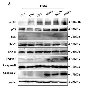
Application: WB Species: mouse Sample: testis

Application: WB Species: Mouse Sample:

Application: WB Species: Human Sample: AT2 cells
Application: WB Species: human Sample: LS174T cells

Application: WB Species: Mouse Sample: GCs
限制条款
产品的规格、报价、验证数据请以官网为准,官网链接:www.affbiotech.com | www.affbiotech.cn(简体中文)| www.affbiotech.jp(日本語)产品的数据信息为Affinity所有,未经授权不得收集Affinity官网数据或资料用于商业用途,对抄袭产品数据的行为我们将保留诉诸法律的权利。
产品相关数据会因产品批次、产品检测情况随时调整,如您已订购该产品,请以订购时随货说明书为准,否则请以官网内容为准,官网内容有改动时恕不另行通知。
Affinity保证所销售产品均经过严格质量检测。如您购买的商品在规定时间内出现问题需要售后时,请您在Affinity官方渠道提交售后申请。产品仅供科学研究使用。不用于诊断和治疗。
产品未经授权不得转售。
Affinity Biosciences将不会对在使用我们的产品时可能发生的专利侵权或其他侵权行为负责。Affinity Biosciences, Affinity Biosciences标志和所有其他商标所有权归Affinity Biosciences LTD.