产品: Crk p38 抗体
货号: AF6323
描述: Rabbit polyclonal antibody to Crk p38
应用: WB IHC IF/ICC
反应: Human, Mouse, Rat, Monkey
预测: Pig, Bovine, Horse, Sheep, Dog, Chicken, Xenopus
分子量: 40kDa; 34kD(Calculated).
蛋白号: P46108
RRID: AB_2835181

浏览相似产品>>

   规格 价格 库存
 50ul RMB¥ 1250 现货
 100ul RMB¥ 2300 现货
 200ul RMB¥ 3000 现货

货期: 当天发货

联系销售

产品描述

来源:
Rabbit
应用:
WB 1:500-1:2000, IHC 1:50-1:200, IF/ICC 1:100-1:500
*The optimal dilutions should be determined by the end user.
*Tips:

WB: 适用于变性蛋白样本的免疫印迹检测. IHC: 适用于组织样本的石蜡(IHC-p)或冰冻(IHC-f)切片样本的免疫组化/荧光检测. IF/ICC: 适用于细胞样本的荧光检测. ELISA(peptide): 适用于抗原肽的ELISA检测.

反应:
Human,Mouse,Rat,Monkey
预测:
Pig(100%), Bovine(100%), Horse(100%), Sheep(100%), Dog(100%), Chicken(100%), Xenopus(100%)
克隆:
Polyclonal
特异性:
Crk p38 Antibody detects endogenous levels of total Crk p38.
RRID:
AB_2835181
引用格式: Affinity Biosciences Cat# AF6323, RRID:AB_2835181.
偶联:
Unconjugated.
纯化:
The antiserum was purified by peptide affinity chromatography using SulfoLink™ Coupling Resin (Thermo Fisher Scientific).
保存:
Rabbit IgG in phosphate buffered saline , pH 7.4, 150mM NaCl, 0.02% sodium azide and 50% glycerol. Store at -20 °C. Stable for 12 months from date of receipt.
别名:

展开/折叠

Adapter molecule crk; Avian sarcoma virus CT10 (v crk) oncogene homolog; CRK; CRK isoform 2; CRK isoform II; CRK_HUMAN; CRKII; FLJ38130; OTTHUMP00000115366; OTTHUMP00000198330; p38; Proto oncogene C crk; Proto-oncogene C-crk; v crk avian sarcoma virus CT10 oncogene homolog; v crk sarcoma virus CT10 oncogene homolog; v crk sarcoma virus CT10 oncogene homolog (avian);

抗原和靶标

免疫原:
Uniprot:
基因/基因ID:
描述:
Crk an adaptor protein with an SH2-SH3-SH3 domain structure. Recruits cytoplasmic proteins through SH2-phospho-tyrosine interaction. Phosphorylated by Abl, IGF-IR and EGFR.
序列:
MAGNFDSEERSSWYWGRLSRQEAVALLQGQRHGVFLVRDSSTSPGDYVLSVSENSRVSHYIINSSGPRPPVPPSPAQPPPGVSPSRLRIGDQEFDSLPALLEFYKIHYLDTTTLIEPVSRSRQGSGVILRQEEAEYVRALFDFNGNDEEDLPFKKGDILRIRDKPEEQWWNAEDSEGKRGMIPVPYVEKYRPASASVSALIGGNQEGSHPQPLGGPEPGPYAQPSVNTPLPNLQNGPIYARVIQKRVPNAYDKTALALEVGELVKVTKINVSGQWEGECNGKRGHFPFTHVRLLDQQNPDEDFS

种属预测

种属预测:

score>80的预测可信度较高,可尝试用于WB检测。*预测模型主要基于免疫原序列比对,结果仅作参考,不作为质保凭据。

Species
Results
Score
Pig
100
Horse
100
Bovine
100
Sheep
100
Dog
100
Xenopus
100
Chicken
100
Zebrafish
78
Rabbit
0
Model Confidence:
High(score>80) Medium(80>score>50) Low(score<50) No confidence

翻译修饰 - P46108 作为底物

Site PTM Type Enzyme
A2 Acetylation
S40 Phosphorylation
S41 Phosphorylation Q13153 (PAK1)
T42 Phosphorylation
S43 Phosphorylation
Y47 Phosphorylation
S74 Phosphorylation
S83 Phosphorylation
S85 Phosphorylation
S96 Phosphorylation
Y108 Phosphorylation
T111 Phosphorylation
T112 Phosphorylation
T113 Phosphorylation
S121 Phosphorylation
R122 Methylation
S125 Phosphorylation
Y136 Phosphorylation
K154 Acetylation
S175 Phosphorylation
Y190 Phosphorylation
S194 Phosphorylation
S196 Phosphorylation
S198 Phosphorylation
S208 Phosphorylation
Y221 Phosphorylation P00519 (ABL1) , P00533 (EGFR) , A0A173G4P4 (Abl fusion) , P08069 (IGF1R) , P42684 (ABL2)
S225 Phosphorylation
T228 Phosphorylation
Y239 Phosphorylation P00519 (ABL1)
Y251 Phosphorylation P00519 (ABL1)
K253 Acetylation
T254 Phosphorylation
S304 Phosphorylation

翻译修饰 - P46108 作为激酶

Substrate Site Source
Q05397-1 (PTK2) Y397 Uniprot

研究背景

功能:

Regulates cell adhesion, spreading and migration. Mediates attachment-induced MAPK8 activation, membrane ruffling and cell motility in a Rac-dependent manner. Involved in phagocytosis of apoptotic cells and cell motility via its interaction with DOCK1 and DOCK4. May regulate the EFNA5-EPHA3 signaling.

翻译修饰:

Phosphorylation of Crk-II (40 kDa) gives rise to a 42 kDa form. Isoform Crk-II is phosphorylated by KIT.

Phosphorylated on Tyr-221 upon cell adhesion. Results in the negative regulation of the association with SH2- and SH3-binding partners, possibly by the formation of an intramolecular interaction of phosphorylated Tyr-221 with the SH2 domain. This leads finally to the down-regulation of the Crk signaling pathway.

Proline isomerization at Pro-237 by PPIA acts as a switch between two conformations: an autoinhibitory conformation in the cis form, where the tandem SH3 domains interact intramolecularly, and an activated conformation in the trans form.

细胞定位:

Cytoplasm. Cell membrane.
Note: Translocated to the plasma membrane upon cell adhesion.

Extracellular region or secreted Cytosol Plasma membrane Cytoskeleton Lysosome Endosome Peroxisome ER Golgi apparatus Nucleus Mitochondrion Manual annotation Automatic computational assertionSubcellular location
亚基结构:

Interacts with ABL1, C3G, DOCK3, DOCK5, MAP4K1, MAPK8 and SOS via its first SH3 domain. Interacts (via SH2 domain) with BCAR1, CBL, CBLB, PXN, IRS4 and GAB1 upon stimulus-induced tyrosine phosphorylation. Interacts (via SH2 domain) with several tyrosine-phosphorylated growth factor receptors such as EGFR and INSR. Interacts with FLT1 (tyrosine-phosphorylated). Interacts with DOCK1 and DOCK4. Interacts with SHB. Interacts with PEAK1. Interacts with FASLG. Isoform Crk-II interacts with KIT. Interacts with EPHA3; upon activation of EPHA3 by the ligand EFNA5 and EPHA3 tyrosine kinase activity-dependent. Interacts with EPHA3 (phosphorylated); mediates EFNA5-EPHA3 signaling through RHOA GTPase activation. Interacts with FLT4 (tyrosine-phosphorylated). Isoform Crk-II (via SH2 domain) interacts with PDGFRA (tyrosine phosphorylated) and PDGFRB (tyrosine phosphorylated). Part of a collagen stimulated complex involved in cell migration composed of CDC42, CRK, TNK2 and p130cas/BCAR1. Interacts (via SH2 domain) with the 'Tyr-9' phosphorylated form of PDPK1. Interacts with CBLC. Found in a complex with ABL1, ABL2, CRK and UNC119; leading to the inhibition of CRK phosphorylation by ABL kinases.

蛋白家族:

The C-terminal SH3 domain function as a negative modulator for transformation and the N-terminal SH3 domain appears to function as a positive regulator for transformation.

The SH2 domain mediates interaction with tyrosine phosphorylated proteins. Mediates interaction with SHB.

Belongs to the CRK family.

研究领域

· Cellular Processes > Cellular community - eukaryotes > Focal adhesion.   (View pathway)

· Cellular Processes > Cell motility > Regulation of actin cytoskeleton.   (View pathway)

· Environmental Information Processing > Signal transduction > MAPK signaling pathway.   (View pathway)

· Environmental Information Processing > Signal transduction > ErbB signaling pathway.   (View pathway)

· Environmental Information Processing > Signal transduction > Rap1 signaling pathway.   (View pathway)

· Human Diseases > Infectious diseases: Bacterial > Bacterial invasion of epithelial cells.

· Human Diseases > Infectious diseases: Bacterial > Shigellosis.

· Human Diseases > Cancers: Overview > Pathways in cancer.   (View pathway)

· Human Diseases > Cancers: Overview > MicroRNAs in cancer.

· Human Diseases > Cancers: Specific types > Renal cell carcinoma.   (View pathway)

· Human Diseases > Cancers: Specific types > Chronic myeloid leukemia.   (View pathway)

· Organismal Systems > Immune system > Chemokine signaling pathway.   (View pathway)

· Organismal Systems > Immune system > Fc gamma R-mediated phagocytosis.   (View pathway)

· Organismal Systems > Nervous system > Neurotrophin signaling pathway.   (View pathway)

· Organismal Systems > Endocrine system > Insulin signaling pathway.   (View pathway)

文献引用

1). Protective effect of remdesivir against pulmonary fibrosis in mice. Frontiers in Pharmacology (PubMed: 34512328) [IF=5.6]

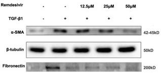
Application: WB    Species: Mice    Sample: lung tissues

FIGURE 6 Remdesivir inhibits TGF-β1-induced activation of Smad and non-Smad signaling pathway in lung fibroblasts (A) Luciferase assays of CAGA-NIH3T3 cells. Cells were pretreated with Remdesivir (0–50 μM) for 30 min and then incubated with TGF-β1 (5 ng ml−1) for 24 h, then analyzed with luciferase assay. SB431542 is a TGF-β1/Smad pathway inhibitor and serves as a positive control (B) NIH-3T3 cells were co-treated with TGF-β1 (5 ng ml−1) and Remdesivir (12.5, 25, 50 μM) for 1 h. P-Smad3 and Smad3 were assessed using western blot. GAPDH was used as the internal control (C) PPF cells were co-treated with TGF-β1 (5 ng ml−1) and Remdesivir (12.5, 25, 50 μM) for 1 h. P-Smad3 and Smad3 were assessed using western blot. GAPDH was used as the internal control (D) NIH-3T3 cells were co-treated with TGF-β1 (5 ng ml−1) and Remdesivir (12.5, 25, 50 μM) for 1 h and the phosphorylation levels of P-38, JNK, ERK and Akt were analyzed by Western blot. β-tubulin was used as a loading control in grayscale analysis. Scale bar = 60 μm. Data was presented as the means ± SD, n = 3. * p < 0.05, ** p < 0.01, *** p < 0.001, **** p < 0.0001.

限制条款

产品的规格、报价、验证数据请以官网为准,官网链接:www.affbiotech.com | www.affbiotech.cn(简体中文)| www.affbiotech.jp(日本語)

产品的数据信息为Affinity所有,未经授权不得收集Affinity官网数据或资料用于商业用途,对抄袭产品数据的行为我们将保留诉诸法律的权利。

产品相关数据会因产品批次、产品检测情况随时调整,如您已订购该产品,请以订购时随货说明书为准,否则请以官网内容为准,官网内容有改动时恕不另行通知。

Affinity保证所销售产品均经过严格质量检测。如您购买的商品在规定时间内出现问题需要售后时,请您在Affinity官方渠道提交售后申请。

产品仅供科学研究使用。不用于诊断和治疗。 

产品未经授权不得转售。

Affinity Biosciences将不会对在使用我们的产品时可能发生的专利侵权或其他侵权行为负责。Affinity Biosciences, Affinity Biosciences标志和所有其他商标所有权归Affinity Biosciences LTD.