产品: STAT1 抗体
货号: AF6300
描述: Rabbit polyclonal antibody to STAT1
应用: WB IHC IF/ICC IP
文献验证: WB, IHC
反应: Human, Mouse, Rat, Monkey
预测: Pig, Bovine, Horse, Rabbit, Dog, Chicken, Xenopus
蛋白号: P42224
RRID: AB_2835149

浏览相似产品>>

   规格 价格 库存
 50ul RMB¥ 1250 现货
 100ul RMB¥ 2300 现货
 200ul RMB¥ 3000 现货

货期: 当天发货

联系销售

产品描述

来源:
Rabbit
应用:
WB 1:500-1:2000, IHC 1:50-1:200, IP, IF/ICC 1:100-1:500
*The optimal dilutions should be determined by the end user.
*Tips:

WB: 适用于变性蛋白样本的免疫印迹检测. IHC: 适用于组织样本的石蜡(IHC-p)或冰冻(IHC-f)切片样本的免疫组化/荧光检测. IF/ICC: 适用于细胞样本的荧光检测. ELISA(peptide): 适用于抗原肽的ELISA检测.

反应:
Human, Mouse, Rat, Monkey
克隆:
Polyclonal
特异性:
STAT1 Antibody detects endogenous levels of total STAT1.
RRID:
AB_2835149
引用格式: Affinity Biosciences Cat# AF6300, RRID:AB_2835149.
偶联:
Unconjugated.
纯化:
The antiserum was purified by peptide affinity chromatography using SulfoLink™ Coupling Resin (Thermo Fisher Scientific).
保存:
Rabbit IgG in phosphate buffered saline , pH 7.4, 150mM NaCl, 0.02% sodium azide and 50% glycerol. Store at -20 °C. Stable for 12 months from date of receipt.
别名:

展开/折叠

CANDF7; DKFZp686B04100; ISGF 3; ISGF3; OTTHUMP00000163552; OTTHUMP00000165046; OTTHUMP00000165047; OTTHUMP00000205845; Signal transducer and activator of transcription 1; Signal transducer and activator of transcription 1, 91kDa; Signal transducer and activator of transcription 1-alpha/beta; Stat1; STAT1_HUMAN; STAT91; Transcription factor ISGF-3 components p91/p84;

抗原和靶标

免疫原:

A synthesized peptide derived from human STAT1, corresponding to a region within C-terminal amino acids.

基因/基因ID:
描述:
The protein encoded by this gene is a member of the STAT protein family. In response to cytokines and growth factors, STAT family members are phosphorylated by the receptor associated kinases, and then form homo- or heterodimers that translocate to the cell nucleus where they act as transcription activators.

研究领域

· Cellular Processes > Cell growth and death > Necroptosis.   (View pathway)

· Environmental Information Processing > Signal transduction > Jak-STAT signaling pathway.   (View pathway)

· Human Diseases > Infectious diseases: Parasitic > Leishmaniasis.

· Human Diseases > Infectious diseases: Parasitic > Toxoplasmosis.

· Human Diseases > Infectious diseases: Bacterial > Tuberculosis.

· Human Diseases > Infectious diseases: Viral > Hepatitis C.

· Human Diseases > Infectious diseases: Viral > Hepatitis B.

· Human Diseases > Infectious diseases: Viral > Measles.

· Human Diseases > Infectious diseases: Viral > Influenza A.

· Human Diseases > Infectious diseases: Viral > Human papillomavirus infection.

· Human Diseases > Infectious diseases: Viral > Herpes simplex infection.

· Human Diseases > Cancers: Overview > Pathways in cancer.   (View pathway)

· Human Diseases > Cancers: Specific types > Pancreatic cancer.   (View pathway)

· Human Diseases > Immune diseases > Inflammatory bowel disease (IBD).

· Organismal Systems > Immune system > Chemokine signaling pathway.   (View pathway)

· Organismal Systems > Development > Osteoclast differentiation.   (View pathway)

· Organismal Systems > Immune system > Toll-like receptor signaling pathway.   (View pathway)

· Organismal Systems > Immune system > NOD-like receptor signaling pathway.   (View pathway)

· Organismal Systems > Immune system > Th1 and Th2 cell differentiation.   (View pathway)

· Organismal Systems > Immune system > Th17 cell differentiation.   (View pathway)

· Organismal Systems > Endocrine system > Prolactin signaling pathway.   (View pathway)

· Organismal Systems > Endocrine system > Thyroid hormone signaling pathway.   (View pathway)

文献引用

1). Programmed BRD9 Degradation and Hedgehog Signaling Activation via Silk-Based Core-Shell Microneedles Promote Diabetic Wound Healing. Advanced science (Weinheim, Baden-Wurttemberg, Germany), 2024 (PubMed: 39413023) [IF=15.1]

2). Calcium silicate-human serum albumin composite hydrogel decreases random pattern skin flap necrosis by attenuating vascular endothelial cell apoptosis and inflammation. Chemical Engineering Journal, 2021 [IF=13.3]

3). A Constant Filgotinib Delivery Adhesive Platform Based on Polyethylene Glycol (PEG) Hydrogel for Accelerating Wound Healing via Restoring Macrophage Mitochondrial Homeostasis. Small (Weinheim an der Bergstrasse, Germany), 2024 (PubMed: 39679768) [IF=13.0]

4). Targeting CDCP1 boost CD8+ T cells-mediated cytotoxicity in cervical cancer via the JAK/STAT signaling pathway. Journal for immunotherapy of cancer, 2024 (PubMed: 39455095) [IF=10.9]

5). Aligned electrospun poly(l-lactide) nanofibers facilitate wound healing by inhibiting macrophage M1 polarization via the JAK-STAT and NF-κB pathways. JOURNAL OF NANOBIOTECHNOLOGY, 2022 (PubMed: 35883095) [IF=10.2]

Application: WB    Species: Mice    Sample:

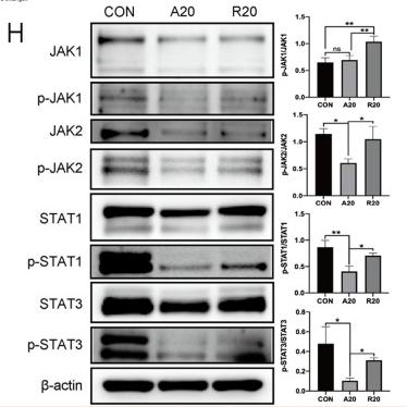
Fig. 3 The underlying mechanism by which aligned fibers affected macrophage polarization. A Venn diagram showing differentially expressed genes. B KEGG pathway analysis between the A20 and R20 groups. C Heatmap of differentially expressed genes among the three groups. D Heatmap of macrophage polarization-related genes between the A20 and R20 groups. E Volcano diagram of differentially expressed genes. F Western blot analysis of the NF-κB signaling pathway. G Immunofluorescence staining showing the nuclear translocation of NF-κB p65. The nucleus is stained blue, and NF-κB p65 protein is stained red. H Western blot images and semiquantitative analysis of the JAK-STAT signaling pathway (*p < 0.05, **p < 0.01, n = 3)

6). Tofacitinib Promotes Functional Recovery after Spinal Cord Injury by Regulating Microglial Polarization via JAK/STAT Signaling Pathway. International Journal of Biological Sciences, 2023 (PubMed: 37781508) [IF=8.2]

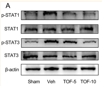
Application: WB    Species: Rat    Sample: spinal cord

Figure 8 TOF normalizes the activated STAT signaling in injured spinal cord and ex vivo microglia under inflammatory conditions. (A) Western blot analysis performed for p-STAT1, STAT1, p-STAT3 and STAT3 expression in spinal cord obtained on day 7 post-injury. (B) Western blot analysis of p-STAT1, STAT1, p-STAT3 and STAT3 expression in primary microglia. (C) Quantification of the ratio of p-STAT1/ STAT1 and p-STAT3/ STAT3 (The values are presented as mean ± SD; *p

7). Lactate Facilitates Pancreatic Repair Following Acute Pancreatitis by Promoting Reparative Macrophage Polarization. Cellular and Molecular Gastroenterology and Hepatology, 2025 [IF=7.4]

Application: WB    Species: Mouse    Sample:

Figure 12. Lactate inhibits the JAK2-STAT1 signaling pathway in macrophages through the GPR132 receptor, facilitating a switch in macrophage phenotype. (A) KEGG pathway analysis was conducted on significantly differentially expressed genes in pancreatic macrophages during recovery following AP in WT mice, alongside the mRNA expression levels of Gpr132, n = 3/group. (B) qPCR analysis of Gpr132 mRNA expression in macrophages under lactate and GPR132 antagonist (0.075 μM, 0.15 μM, or 0.3 μM) treatment, Lactate (Lac) = 15 mM, n = 3–4/group. (C–D) WB analysis was performed to assess the expression levels of β-actin, STAT1, P-STAT1, JAK2, P-JAK2 (C), with the relative quantification (D), n = 3/group, Gpr = 0.075 μM. (E) qPCR detection of pro-inflammatory genes (Cxcl10, Ccl12, Il6) expression levels in macrophages following intervention with a GPR132 inhibitor, n = 3–4/group. (F) qPCR analysis of repair genes (Lrg1, Retnla, Vegfα) expression levels in macrophages post-intervention with a GPR132 inhibitor, n = 3–4/group. Data are expressed as mean ± SEM. Statistical significance is indicated as follows: ∗P < .05; ∗∗P < .01; ∗∗∗P < .001; and ∗∗∗∗P < .0001.

8). Phillygenin inhibited M1 macrophage polarization and reduced hepatic stellate cell activation by inhibiting macrophage exosomal miR-125b-5p. BIOMEDICINE & PHARMACOTHERAPY, 2023 (PubMed: 36652738) [IF=6.9]

Application: WB    Species: Mouse    Sample: RAW264.7 cells

Fig. 2. PHI inhibited the JAK/STAT and Notch1 signaling pathways in RAW264.7 cells. (a) The 3D and 2D models of molecular docking of PHI and JAK1. (b) The 3D and 2D models of molecular docking of PHI and JAK2. (c) The 3D and 2D models of molecular docking of PHI and Notch1. (d-g) The expression of JAK1, JAK2, STAT1 and Notch1 mRNA in RAW264.7 cells after treatment with LPS/IFNγ and PHI for 12 h was detected by RT-qPCR (n = 3). (h) The expression of JAK1, JAK2, p-JAK1, p-JAK2, STAT1, p-STAT1 and Notch1 proteins in RAW264.7 cells after treatment with LPS/IFNγ and PHI for 12 h was detected by western blotting. (i-l) The relative quantification of p-JAK1/JAK1, p-JAK2/JAK2, p-STAT1/STAT1, and Notch1 protein expression in western blotting results was analyzed by ImageJ software (n = 3). Results are presented as mean ± SD. ###P 

9). Licorice-saponin A3 is a broad-spectrum inhibitor for COVID-19 by targeting viral spike and anti-inflammation. Journal of pharmaceutical analysis, 2023 (PubMed: 37363744) [IF=6.1]

10). Poly(ADP-ribose) polymerase family member 14 promotes functional recovery after spinal cord injury through regulating microglia M1/M2 polarization via STAT1/6 pathway. Neural Regeneration Research, 2023 (PubMed: 36751810) [IF=5.9]

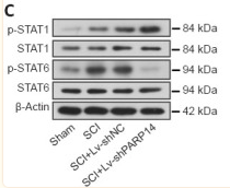
Application: WB    Species: Mouse    Sample:

Figure 5 PARP14 deficiency activates the STAT1 pathway but blocks the STAT6 pathway in mice 7 days post-SCI. (A) Representative images and quantitative analysis showing p-STAT1 (Try701)+/Iba1+ immunofluorescence staining. Lv-shPARP14 injection further promoted SCI-induced STAT1 pathway activation. White arrows indicate p-STAT1 (Try701)+ (green, FITC-labeled)/Iba1+ (red, Cy3-labeled, microglia marker) cells. (B) Representative images and quantitative analysis showing p-STAT6 (Tyr641)+/Iba1+ immunofluorescence staining. Lv-shPARP14 injection inhibited SCI-induced STAT6 pathway activation. White arrows indicated p-STAT6 (Tyr641)+ (green, FITC-labeled)/Iba1+ (red, Cy3-labeled) cells. Scale bars: 50 µm. (C) Relative protein levels of p-STAT1 (Try701), and p-STAT6 (Tyr641) in each group were detected by western blot analysis. p-STAT1 (Try701) expression was increased by Lv-shPARP14 injection, but p-STAT6 (Tyr641) expression was decreased by PARP14 silencing. Values are shown as mean ± SD (n = 6). **P < 0.01 (one-way analysis of variance followed by Tukey’s post hoc test). Images were taken from the gray matter ventral horn at the injury site. Spinal cord tissues from the injury site were used for western blot detection. DAPI: 4′,6-Diamidino-2-phenylindole; Iba1: ionized calcium-binding adaptor molecule 1; PARP14: poly(ADP-ribose)polymerase, member 14; SCI: spinal cord injury; STAT1: signal transducer and activator of transcription.

加载更多

限制条款

产品的规格、报价、验证数据请以官网为准,官网链接:www.affbiotech.com | www.affbiotech.cn(简体中文)| www.affbiotech.jp(日本語)

产品的数据信息为Affinity所有,未经授权不得收集Affinity官网数据或资料用于商业用途,对抄袭产品数据的行为我们将保留诉诸法律的权利。

产品相关数据会因产品批次、产品检测情况随时调整,如您已订购该产品,请以订购时随货说明书为准,否则请以官网内容为准,官网内容有改动时恕不另行通知。

Affinity保证所销售产品均经过严格质量检测。如您购买的商品在规定时间内出现问题需要售后时,请您在Affinity官方渠道提交售后申请。

产品仅供科学研究使用。不用于诊断和治疗。 

产品未经授权不得转售。

Affinity Biosciences将不会对在使用我们的产品时可能发生的专利侵权或其他侵权行为负责。Affinity Biosciences, Affinity Biosciences标志和所有其他商标所有权归Affinity Biosciences LTD.