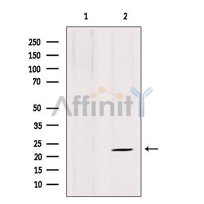
产品: ARL8B 抗体
货号: DF12350
描述: Rabbit polyclonal antibody to ARL8B
应用: WB IHC
文献验证: WB
反应: Human, Mouse
预测: Pig, Bovine, Horse, Sheep, Rabbit, Dog, Chicken
分子量: 22 kDa; 22kD(Calculated).
蛋白号: Q9NVJ2
RRID: AB_2845155

浏览相似产品>>

   规格 价格 库存
 50ul RMB¥ 1250 现货
 100ul RMB¥ 2300 现货
 200ul RMB¥ 3000 现货

货期: 当天发货

联系销售

产品描述

来源:
Rabbit
应用:
WB 1:500-1:2000, IHC 1:50-1:200
*The optimal dilutions should be determined by the end user.
*Tips:

WB: 适用于变性蛋白样本的免疫印迹检测. IHC: 适用于组织样本的石蜡(IHC-p)或冰冻(IHC-f)切片样本的免疫组化/荧光检测. IF/ICC: 适用于细胞样本的荧光检测. ELISA(peptide): 适用于抗原肽的ELISA检测.

反应:
Human, Mouse
预测:
Pig(100%), Bovine(%), Horse(%), Sheep(%), Rabbit(%), Dog(%), Chicken(%)
克隆:
Polyclonal
特异性:
ARL8B Antibody detects endogenous levels of total ARL8B.
RRID:
AB_2845155
引用格式: Affinity Biosciences Cat# DF12350, RRID:AB_2845155.
偶联:
Unconjugated. 130
纯化:
The antiserum was purified by peptide affinity chromatography using SulfoLink™ Coupling Resin (Thermo Fisher Scientific).
保存:
Rabbit IgG in phosphate buffered saline , pH 7.4, 150mM NaCl, 0.02% sodium azide and 50% glycerol. Store at -20 °C. Stable for 12 months from date of receipt.
别名:

展开/折叠

ADP ribosylation factor like 10C; ADP-ribosylation factor-like protein 10C; ADP-ribosylation factor-like protein 8B; ARL10C; ARL8B; ARL8B_HUMAN; FLJ10702; Gie1; Novel small G protein indispensable for equal chromosome segregation 1;

抗原和靶标

免疫原:
Uniprot:
基因/基因ID:
表达:
Q9NVJ2 ARL8B_HUMAN:

Ubiquitously expressed.

序列:
MLALISRLLDWFRSLFWKEEMELTLVGLQYSGKTTFVNVIASGQFSEDMIPTVGFNMRKVTKGNVTIKIWDIGGQPRFRSMWERYCRGVNAIVYMIDAADREKIEASRNELHNLLDKPQLQGIPVLVLGNKRDLPNALDEKQLIEKMNLSAIQDREICCYSISCKEKDNIDITLQWLIQHSKSRRS

种属预测

种属预测:

score>80的预测可信度较高,可尝试用于WB检测。*预测模型主要基于免疫原序列比对,结果仅作参考,不作为质保凭据。

Species
Results
Score
Pig
100
Horse
100
Bovine
100
Sheep
100
Dog
100
Chicken
100
Rabbit
100
Xenopus
0
Zebrafish
0
Model Confidence:
High(score>80) Medium(80>score>50) Low(score<50) No confidence

研究背景

功能:

Plays a role in lysosome motility. In neurons, mediates the anterograde axonal long-range transport of presynaptic lysosome-related vesicles required for presynaptic biogenesis and synaptic function (By similarity). May play a role in chromosome segregation.

细胞定位:

Late endosome membrane. Lysosome membrane. Cytoplasm>Cytoskeleton>Spindle. Cell projection>Axon. Cell junction>Synapse.
Note: Localizes with microtubules at the spindle mid-zone during mitosis.

Extracellular region or secreted Cytosol Plasma membrane Cytoskeleton Lysosome Endosome Peroxisome ER Golgi apparatus Nucleus Mitochondrion Manual annotation Automatic computational assertionSubcellular location
组织特异性:

Ubiquitously expressed.

亚基结构:

Interacts with tubulin Ref.16). Interacts with BORCS5; recruits ARL8B to lysosomes.

蛋白家族:

Belongs to the small GTPase superfamily. Arf family.

文献引用

1). The role of lysosomes as intermediates in betacoronavirus PHEV egress from nerve cells. Journal of virology, 2023 (PubMed: 38009916) [IF=4.0]

Application: WB    Species: Mouse    Sample:

Fig 3 PHEV uses an Arl8b-dependent lysosomal exocytic pathway to egress. (A) Colocalization of PHEV and surface LAMP1. Mock- or PHEV-infected cells at 48 hpi were immunostained with anti-LAMP1 (red) and anti-PHEV (green) antibodies. Scale bar, 10 µm. (B) The surface LAMP1 levels on mock- and PHEV-infected cells from the experiment whose results are shown in panel A were quantified by using Image J. (C) The protein levels of Pro-CTSD, Pro-CTSB, Mat-CTSD, Mat-CTSB, and GAPDH at 24 and 48 hpi were analyzed by western blot, respectively. (D) Mock- or PHEV-infected cells at 48 hpi were immunostained with anti-LAMP1 (red), anti-Arl8b (green), and anti-PHEV (teal) antibodies. Scale bar, 10 µm. (E) The protein levels of Arl8b, PHEV, and GAPDH were analyzed by western blot in PHEV-infected Arl8b small interfering RNA (siRNA)-treated cells or PHEV-infected-non-target siRNA-treated cells. (F) The PHEV N genomic RNA was determined using qPCR in Arl8b siRNA-treated cells and non-target siRNA-treated cells. The data were normalized to the non-target siRNA-treated cells. Representative blots and images are shown. Data are shown as mean ± SD. P values were considered significant when P < 0.05 and denoted as

Application: IF/ICC    Species: Mouse    Sample:

Fig 3 PHEV uses an Arl8b-dependent lysosomal exocytic pathway to egress. (A) Colocalization of PHEV and surface LAMP1. Mock- or PHEV-infected cells at 48 hpi were immunostained with anti-LAMP1 (red) and anti-PHEV (green) antibodies. Scale bar, 10 µm. (B) The surface LAMP1 levels on mock- and PHEV-infected cells from the experiment whose results are shown in panel A were quantified by using Image J. (C) The protein levels of Pro-CTSD, Pro-CTSB, Mat-CTSD, Mat-CTSB, and GAPDH at 24 and 48 hpi were analyzed by western blot, respectively. (D) Mock- or PHEV-infected cells at 48 hpi were immunostained with anti-LAMP1 (red), anti-Arl8b (green), and anti-PHEV (teal) antibodies. Scale bar, 10 µm. (E) The protein levels of Arl8b, PHEV, and GAPDH were analyzed by western blot in PHEV-infected Arl8b small interfering RNA (siRNA)-treated cells or PHEV-infected-non-target siRNA-treated cells. (F) The PHEV N genomic RNA was determined using qPCR in Arl8b siRNA-treated cells and non-target siRNA-treated cells. The data were normalized to the non-target siRNA-treated cells. Representative blots and images are shown. Data are shown as mean ± SD. P values were considered significant when P < 0.05 and denoted as

限制条款

产品的规格、报价、验证数据请以官网为准,官网链接:www.affbiotech.com | www.affbiotech.cn(简体中文)| www.affbiotech.jp(日本語)

产品的数据信息为Affinity所有,未经授权不得收集Affinity官网数据或资料用于商业用途,对抄袭产品数据的行为我们将保留诉诸法律的权利。

产品相关数据会因产品批次、产品检测情况随时调整,如您已订购该产品,请以订购时随货说明书为准,否则请以官网内容为准,官网内容有改动时恕不另行通知。

Affinity保证所销售产品均经过严格质量检测。如您购买的商品在规定时间内出现问题需要售后时,请您在Affinity官方渠道提交售后申请。

产品仅供科学研究使用。不用于诊断和治疗。 

产品未经授权不得转售。

Affinity Biosciences将不会对在使用我们的产品时可能发生的专利侵权或其他侵权行为负责。Affinity Biosciences, Affinity Biosciences标志和所有其他商标所有权归Affinity Biosciences LTD.