产品: 磷酸化 Histone H3 (Ser11) 抗体
货号: AF3358
描述: Rabbit polyclonal antibody to Phospho-Histone H3 (Ser11)
应用: WB IHC IF/ICC
文献验证: WB, IF/ICC
反应: Human, Mouse, Rat, Spodoptera frugiperda
预测: Bovine
蛋白号: P68431 | Q71DI3 | P84243
RRID: AB_2834773

浏览相似产品>>

   规格 价格 库存
 50ul RMB¥ 1300 现货
 100ul RMB¥ 2400 现货
 200ul RMB¥ 3200 现货

货期: 当天发货

联系销售

产品描述

来源:
Rabbit
应用:
WB 1:500-1:2000, IHC 1:50-1:200, IF/ICC 1:100-1:500
*The optimal dilutions should be determined by the end user.
*Tips:

WB: 适用于变性蛋白样本的免疫印迹检测. IHC: 适用于组织样本的石蜡(IHC-p)或冰冻(IHC-f)切片样本的免疫组化/荧光检测. IF/ICC: 适用于细胞样本的荧光检测. ELISA(peptide): 适用于抗原肽的ELISA检测.

反应:
Human, Mouse, Rat, Spodoptera frugiperda
克隆:
Polyclonal
特异性:
Phospho-Histone H3 (Ser11) Antibody detects endogenous levels of Histone H3 only when phosphorylated at Serine 11.
RRID:
AB_2834773
引用格式: Affinity Biosciences Cat# AF3358, RRID:AB_2834773.
偶联:
Unconjugated.
纯化:
The antibody is from purified rabbit serum by affinity purification via sequential chromatography on phospho-peptide and non-phospho-peptide affinity columns.
保存:
Rabbit IgG in phosphate buffered saline , pH 7.4, 150mM NaCl, 0.02% sodium azide and 50% glycerol. Store at -20 °C. Stable for 12 months from date of receipt.
别名:

展开/折叠

H3 histone family, member A; H3/A; H31_HUMAN; H3FA; Hist1h3a; HIST1H3B; HIST1H3C; HIST1H3D; HIST1H3E; HIST1H3F; HIST1H3G; HIST1H3H; HIST1H3I; HIST1H3J; histone 1, H3a; Histone cluster 1, H3a; Histone H3.1; Histone H3/a; Histone H3/b; Histone H3/c; Histone H3/d; Histone H3/f; Histone H3/h; Histone H3/i; Histone H3/j; Histone H3/k; Histone H3/l; ;H3.3A; HIST1 cluster, H3E; H3 histone family, member A; H3.1; H3/l; H3F3; H3FF; H3FJ; H3FL; Histone gene cluster 1, H3 histone family, member E; histone H3.1t; Histone H3/o; FLJ92264; H 3; H3; H3 histone family, member B; H3 histone family, member C; H3 histone family, member D; H3 histone family, member F; H3 histone family, member H; H3 histone family, member I; H3 histone family, member J; H3 histone family, member K; H3 histone family, member L; H3 histone family, member T; H3 histone, family 3A; H3/A; H3/b; H3/c; H3/d; h3/f; H3/h; H3/i; H3/j; H3/k; H3/t; H31_HUMAN; H3F1K; H3F3A; H3FA; H3FB; H3FC; H3FD; H3FH; H3FI; H3FK; HIST1 cluster, H3A; HIST1 cluster, H3B; HIST1 cluster, H3C; HIST1 cluster, H3D; HIST1 cluster, H3F; HIST1 cluster, H3G; HIST1 cluster, H3H; HIST1 cluster, H3I; HIST1 cluster, H3J; HIST1H3A; HIST1H3B; HIST1H3C; HIST1H3D; HIST1H3E; HIST1H3F; HIST1H3G; HIST1H3H; HIST1H3I; HIST1H3J; HIST3H3; Histone 1, H3a; Histone 1, H3b; Histone 1, H3c; Histone 1, H3d; Histone 1, H3e; Histone 1, H3f; Histone 1, H3g; Histone 1, H3h; Histone 1, H3i; Histone 3, H3; histone cluster 1 H3 family member a; histone cluster 1 H3 family member b; histone cluster 1 H3 family member c; histone cluster 1 H3 family member d; histone cluster 1 H3 family member e; histone cluster 1 H3 family member f; histone cluster 1 H3 family member g; histone cluster 1 H3 family member h; histone cluster 1 H3 family member i; histone cluster 1 H3 family member j; Histone cluster 1, H3a; Histone cluster 1, H3b; Histone cluster 1, H3c; Histone cluster 1, H3d; Histone cluster 1, H3e; Histone cluster 1, H3f; Histone cluster 1, H3g; Histone cluster 1, H3i; Histone cluster 1, H3j; Histone gene cluster 1, H3 histone family, member A; Histone gene cluster 1, H3 histone family, member B; Histone gene cluster 1, H3 histone family, member C; Histone gene cluster 1, H3 histone family, member D; Histone gene cluster 1, H3 histone family, member F; Histone gene cluster 1, H3 histone family, member G; Histone gene cluster 1, H3 histone family, member H; Histone gene cluster 1, H3 histone family, member I; Histone gene cluster 1, H3 histone family, member J; Histone gene cluster 1, H3A; Histone gene cluster 1, H3B; Histone gene cluster 1, H3C; Histone gene cluster 1, H3D; Histone gene cluster 1, H3E; Histone gene cluster 1, H3F; Histone gene cluster 1, H3G; Histone gene cluster 1, H3H; Histone gene cluster 1, H3I; Histone gene cluster 1, H3J; Histone H 3; Histone H3.1; Histone H3.2; Histone H3.3; Histone H3/a; Histone H3/b; Histone H3/c; Histone H3/d; Histone H3/f; Histone H3/h; Histone H3/i; Histone H3/j; Histone H3/k; Histone H3/l; Histone H3/m; H3 histone family 3A; H3 histone family 3B; H3 histone, family 3B (H3.3B); H3.3; H3.3A; H3.3B; H33_HUMAN; H3F3; H3F3A; H3f3b; Histone H3.3; Histone H3.3Q; Histone H3.A; Histone H3.B; MGC87782; MGC87783;

抗原和靶标

免疫原:

A synthesized peptide derived from human Histone H3 around the phosphorylation site of Ser11.

基因/基因ID:
描述:
H3F3A Variant histone H3 which replaces conventional H3 in a wide range of nucleosomes in active genes. Constitutes the predominant form of histone H3 in non-dividing cells and is incorporated into chromatin independently of DNA synthesis.

研究领域

· Human Diseases > Substance dependence > Alcoholism.

· Human Diseases > Cancers: Overview > Transcriptional misregulation in cancer.

· Human Diseases > Immune diseases > Systemic lupus erythematosus.

文献引用

1). Prpf31 is essential for the survival and differentiation of retinal progenitor cells by modulating alternative splicing. NUCLEIC ACIDS RESEARCH, 2021 (PubMed: 33476374) [IF=16.6]

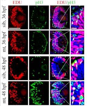
Application: IF/ICC    Species: zebrafish    Sample: EDU (S-phase cells) and pH3 (M-phase cells)

Figure 4. Prpf31 deficiency causes abnormal spindle structure and mitotic arrest. (A) Double staining of EDU (S-phase cells) and pH3 (M-phase cells) in the retinal sections of WT siblings and prpf31−/− mutants at 36 and 48 hpf. The number of M-phase cells were significantly increased in the prpf31−/− mutants compared with siblings, suggesting that RPCs may be arrested in M phase. White arrows, overlapping signals of EDU and pH3. Scale bars: left, 100 μm; right, 10 μm. (B) Quantification of EDU+, pH3+ and EDU+ / pH3+ cells shown in (A). n = 6 for each panel. (C) In vivo imaging of the H2A-mCherry labeled chromosomes showed the mitotic progression of RPCs at 36 and 48 hpf. The time point of nuclear envelope breakdown (NEBD) was set as the start of mitosis. Scale bar, 10 μm. (D, E) Quantification of the time from NEBD to anaphase in RPCs at 36 and 48 hpf. 15 cells from more than five embryos were observed for each group. For abnormally divided cells, the longest observation time was 150 min. Scale bar, 10 μm. (F) The spindle and nuclei of RPCs from mutants and WT siblings were stained with anti-α-tubulin (green) antibody and PI (red), respectively. The different types of spindle anomalies are displayed in the panels (Abnormal 1–4). Scale bar, 10 μm. (G) Quantitative analysis of the RPCs numbers in each of the phases of mitosis in sibling and prpf31 mutant embryos at 36 and 48 hpf.

2). PpV, acting via the JNK pathway, represses apoptosis during normal development of Drosophila wing. APOPTOSIS, 2018 (PubMed: 30159848) [IF=6.1]

3). Securinine inhibits the tumor growth of human bladder cancer cells by suppressing Wnt/β-catenin signaling pathway and activating p38 and JNK signaling pathways. Biochemical pharmacology, 2024 (PubMed: 38484850) [IF=5.3]

Application: WB    Species: Human    Sample: BC cells

Fig. 1. SEC inhibits the proliferation and induces cell cycle arrest of BC cells. (A) The chemical structure of SEC. (B) The effect of SEC on the viability of BC cells (Crystal violet staining). (C) The effect of SEC on the viability of BC cells (MTT). (D) The effect of SEC on the colony-forming ability of BC cells (Colony formation assay). (E) The effect of SEC on the protein level of PCNA and c-Myc of BC cells (Western blot). (F) The effect of SEC on the cell cycle of BC cells (Flow cytometry). (G) The effect of SEC on the protein levels of CDK2, Cyclin A, Cyclin B1 and P-Histone H3 of BC cells (Western blot). Data are shown as mean ± SD from three independent experiments. *P < 0.05, **P < 0.01, ***P < 0.001, vs. control group. #P < 0.05, ##P < 0.01, ###P < 0.001. (For interpretation of the references to colour in this figure legend, the reader is referred to the web version of this article.)

4). Deficiency of copper responsive gene stmn4 induces retinal developmental defects. Cell biology and toxicology, 2024 (PubMed: 38252267) [IF=5.3]

Application: IF/ICC    Species: Zebrafish    Sample:

Fig. 5 RPCs in stmn4−/− mutants arrested in M-phase and showed abnormal mitosis. (A) Brdu cell proliferation assays in WT and stmn4−/− zebrafish embryos at 48 hpf (A1 − A8) and the calculation data (A9). (B) PH3 cell proliferation assays in WT and stmn4−/− zebrafish embryos at 48 hpf (B1 − B6) and the calculation data (B7). (C) TUNEL assays in WT and stmn4−/− zebrafish embryos at 48 hpf (C1 − C6) and the calculation data (C7). (D) Immunofluorescence assays for cytoskeleton Tubulin in zebrafish embryo at 48 hpf (D1 − D8) and the calculation data (D9). Scale bar, 50 μm (A1 − A6, B1 − B6, C1 − C6), 25 μm (A7, A8), 10 μm (D1 − D8). ***P 

5). Rictor regulates the vasculogenic mimicry of melanoma via the AKT-MMP-2/9 pathway. Journal of Cellular and Molecular Medicine, 2017 (PubMed: 28699701) [IF=5.3]

Application: WB    Species: human    Sample:

Fig. 3 Knockdown of Rictor inhibits melanoma cells proliferation and blocked the cell cycle in G2/M phase. (A) Cell viability of A375 and MUM-2B cells after Rictor knockdown evaluated by MTT assay (*P < 0.05, **P < 0.01). (B) Cell cycle of A375 and MUM-2B cells after Rictor knockdown examined by FCM. (C) The expression of p-CDK2 and p-Histone H3 induced by knockdown of Rictor.

6). Knockout of mafba Causes Inner-Ear Developmental Defects in Zebrafish via the Impairment of Proliferation and Differentiation of Ionocyte Progenitor Cells. Biomedicines, 2021 (PubMed: 34829928) [IF=4.7]

Application: IF/ICC    Species: Mouse    Sample:

Figure 4. Knockout of mafba reduced the proliferation of epidermal stem cells and the dlc+ ionocyte progenitor cell number. (A) Examples of p63 with dlc or p63 with EDU or dlc with EDU colocalized (arrowhead) or non-colocalized (arrow) cells between sibling and mafba−/− groups are shown in (a–l). The quantitative analyses of the p63+ (marker for epidermal stem cells), dlc−p63+ (marker for keratinocyte precursors), and dlc+p63+ (marker for ionocyte precursors) cells in each group at the bud stage are shown in (m). The quantitative analyses of p63+, dlc−p63+, and dlc+p63+ cells colocalized with EDU (S-phase cells) in sibling and mafba−/− embryos at the bud stage are shown in (n). The n = 13 for each panel. Scale bars: 100 µm. (B) Double staining of p63 and pH3 (M-phase cells) in the siblings and mafba−/− group at the bud stage. Examples of p63 and pH3 colocalized (arrowhead) or non-colocalized (arrow) cells are shown. The quantitative analyses of p63+ cell and p63+ colocalized with pH3-positive cells of sibling and mafba−/− embryos at the bud stage are shown in (i) and (j), respectively. The n = 16 for each panel. Scale bars: 100 µm. (C) The dlc+ ionocyte progenitors’ cell density was reduced in the mafba−/− group (b) as compared to the sibling group (a) at the bud stage. The quantitative analysis of dlc+ ionocyte progenitors’ cell densities are shown in (c). (D) The cell density of foxi3a+ and foxi3b+ ionocyte progenitors are compared between the sibling (a,d) and mafba−/− group (b,e) at the 5-somites stage, respectively. Cell densities of foxi3a+ and foxi3b+ ionocyte progenitors are quantified in (c) and (f), respectively. Scale bars: 100 µm. Data are expressed as mean ± SD; **, p < 0.01; ***, p < 0.001; ****, p < 0.0001.

7). The splicing factor Prpf31 is required for hematopoietic stem and progenitor cell expansion during zebrafish embryogenesis. The Journal of biological chemistry, 2024 (PubMed: 38382674) [IF=4.0]

8). Atp7b deficiency induces zebrafish eye developmental defects. Metallomics : integrated biometal science, 2023 (PubMed: 37070960) [IF=2.9]

9). Schisandrin A and B enhance the dentate gyrus neurogenesis in mouse hippocampus. JOURNAL OF CHEMICAL NEUROANATOMY, 2020 (PubMed: 32027950) [IF=2.7]

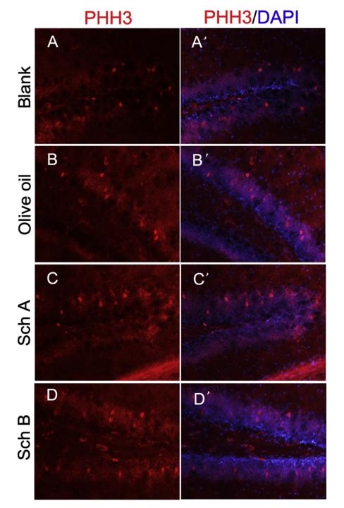
Application: IF/ICC    Species: Mouse    Sample: hippocampus

Fig. 2. Sch A and B promote DG proliferation. (ADe) PHH3 immunostaining in the DG of the Blank, Olive oil, Sch A and Sch B groups respectively. (A'–D') Merged images of PHH3 and DAPI staining. (E) Statistical analysis of the numbers of PHH3+ cells among 4 groups. Data were the mean ± S.D.; * P < 0.05; ** P < 0.01; Scale bar = 50 μm in (A-D').

10). MAD2-p31comet axis deficiency reduces cell proliferation, migration and sensitivity of microtubule-interfering agents in glioma. BIOCHEMICAL AND BIOPHYSICAL RESEARCH COMMUNICATIONS, 2018 (PubMed: 29408509) [IF=2.5]

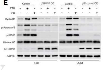
Application: WB    Species: human    Sample: shMAD2-U87 and shMAD2-U251 cells

Figure.4| MAD2 depletion or p31comet overexpression reduces sensitivity of glioma cells to microtubule-interfering agents. (E) The expression levels of cell cycle regulators (Cyclin B1, p-Aurora A/B, p-H3S10 and Histone H3 protein) were analyzed by western blot expression in control or p31comet OE-U87 and p31comet OE-U251 cells in the presence of paclitaxel (100 nM) or vinblastine (100 nM). GPADH was used as internal control in (D) and (E).

限制条款

产品的规格、报价、验证数据请以官网为准,官网链接:www.affbiotech.com | www.affbiotech.cn(简体中文)| www.affbiotech.jp(日本語)

产品的数据信息为Affinity所有,未经授权不得收集Affinity官网数据或资料用于商业用途,对抄袭产品数据的行为我们将保留诉诸法律的权利。

产品相关数据会因产品批次、产品检测情况随时调整,如您已订购该产品,请以订购时随货说明书为准,否则请以官网内容为准,官网内容有改动时恕不另行通知。

Affinity保证所销售产品均经过严格质量检测。如您购买的商品在规定时间内出现问题需要售后时,请您在Affinity官方渠道提交售后申请。

产品仅供科学研究使用。不用于诊断和治疗。 

产品未经授权不得转售。

Affinity Biosciences将不会对在使用我们的产品时可能发生的专利侵权或其他侵权行为负责。Affinity Biosciences, Affinity Biosciences标志和所有其他商标所有权归Affinity Biosciences LTD.