 Antibody.
Lane 1: Pc12 cells(heat-shock treatment), blocked with antigen-specific peptides.
Lane 2: Pc12 cells(heat-shock treatment).
Lane 3: Mcf7 cells(heat-shock treatment).")

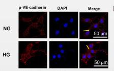
 by IF/ICC. The samples were fixed with PFA and permeabilized in 0.1% Triton X-100,then blocked in 10% serum for 45 minutes at 25°C. Samples were then incubated with primary Ab(AF3265) and mouse anti-beta tubulin Ab(T0023) for 1 hour at 37°C. An AlexaFluor594 conjugated goat anti-rabbit IgG(H+L) Ab(Red) and an AlexaFluor488 conjugated goat anti-mouse IgG(H+L) Ab(Green) were used as the secondary antibody.
The nuclear counter stain is DAPI(blue).")

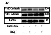
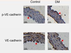
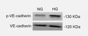
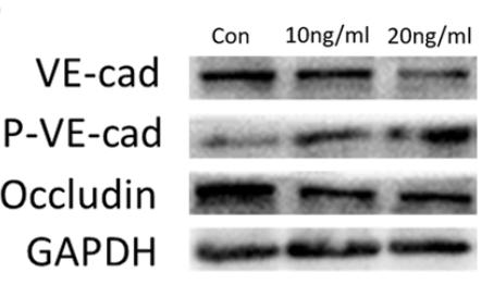
 WB analysis of VLA4, VCAM-1, RAC1, p-PYK2 and p-VE-cad. The protein expression levels of VLA4, VCAM-1, RAC1, p-PYK2 and p-VE-cad were significantly higher in the RHD group than in the control group. (C) RT‒qPCR results for VLA4, VCAM-1 and RAC1. The mRNA expression levels of VLA4, VCAM-1 and RAC1 were significantly higher in the RHD group than in the control group. (D,E) Immunohistochemical results for VLA4, VCAM-1, p-PYK2 and p-VE-cad in heart valves. Magnification, 400 × ; scale bar: 50 µm; arrows point to positive staining. The expression levels of VLA4, VCAM-1, p-PYK2 and p-VE-cad were significantly higher in the RHD group than in the control group. The data are presented as the mean ± SD; *p < 0.05 vs. the control group. VLA4, very late antigen4; VCAM-1, vascular cell adhesion molecule-1; RAC1, RAS-related C3 botulinum substrate 1; p-PYK2, phosphorylated proline-rich tyrosine kinase 2; p-VE-cad, phosphorylated VE cadherin; RHD, rheumatic heart disease.")
 WB analysis of VLA4, VCAM-1, RAC1, p-PYK2 and p-VE-cad. The protein expression levels of VLA4, VCAM-1, RAC1, p-PYK2 and p-VE-cad were significantly higher in the RHD group than in the control group. (C) RT‒qPCR results for VLA4, VCAM-1 and RAC1. The mRNA expression levels of VLA4, VCAM-1 and RAC1 were significantly higher in the RHD group than in the control group. (D,E) Immunohistochemical results for VLA4, VCAM-1, p-PYK2 and p-VE-cad in heart valves. Magnification, 400 × ; scale bar: 50 µm; arrows point to positive staining. The expression levels of VLA4, VCAM-1, p-PYK2 and p-VE-cad were significantly higher in the RHD group than in the control group. The data are presented as the mean ± SD; *p < 0.05 vs. the control group. VLA4, very late antigen4; VCAM-1, vascular cell adhesion molecule-1; RAC1, RAS-related C3 botulinum substrate 1; p-PYK2, phosphorylated proline-rich tyrosine kinase 2; p-VE-cad, phosphorylated VE cadherin; RHD, rheumatic heart disease.")
产品描述
*The optimal dilutions should be determined by the end user. For optimal experimental results, antibody reuse is not recommended.
*Tips:
WB: 适用于变性蛋白样本的免疫印迹检测. IHC: 适用于组织样本的石蜡(IHC-p)或冰冻(IHC-f)切片样本的免疫组化/荧光检测. IF/ICC: 适用于细胞样本的荧光检测. ELISA(peptide): 适用于抗原肽的ELISA检测.
引用格式: Affinity Biosciences Cat# AF3265, RRID:AB_2834691.
展开/折叠
7B 4; 7B4; 7B4 antigen; CADH5_HUMAN; Cadherin 5; Cadherin 5 type 2; Cadherin 5, type 2 (vascular endothelium); Cadherin 5, type 2, VE cadherin (vascular epithelium); cadherin, vascular endothelial; cadherin, vascular endothelial, 1; Cadherin-5; Cadherin5; CD 144; CD144; CD144 antigen; CDH 5; CDH5; CDH5 protein; Endothelial specific cadherin; FLJ17376; OTTHUMP00000174777; Vascular endothelial cadherin; Vascular epithelium cadherin; VE Cad; VE-cadherin; VEC;
抗原和靶标
A synthesized peptide derived from human VE-Cadherin around the phosphorylation site of Tyr731.
研究领域
· Environmental Information Processing > Signaling molecules and interaction > Cell adhesion molecules (CAMs). (View pathway)
· Organismal Systems > Immune system > Leukocyte transendothelial migration. (View pathway)
文献引用

Application: WB Species: Human Sample: HUVECs
Application: WB Species: Human Sample: ECs

Application: WB Species: Mouse Sample: colon tissue

Application: IF/ICC Species: Mouse Sample: colon tissue
Application: WB Species: Human Sample: HMVEC cells

Application: WB Species: Human Sample: HUVECs
Application: IHC Species: Mouse Sample: aortic endothelial cells
Application: WB Species: Mouse Sample: aortic endothelial cells
Application: IF/ICC Species: Mouse Sample: aortic endothelial cells
Application: WB Species: human Sample: HUVECs
限制条款
产品的规格、报价、验证数据请以官网为准,官网链接:www.affbiotech.com | www.affbiotech.cn(简体中文)| www.affbiotech.jp(日本語)产品的数据信息为Affinity所有,未经授权不得收集Affinity官网数据或资料用于商业用途,对抄袭产品数据的行为我们将保留诉诸法律的权利。
产品相关数据会因产品批次、产品检测情况随时调整,如您已订购该产品,请以订购时随货说明书为准,否则请以官网内容为准,官网内容有改动时恕不另行通知。
Affinity保证所销售产品均经过严格质量检测。如您购买的商品在规定时间内出现问题需要售后时,请您在Affinity官方渠道提交售后申请。产品仅供科学研究使用。不用于诊断和治疗。
产品未经授权不得转售。
Affinity Biosciences将不会对在使用我们的产品时可能发生的专利侵权或其他侵权行为负责。Affinity Biosciences, Affinity Biosciences标志和所有其他商标所有权归Affinity Biosciences LTD.