


. After a period of 10-day drug treatments, all mice were sacrificed on the 11 th day for the following tests.
Pathological results were shown by the (a) images as well as (b-g) corresponding quantitative analysis through HE staining, Pizzolato staining, PAS staining, TUNEL
staining, OPN and CD44 immunohistochemistry staining of paraffin embedded kidney sections. (h-j) Renal dysfunction was determined by serum levels of BUN,
creatinine and NAGL among the five groups. (k) Group annotations were applied for all histograms. Data were expressed as the fold changes of experimental group to
NC group or GA group, and were represented as means ± SD. * p < 0.05 vs. NC group; ** p < 0.05 as GA+Kae (25mg/kg) vs. GA group, GA + Kae (50 mg/kg) vs. GA
group, and GA + Kae (25 mg/kg) vs. GA + Kae (50 mg/kg) group, respectively.")
 or adenovirus carrying empty vector (Ad-empty), followed by Ang II treatment (500 nM). Real-time PCR
and Western blotting analysis of ACTA2 (A-B), MYH11 (C-D), SPP1 (E-F), and vimentin
(G-H). β-actin was used as the internal reference. (I) Immunofluorescence staining for
ACTA2 in mAoSMCs. Data are present as mean ± SD (n = 3), and analyzed by one-way
ANOVA test. *** P < 0.001 vs. Control group; ^^ P < 0.01, ^^^ P < 0.001 vs. Ang II +
Ad-empty group. IGFBP3, insulin like growth factor binding protein 3; Ang II, angiotension
II; mAoSMCs, mouse aortic smooth muscle cells; ACTA2, smooth muscle actin alpha 2;
MYH11, myosin heavy chain 11; SPP1, secreted phosphoprotein 1.")
 Polarized light optical microscopy (arrows). ×20 magnification. (b) Pizzolato staining. Pizzolato staining indicates CaOx crystals (arrows). Scale bar = 50 μm. (c) The proportion of the crystal deposition area in the kidney and the proportion of crystal deposition areas in the corticomedullary border. (d) Immunohistochemical distribution of crystal-related gene OPN and crystal adhesion-related gene CD44. Scale bar = 50 μm. (e) The proportion of the IHC-positive area. Gly: glyoxylic acid; ROSI: rosiglitazone; CaOx: calcium oxalate. ∗p < 0.05; ∗∗p < 0.01.")
 Immunofluorescent images of Col-I and OPN expressed by BMSCs cultured on the different microcarriers for 14 days which were observed under CLSM (Scale bar = 50 μm); (B,C) qRT-PCR analysis of Col-I and OPN. (*p < 0.05, **p < 0.01, n = 3).")
 Representative images of wound healing assays in the normoxic or hypoxic group. Scale bar, 1 mm. (B) Percentages of wound closure from three independent experiments are quantified. (C) Representative images of migratory cells stained with crystal violet. (D) Statistical quantification of the number of migratory cells from three independent experiments are shown. Scale bar, 100 µm. (E) Cell viability of HDPCs was detected using Cell Counting Kit-8 analysis. (F) ALP staining and (G) activity in the normoxic or hypoxic group on day 3. Scale bar, 200 µm. (H) Representative western blotting images showing the protein expression levels of RUNX2, Col I, OSX and OPN in the normoxic or hypoxic group on day 3, (I) which were quantified. Results are presented as the means ± SD from ≥ three independent experiments. **P<0.01 and ***P<0.001 vs. normoxia. Nor, normoxia; Hypo, hypoxia; OD, optical density; ALP, alkaline phosphatase; RUNX2, runt-related transcription factor 2; Col I, collagen type I; OSX, osterix; OPN, osteopontin.")

 treated VSMCs. The relative protein expressions in Ang II-treated VSMCs with presence of C/EBPα overexpression or knockdown were analyzed by Western blot. GAPDH was used as an internal control. Data are presented as means ± SEM. (a, b) Evaluation of the influence of C/EBPα on the expression of α-SMA, SM-MHC, and OPN proteins. (c, d) Evaluation of the influence of C/EBPα on the expression of PIK3C2A protein. (e, f) Evaluation of the influence of C/EBPα on the expression of Beclin-1, p62, LC3II, and LC3I proteins. (g, h) Evaluation of the influence of C/EBPα on the expression of MMP-2 and MMP-9 proteins. (i) Intracellular ROS was measured by flow cytometry using DCFH-DA probe with presence of C/EBPα overexpression or knockdown. Control: control group; model: treated with 1 μM Ang II; vector: treated with 1 μM Ang II in vector transfected VSMCs; pcC/EBPα: treated with 1 μM Ang II in C/EBPα-overexpressed VSMCs; siNC: treated with 1 μM Ang II in siRNA NC-transfected VSMCs; siC/EBPα: treated with 1 μM Ang II in C/EBPα-knockdown VSMCs. ∗P < 0.05 vs. control,")
 Evaluation of cell invasion among different groups using crystal violet staining. (B) Comparison of cell invasion among different groups is presented. (C) Protein expression levels of α-SMA and OPN in different groups. Comparison of the (D) α-SMA and (E) OPN protein expression levels among different groups. *P")
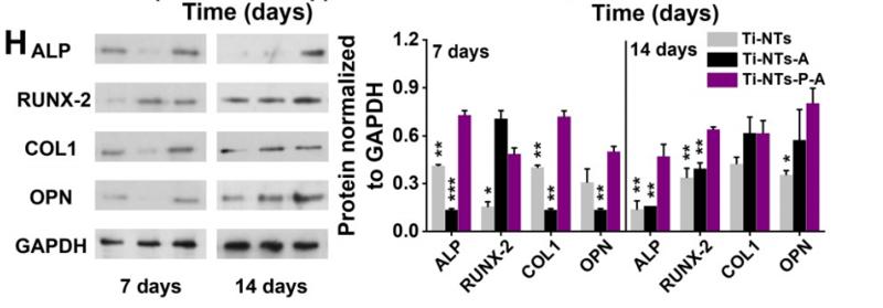
 ALP activity in the femoral heads. (b) mRNA levels of OCN, COL I, OPN, and Runx2 in the femoral heads. (c) Expression levels of OCN, COL I, OPN, and Runx2 in the femoral heads. (d) Representative images of OCN staining and COL I staining (scale bar, 200 μm) in the femoral heads. ∗, p < 0.05; ∗∗, p < 0.01; ns, no significant. ALP, alkaline phosphatase; OCN, osteocalcin; COL I, type I collagen; OPN, osteopontin; Runx2, Runt-related transcription factor 2.")
 Representative pictures of rat knee tissue sections stained with H & E, Saffron O/Fast Green and TRAP. (B) Representative images of protein bands in rat tissues. (C) Representative images of protein bands in rat cartilage. (D) The relative expression levels of MMP3 in cartilage. (E) The relative expression levels of MMP13 in cartilage. (F) The relative expression levels of OPN in femur. (G) The relative expression levels of RUNX-2 in femur. *Statistically significant difference (P < 0.05). **Statistically significant difference (P < 0.01). ***Statistically significant difference (P < 0.001).")
 HE and IHC staining of SPP1 in the lungs of rats (scale bar=100 µm). (B) Protein expression of Col I, TGF-β1 and SPP1 was detected by western blotting and quantified. Data are presented as the mean ± standard deviation. n=5 per group. SPP1, secreted phosphoprotein 1; HE, hematoxylin and eosin; IHC, immunohistochemistry; Col I, collagen I; TGF, transforming growth factor.")
 HE and IHC staining of SPP1 in the lungs of rats (scale bar=100 µm). (B) Protein expression of Col I, TGF-β1 and SPP1 was detected by western blotting and quantified. Data are presented as the mean ± standard deviation. n=5 per group. SPP1, secreted phosphoprotein 1; HE, hematoxylin and eosin; IHC, immunohistochemistry; Col I, collagen I; TGF, transforming growth factor.")
 was associated with the metastasis and the survival of NSCLC. A) OPN was high expressed in NSCLC cell lines. B) The expression of VEGF and E-cadherin in NSCLC tissues was positively correlated with the expression of OPN; scale bar: 100 μm. The disease-free survival (C) and overall survival (D) of NSCLC patients were negatively correlated with the expression of OPN.")
 was associated with the metastasis and the survival of NSCLC. A) OPN was high expressed in NSCLC cell lines. B) The expression of VEGF and E-cadherin in NSCLC tissues was positively correlated with the expression of OPN; scale bar: 100 μm. The disease-free survival (C) and overall survival (D) of NSCLC patients were negatively correlated with the expression of OPN.")
 Body weight of mice at sacrifice. n = 11–12 for each group. (b) HE staining of liver sections. (c, g) ORO, (d, h) Nile Red, (e, i–j) HE, and (f, k) Sirius Red staining of aortic root sections. Scale bar: 200 μm. n = 3–4 for each group. (l) Western blotting on (m) CD36, (n) OPN, and (o) TNF-α also indicated AS development. n = 4 per group. Data were presented as mean ± standard deviation in one-way ANOVA with Dunnett post hoc tests. ns: not significant")
 The mRNA expression of Runx2, OPN, and ALP was measured after 3 and 7 days of CTS. (D) The protein expression of Runx2, OPN, and ALP was also measured after 3 and 7 days of CTS. (E) The ALP staining and quantitative analysis was performed after 3 and 7 days of CTS. (F-G) The mRNA expression of miR-187-3p and CNR2 was measured after 3 and 7 days of CTS. (H) The protein expression of CNR2 was also measured after 3 and 7 days of CTS. (I) Immunofluorescence of CNR2 was performed after 7 days of CTS, with a scale bar of 50 μm.*p")
, RunX2 (B), Col-1 (C), and OCN (D). E and F: Western blotting assay used to detect osteogenic-related proteins. All statistical data are represented as mean ± SD (n = 3; ∗∗∗∗P < 0.0001).")

 Expression differences of the five hub genes between IPF and control groups. (B–E) ROC curves of the four hub genes in IPF and control groups. (F) Representative images of immunohistochemical of CXCL12, CTSG, CXCR2, and SPP1 in lung tissues. (G) The areas of the four hub genes. PPV, positive predictive value; NPV, negative predictive value; AUROC, area under the receiver operating characteristic curve. *p < 0.05; **p < 0.01; ***p < 0.001.")

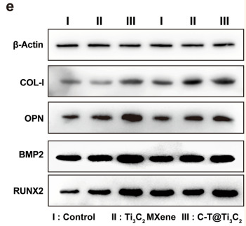
. The full-length gels were included in Additional file 1. The relative expression levels of Smad7 (B), collagen 3 (C), elastase-2B D and osteopontin E were analysed by ImageJ software. *P")
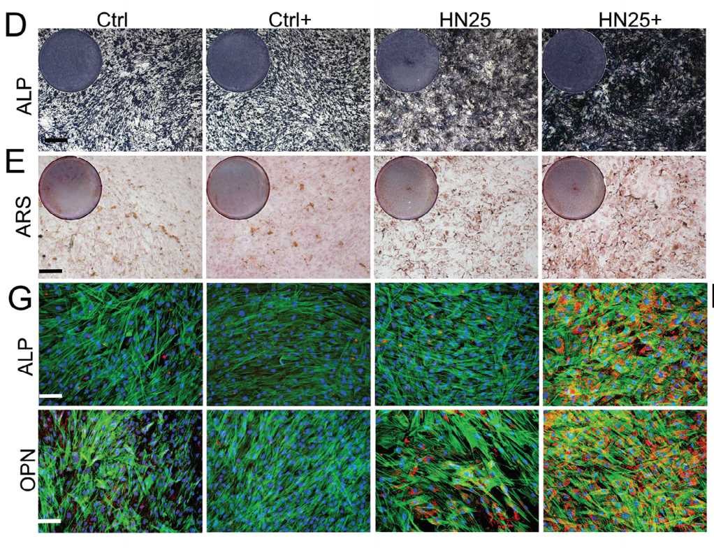
 and (b) Representative images of immunofluorescence costaining RUNX2 (red) and OSX (green) of the bone tissue in each group of implants after 8 weeks and the quantitative analysis of fluorescence intensity. (c and d) Representative images of immunofluorescence costaining β-catenin (red), ALP (green) and AXIN2 (pink) of the bone tissue in each group of implants after 8 weeks and the quantitative analysis of fluorescence intensity. (e and f) Representative images of immunofluorescence costaining Collagen-I (red), OPN (green) and OCN (pink) of the bone tissue in each group of implants after 8 weeks and the quantitative analysis of fluorescence intensity.")
 of rPTX3 treatment. B Cell morphology of MC3T3-E1 after treatment with different doses of rPTX3 (0, 50, 100, 200, 500, and 1000 ng/ml). Scale bar, 100 μm. C WB analysis of osteogenesis markers (ALP, Runx2 and COL1 at day 7, OCN and OPN at day 14) and apoptosis markers (Bcl-2 and Bax at 24 h) in Dex-stimulated MC3T3-E1 treated with or without rPTX3 and related quantification. Actin was used as an internal control. D, E Immunofluorescence staining and quantitative analysis of osteogenesis markers OCN (day 14), Runx2 (day 7). F Cell death/live analysis in Dex stimulated MC3T3-E1 treated with or without rPTX3 and related quantification. G Flow cytometry analysis in Dex stimulated MC3T3-E1 treated with or without rPTX3 and quantification. (Apoptotic cells: Q2 + Q3). H, I ALP staining and ARS staining in Dex stimulated MC3T3-E1 treated with or without rPTX3 and quantification. Control: Standard OIM, n = 3; Dex: Standard OIM co-cultured with dexamethasone (10 μM), n = 3; Dex+rPTX3: Standard OIM co-cultured with dexamethasone (10 μM) and rPTX3 (200 ng/mL), n = 3. Statistical analysis: Dunnett’s post-hoc tests (n = 3 independent experiments). Error bars: standard deviation, SD. The images provided in all figures represent typical examples from each experimental group.")
 of rPTX3 treatment. B Cell morphology of MC3T3-E1 after treatment with different doses of rPTX3 (0, 50, 100, 200, 500, and 1000 ng/ml). Scale bar, 100 μm. C WB analysis of osteogenesis markers (ALP, Runx2 and COL1 at day 7, OCN and OPN at day 14) and apoptosis markers (Bcl-2 and Bax at 24 h) in Dex-stimulated MC3T3-E1 treated with or without rPTX3 and related quantification. Actin was used as an internal control. D, E Immunofluorescence staining and quantitative analysis of osteogenesis markers OCN (day 14), Runx2 (day 7). F Cell death/live analysis in Dex stimulated MC3T3-E1 treated with or without rPTX3 and related quantification. G Flow cytometry analysis in Dex stimulated MC3T3-E1 treated with or without rPTX3 and quantification. (Apoptotic cells: Q2 + Q3). H, I ALP staining and ARS staining in Dex stimulated MC3T3-E1 treated with or without rPTX3 and quantification. Control: Standard OIM, n = 3; Dex: Standard OIM co-cultured with dexamethasone (10 μM), n = 3; Dex+rPTX3: Standard OIM co-cultured with dexamethasone (10 μM) and rPTX3 (200 ng/mL), n = 3. Statistical analysis: Dunnett’s post-hoc tests (n = 3 independent experiments). Error bars: standard deviation, SD. The images provided in all figures represent typical examples from each experimental group.")
 Intracellular cAMP concentration measured by ELISA after 30 min of treatment, normalized to total protein content (n = 3). (B–D) Western blot analysis and quantitative assessment of PKA and CREB phosphorylation levels. (B) Representative Western blot images showing p-PKA (Thr197), total PKA, p-CREB (Ser133), total CREB, and GAPDH expression. (C) Quantitative analysis of p-PKA/total PKA ratio. (D) Quantitative analysis of p-CREB/total CREB ratio. (E) qRT-PCR analysis of osteogenic-related gene mRNA expression after 7 days of osteogenic induction. Relative expression levels of Runx2, Osx, OPN (Spp1), OCN (Bglap), and CyclinD1 normalized to Gapdh internal control using 2^-ΔΔCt^ method (n = 3). (F) Representative Western blot images showing expression of osteogenic proteins Runx2, Osx, OPN, OCN, CyclinD1, and GAPDH loading control. (G) Quantitative analysis of protein expression levels normalized to GAPDH (n = 3).")
 qRT-PCR analysis of osteogenic transcription factors (Runx2 and Osterix) and marker genes (OPN and OCN), along with cell cycle gene (CyclinD1), after 7 days of osteogenic induction. Data normalized to GAPDH and expressed relative to control. (B, C) Western blot analysis and quantification of osteogenic proteins and CyclinD1 expression. Data represent mean ± SD. *P < 0.05.")
 images showing fluorescence-labelled (A) OPN in the BMSCs (scale bar = 200 μm), and the Q-PCR data (B) revealed the expression of osteogenic differentiation-related genes in BMSCs cultured on the GaPP@FSB scaffold. Additionally, the CLSM images also showed the fluorescence-labelled (C) Tnmd in TSPCs (bar 100 μm), and Q-PCR data (D) revealed the expression of tenogenic differentiation-related genes in TSPCs cultured on the GaPP@FSB scaffold. The investigation approach for the influence of scaffolds on chondrocytes is uniform. (E) CLSM reveals the level of ACAN, a chondrocyte matrix-related proteoglycan, and (F) the Q-PCR results demonstrate the expression of genes related to chondrogenic differentiation. Data are presented as mean values ± SD, *p")
 and PSAP/GPR37L1(c). d, e Spatial plots showing the expression of SPP1, CD44 (d) and SPP1/CD44 (e). f Representative images of multiplex immunofluorescence staining indicating SPP1+ and CD44+ cells in VS. Scale bar, 20 and 50 μm.")
 IHC staining of SPP1 in adjacent normal tissues of papillary thyroid carcinoma (×100). (B) IHC staining of SPP1 in papillary thyroid carcinoma tissues (×100). (C) The expression level of SPP1 in papillary thyroid carcinoma tissues was higher than in normal tissues. (D) The ROC curves for tumor-capsule distance status, MPV, PDW, RDW-CV, P-LCR, NPR, MCH, MCV, and the prediction model. FPR, false positive rate; IHC, immunochemical; MCH, mean corpuscular-hemoglobin; MCV, mean corpuscular volume; MPV, mean platelet volume; NPR, neutrophil-platelet ratio; PDW, platelet distribution width; P-LCR, large platelet ratio; RDW-CV, red blood cell distribution width variation coefficient; ROC, receiver operating characteristic; SPP1, secreted phosphoprotein 1; TPR, true positive rate.")
 and cortical bone mineral density (Ct. BMD) (n=5 per genotype). C Statical analysis of micro-CT of bone volume ratio (BV/TV) and bone mineral density (BMD) of femoral head (n=5 per genotype). D Statical analysis of micro-CT of trabecular bone volume ratio (BV/TV), trabecular thickness (Tb. Th), trabecular number (Tb. N), trabecular spacing (Tb. Sp), bone mineral density (BMD), structure model index (SMI) and connectivity density (Conn.D) (n=5 per genotype). E Representative H&E and Masson trichrome staining images of the femur sections from 12-wk-old male OsxCre; Igf2fl/- mice and littermate controls. Scale bar=500μm. F Representative Tartrate-resistant acid phosphatase (TRAP) staining images of the femur sections from 12-wk-old male OsxCre; Igf2fl/- mice and littermate controls. Scale bar=200μm. G Quantification of osteoblast number /bone surface (N.Ob/BS) and osteoclast number /bone surface (N.Oc/BS) of the femur sections from 12-wk-old male OsxCre; Igf2fl/- mice and littermate controls according to E&F (n=5 per genotype). H Representative Immunofluorescence staining and its magnifying views of osteocalcin (OCN) and osteopontin (OPN) of the femur sections from 12-wk-old male OsxCre; Igf2fl/- mice and littermate controls. Scale bar=500μm. I Representative Alkaline Phosphatase (ALP) staining images of the femur sections from 12-wk-old male OsxCre; Igf2fl/- mice and littermate controls. Scale bar=200μm. J Statical analysis of the mean fluorescent intensity (MFI) of H (Ctrl set as 1, n=3 per genotype). K Statical analysis of ALP staining area of I (Ctrl set as 1, n=3 per genotype). L, M Calcein double labeling in trabecular and cortical bones and representative images (L) and statical analysis of each group (M, n=4 per genotype) were shown. Scale bar=50μm. N Serum ELISA of pro-peptide of type I procollagen (PINP), C-terminal telopeptides of type I collagen (CTX-I), receptor activator NF-kappa B ligand (RANKL) and osteoprotegerin (OPG) (n=5 per genotype). Data are presented as mean ± SEM Statistical significance is denoted as follows: *p < 0.05, **p < 0.01, ***p < 0.001.")
产品描述
*The optimal dilutions should be determined by the end user. For optimal experimental results, antibody reuse is not recommended.
*Tips:
WB: 适用于变性蛋白样本的免疫印迹检测. IHC: 适用于组织样本的石蜡(IHC-p)或冰冻(IHC-f)切片样本的免疫组化/荧光检测. IF/ICC: 适用于细胞样本的荧光检测. ELISA(peptide): 适用于抗原肽的ELISA检测.
引用格式: Affinity Biosciences Cat# AF0227, RRID:AB_2833402.
展开/折叠
BNSP; Bone sialoprotein 1; BSP I; BSPI; Early T lymphocyte activation 1; ETA 1; ETA1; MGC110940; Nephropontin; OPN; Osteopontin; osteopontin/immunoglobulin alpha 1 heavy chain constant region fusion protein; OSTP_HUMAN; PSEC0156; secreted phosphoprotein 1 (osteopontin, bone sialoprotein I, early T-lymphocyte activation 1); Secreted phosphoprotein 1; SPP 1; SPP-1; SPP1; SPP1/CALPHA1 fusion; Urinary stone protein; Uropontin;
抗原和靶标
A synthesized peptide derived from human Osteopontin, corresponding to a region within C-terminal amino acids.
研究领域
· Cellular Processes > Cellular community - eukaryotes > Focal adhesion. (View pathway)
· Environmental Information Processing > Signal transduction > PI3K-Akt signaling pathway. (View pathway)
· Environmental Information Processing > Signal transduction > Apelin signaling pathway. (View pathway)
· Environmental Information Processing > Signaling molecules and interaction > ECM-receptor interaction. (View pathway)
· Human Diseases > Infectious diseases: Viral > Human papillomavirus infection.
· Organismal Systems > Immune system > Toll-like receptor signaling pathway. (View pathway)
文献引用
Application: IF/ICC Species: Rat Sample: Bone
Application: WB Species: Human Sample: Human bone mesenchymal stem cells(hBMSCs)

Application: WB Species: Mouse Sample: BMDMs

Application: IHC Species: Rat Sample:

Application: IHC Species: Rat Sample:
Application: IF/ICC Species: Rat Sample: BMMSCs
Application: WB Species: Rat Sample: BMMSCs
限制条款
产品的规格、报价、验证数据请以官网为准,官网链接:www.affbiotech.com | www.affbiotech.cn(简体中文)| www.affbiotech.jp(日本語)产品的数据信息为Affinity所有,未经授权不得收集Affinity官网数据或资料用于商业用途,对抄袭产品数据的行为我们将保留诉诸法律的权利。
产品相关数据会因产品批次、产品检测情况随时调整,如您已订购该产品,请以订购时随货说明书为准,否则请以官网内容为准,官网内容有改动时恕不另行通知。
Affinity保证所销售产品均经过严格质量检测。如您购买的商品在规定时间内出现问题需要售后时,请您在Affinity官方渠道提交售后申请。产品仅供科学研究使用。不用于诊断和治疗。
产品未经授权不得转售。
Affinity Biosciences将不会对在使用我们的产品时可能发生的专利侵权或其他侵权行为负责。Affinity Biosciences, Affinity Biosciences标志和所有其他商标所有权归Affinity Biosciences LTD.