 Antibody at 1/1000 dilution.
5ug/NC membrane strip.
Exposure for 2min with Affinity™ ECL Kit(#KF8003).
Bands result from membrane strip incubation.")

. HG-HF, high glucose-high fat; p-, phosphorylated.")
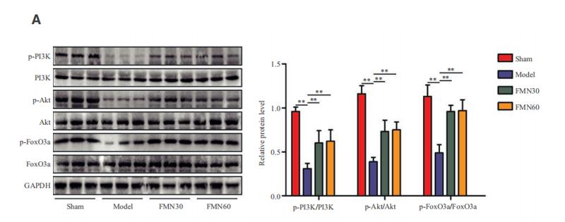
 Representative immunoblotting images and quantitative data for p-PI3K, p-Akt, p-FoxO3ɑ, MAFbx, MuRF-1, PI3K, Akt, FoxO3ɑ and GAPDH in gastrocnemius muscles at Day 14 after stress (n = 4). Data are mean ± SEM, and p-values were determined by a one-way ANOVA followed by Bonferroni post hoc tests. CW: CTSS+/+ control mice; CK: CTSS−/− control mice; SW: 14-day-stressed CTSS+/+ mice; SK: 14-day-stressed CTSS−/− mice. *p")
 Comparison and the expression levels of PI3K/Akt/FOXO3a signaling pathway related proteins in A549 and A549/Taxol cells were determined by Western blot, in the presence and absence of DHW-221 and GDC-0980. Statistical comparisons were performed with unpaired Student’s t-test (n = 3) in Panel (A). *p < 0.05, **p < 0.01 versus A549. Statistical comparisons were performed with one-way ANOVA followed by Dunnett’s post-hoc test for multiple comparisons in Panel (B) (n = 3). *p < 0.05, **p < 0.01, ***p < 0.001 versus control. (C, E) The FOXO3a and p-FOXO3a expressions in the nuclear and cytoplasmic fractions of A549/Taxol cells were detected by Western blot. Proliferating cell nuclear antigen (PCNA) was selected as nucleoprotein internal control. The quantitative results were shown in Panel (E). (D) Immunofluorescence staining of FOXO3a in A549/Taxol cells was carried out to evaluate the effect of DHW-221 on FOXO3a nuclear translocation. Scale bar = 20 µm. The histograms indicated the percentage of the cells in each condition exhibiting FOXO3a nuclear mean fluorescence intensity (positive cells, green fluorescence) by ImageJ. (F) FOXO3a degradation in A549/Taxol cells with or without DHW-221 co-treatment in different time spot when protein biosynthesis was blocked with 20 µM cycloheximide (CHX). FOXO3a stability was analyzed relative to control by ImageJ software. (G) FOXO3a proteins levels in the presence of MG132 (0.20 μM) or 2.40 μM DHW-221-treated A549/Taxol cells at 24 h. Statistical comparisons were performed with one-way ANOVA followed by Dunnett’s post-hoc test for multiple comparisons (n = 3). Data were presented as mean ± SD. *p < 0.05, **p < 0.01, ***p < 0.001 versus control.")
 The docking stick model of griffithazanone A combining with the PHE-254/PRO-241/GLU-247/GLN-252/ARG-250 domain of PIM1. Molecular docking score achieved −7.69. (b, c) Cellular thermal shift assay (CETSA): Used PBS as a negative control, quantitatively analyzed of the binding of griffithazanone A and PIM1 in A549 cells at different temperatures, and evaluated the expression using grayscale analysis. (d–g) Western blot analysis: p-ASK1, ASK1, p-JNK, JNK, p-p38, p38, p-BAD, BAD, p-foxo3a, foxo3a, Bax, Bcl-2, caspase 3 and cleaved-caspase 3 expression and its gray level in A549 cells. Take Tubulin as the internal parameter. (h) qRT-PCR showed the mRNA expression levels of Bax and Bcl-2 in A549 cells. (i) A549 cells were incubated with griffithazanone A (0.5 1 and 2 μM), and then detect the ROS level using a fluorescence enzyme-linked immunosorbent assay.")

产品描述
*The optimal dilutions should be determined by the end user. For optimal experimental results, antibody reuse is not recommended.
*Tips:
WB: 适用于变性蛋白样本的免疫印迹检测. IHC: 适用于组织样本的石蜡(IHC-p)或冰冻(IHC-f)切片样本的免疫组化/荧光检测. IF/ICC: 适用于细胞样本的荧光检测. ELISA(peptide): 适用于抗原肽的ELISA检测.
引用格式: Affinity Biosciences Cat# AF3020, RRID:AB_2834427.
展开/折叠
AF6q21; AF6q21 protein; DKFZp781A0677; FKHR2; FKHRL 1; FKHRL1; FKHRL1P2; Forkhead (Drosophila) homolog (rhabdomyosarcoma) like 1; Forkhead box O3; Forkhead box O3A; Forkhead box protein O3; Forkhead box protein O3A; Forkhead Drosophila homolog of in rhabdomyosarcoma like 1; Forkhead homolog (rhabdomyosarcoma) like 1; Forkhead in rhabdomyosarcoma like 1; Forkhead in rhabdomyosarcoma-like 1; FOX O3A; FOXO2; foxo3; FOXO3_HUMAN; FOXO3A; MGC12739; MGC31925;
抗原和靶标
A synthesized peptide derived from human FOXO3A around the phosphorylation site of Ser253.
研究领域
· Cellular Processes > Cell growth and death > Cellular senescence. (View pathway)
· Environmental Information Processing > Signal transduction > FoxO signaling pathway. (View pathway)
· Environmental Information Processing > Signal transduction > PI3K-Akt signaling pathway. (View pathway)
· Environmental Information Processing > Signal transduction > AMPK signaling pathway. (View pathway)
· Human Diseases > Drug resistance: Antineoplastic > EGFR tyrosine kinase inhibitor resistance.
· Human Diseases > Cancers: Specific types > Endometrial cancer. (View pathway)
· Human Diseases > Cancers: Specific types > Non-small cell lung cancer. (View pathway)
· Organismal Systems > Immune system > Chemokine signaling pathway. (View pathway)
· Organismal Systems > Aging > Longevity regulating pathway. (View pathway)
· Organismal Systems > Aging > Longevity regulating pathway - multiple species. (View pathway)
· Organismal Systems > Nervous system > Neurotrophin signaling pathway. (View pathway)
· Organismal Systems > Endocrine system > Prolactin signaling pathway. (View pathway)
文献引用
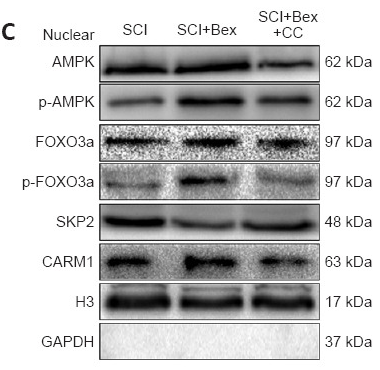
Application: WB Species: Mouse Sample: spinal cord
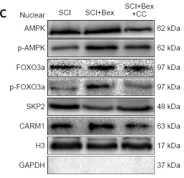
Application: WB Species: Mouse Sample: spinal cord

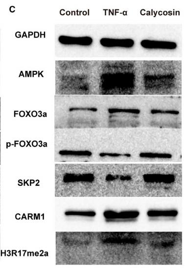
Application: WB Species: Mouse Sample: breast cancer cells

Application: WB Species: Mouse Sample:
Application: WB Species: mouse Sample: C2C12 cells
Application: WB Species: Mouse Sample: C2C12 myoblasts

Application: WB Species: Rat Sample: spinal cord
限制条款
产品的规格、报价、验证数据请以官网为准,官网链接:www.affbiotech.com | www.affbiotech.cn(简体中文)| www.affbiotech.jp(日本語)产品的数据信息为Affinity所有,未经授权不得收集Affinity官网数据或资料用于商业用途,对抄袭产品数据的行为我们将保留诉诸法律的权利。
产品相关数据会因产品批次、产品检测情况随时调整,如您已订购该产品,请以订购时随货说明书为准,否则请以官网内容为准,官网内容有改动时恕不另行通知。
Affinity保证所销售产品均经过严格质量检测。如您购买的商品在规定时间内出现问题需要售后时,请您在Affinity官方渠道提交售后申请。产品仅供科学研究使用。不用于诊断和治疗。
产品未经授权不得转售。
Affinity Biosciences将不会对在使用我们的产品时可能发生的专利侵权或其他侵权行为负责。Affinity Biosciences, Affinity Biosciences标志和所有其他商标所有权归Affinity Biosciences LTD.