产品: Histone H3 抗体
货号: AF0863
描述: Rabbit polyclonal antibody to Histone H3
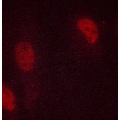
应用: WB IHC IF/ICC
文献验证: WB, IF/ICC
反应: Human, Mouse, Rat, Fish
预测: Bovine
蛋白号: P68431 | Q71DI3 | P84243
RRID: AB_2810277

浏览相似产品>>

   规格 价格 库存
 50ul RMB¥ 900 现货
 100ul RMB¥ 1500 现货
 200ul RMB¥ 2000 现货

货期: 当天发货

联系销售

产品描述

来源:
Rabbit
应用:
WB 1:500-1:2000, IHC 1:50-1:200, IF/ICC 1:100-1:500
*The optimal dilutions should be determined by the end user.
*Tips:

WB: 适用于变性蛋白样本的免疫印迹检测. IHC: 适用于组织样本的石蜡(IHC-p)或冰冻(IHC-f)切片样本的免疫组化/荧光检测. IF/ICC: 适用于细胞样本的荧光检测. ELISA(peptide): 适用于抗原肽的ELISA检测.

反应:
Human, Mouse, Rat, Fish
克隆:
Polyclonal
特异性:
Histone H3 Antibody detects endogenous levels of Histone H3.
RRID:
AB_2810277
引用格式: Affinity Biosciences Cat# AF0863, RRID:AB_2810277.
偶联:
Unconjugated.
纯化:
The antiserum was purified by peptide affinity chromatography using SulfoLink™ Coupling Resin (Thermo Fisher Scientific).
保存:
Rabbit IgG in phosphate buffered saline , pH 7.4, 150mM NaCl, 0.02% sodium azide and 50% glycerol. Store at -20 °C. Stable for 12 months from date of receipt.
别名:

展开/折叠

H3 histone family, member A; H3/A; H31_HUMAN; H3FA; Hist1h3a; HIST1H3B; HIST1H3C; HIST1H3D; HIST1H3E; HIST1H3F; HIST1H3G; HIST1H3H; HIST1H3I; HIST1H3J; histone 1, H3a; Histone cluster 1, H3a; Histone H3.1; Histone H3/a; Histone H3/b; Histone H3/c; Histone H3/d; Histone H3/f; Histone H3/h; Histone H3/i; Histone H3/j; Histone H3/k; Histone H3/l; ;H3.3A; HIST1 cluster, H3E; H3 histone family, member A; H3.1; H3/l; H3F3; H3FF; H3FJ; H3FL; Histone gene cluster 1, H3 histone family, member E; histone H3.1t; Histone H3/o; FLJ92264; H 3; H3; H3 histone family, member B; H3 histone family, member C; H3 histone family, member D; H3 histone family, member F; H3 histone family, member H; H3 histone family, member I; H3 histone family, member J; H3 histone family, member K; H3 histone family, member L; H3 histone family, member T; H3 histone, family 3A; H3/A; H3/b; H3/c; H3/d; h3/f; H3/h; H3/i; H3/j; H3/k; H3/t; H31_HUMAN; H3F1K; H3F3A; H3FA; H3FB; H3FC; H3FD; H3FH; H3FI; H3FK; HIST1 cluster, H3A; HIST1 cluster, H3B; HIST1 cluster, H3C; HIST1 cluster, H3D; HIST1 cluster, H3F; HIST1 cluster, H3G; HIST1 cluster, H3H; HIST1 cluster, H3I; HIST1 cluster, H3J; HIST1H3A; HIST1H3B; HIST1H3C; HIST1H3D; HIST1H3E; HIST1H3F; HIST1H3G; HIST1H3H; HIST1H3I; HIST1H3J; HIST3H3; Histone 1, H3a; Histone 1, H3b; Histone 1, H3c; Histone 1, H3d; Histone 1, H3e; Histone 1, H3f; Histone 1, H3g; Histone 1, H3h; Histone 1, H3i; Histone 3, H3; histone cluster 1 H3 family member a; histone cluster 1 H3 family member b; histone cluster 1 H3 family member c; histone cluster 1 H3 family member d; histone cluster 1 H3 family member e; histone cluster 1 H3 family member f; histone cluster 1 H3 family member g; histone cluster 1 H3 family member h; histone cluster 1 H3 family member i; histone cluster 1 H3 family member j; Histone cluster 1, H3a; Histone cluster 1, H3b; Histone cluster 1, H3c; Histone cluster 1, H3d; Histone cluster 1, H3e; Histone cluster 1, H3f; Histone cluster 1, H3g; Histone cluster 1, H3i; Histone cluster 1, H3j; Histone gene cluster 1, H3 histone family, member A; Histone gene cluster 1, H3 histone family, member B; Histone gene cluster 1, H3 histone family, member C; Histone gene cluster 1, H3 histone family, member D; Histone gene cluster 1, H3 histone family, member F; Histone gene cluster 1, H3 histone family, member G; Histone gene cluster 1, H3 histone family, member H; Histone gene cluster 1, H3 histone family, member I; Histone gene cluster 1, H3 histone family, member J; Histone gene cluster 1, H3A; Histone gene cluster 1, H3B; Histone gene cluster 1, H3C; Histone gene cluster 1, H3D; Histone gene cluster 1, H3E; Histone gene cluster 1, H3F; Histone gene cluster 1, H3G; Histone gene cluster 1, H3H; Histone gene cluster 1, H3I; Histone gene cluster 1, H3J; Histone H 3; Histone H3.1; Histone H3.2; Histone H3.3; Histone H3/a; Histone H3/b; Histone H3/c; Histone H3/d; Histone H3/f; Histone H3/h; Histone H3/i; Histone H3/j; Histone H3/k; Histone H3/l; Histone H3/m; H3 histone family 3A; H3 histone family 3B; H3 histone, family 3B (H3.3B); H3.3; H3.3A; H3.3B; H33_HUMAN; H3F3; H3F3A; H3f3b; Histone H3.3; Histone H3.3Q; Histone H3.A; Histone H3.B; MGC87782; MGC87783;

抗原和靶标

免疫原:

A synthesized peptide derived from human Histone H3.

基因/基因ID:
描述:
H3 Core component of nucleosome. Nucleosomes wrap and compact DNA into chromatin, limiting DNA accessibility to the cellular machineries which require DNA as a template. Histones thereby play a central role in transcription regulation, DNA repair, DNA replication and chromosomal stability. DNA accessibility is regulated via a complex set of post-translational modifications of histones, also called histone code, and nucleosome remodeling. The nucleosome is a histone octamer containing two molecules each of H2A, H2B, H3 and H4 assembled in one H3-H4 heterotetramer and two H2A-H2B heterodimers.

研究领域

· Human Diseases > Substance dependence > Alcoholism.

· Human Diseases > Cancers: Overview > Transcriptional misregulation in cancer.

· Human Diseases > Immune diseases > Systemic lupus erythematosus.

文献引用

1). ETV2 regulating PHD2-HIF-1α axis controls metabolism reprogramming promotes vascularized bone regeneration. Bioactive materials, 2024 (PubMed: 38549772) [IF=18.9]

2). Psychologic Stress Drives Progression of Malignant Tumors via DRD2/HIF1α Signaling. CANCER RESEARCH, 2021 (PubMed: 34321238) [IF=12.5]

Application: WB    Species: mouse    Sample: B16F10 and 4T1 tumor

Fig. 1 |DRD2 may promote the malignant progression of melanoma and breast cancer under psychologic stress condition. J, Western blot analysis of DRD2 expression levels in the nuclei of B16F10 and 4T1 tumor tissues with or without stress stimulation.

3). Salidroside can target both P4HB-mediated inflammation and melanogenesis of the skin. Theranostics, 2020 (PubMed: 33042273) [IF=12.4]

Application: WB    Species: human    Sample: A375 cells

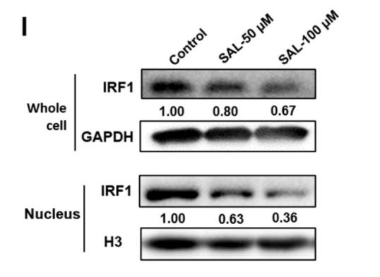
Figure 4. P4HB regulates the ubiquitination degradation of IRF1. A. Changes in TYR activity in P4HB overexpression or knockdown A375 cells. B. Changes in TYR mRNA expression in Sal treated, P4HB overexpression, and P4HB knockdown A375 cells. C. Western blot analysis of TYR expression in P4HB overexpression or knockdown A375 cells. D. Proteins have interactions with P4HB and USF1 analyzed by FpClass. E. Interaction of P4HB and IRF1 in A375 cells detected by PLA. F. Western blot analysis of the expression of IRF1 and P4HB in P4HB overexpression or knockdown A375 cells. G. Western blot analysis of IRF1 expression in the nucleus of A375 cells after P4HB overexpression or knockdown. H. Effect of SAL on the ubiquitination of IRF1 as determined by Western blot. I. Western blot analysis of IRF1 expression in whole cells and nucleus of SAL-treated A375 cells. Data are expressed as mean ± SD (*P < 0.05, **P < 0.01).

4). Lactate drives epithelial-mesenchymal transition in diabetic kidney disease via the H3K14la/KLF5 pathway. Redox biology, 2024 (PubMed: 38925041) [IF=10.7]

Application: WB    Species: Mouse    Sample:

Fig. 5. Histone lactylaction is increased in db/db mice and AGEs-induced mTEC. (A) Immunoblot analysis of Pan kla expression in db/db mice. (B) The Pan kla immunoblots in AGE-induced mTEC. (C) The Pan kla and Pan kac immunoblots in AGE-induced mTEC at 0, 4, 8, 12, 24, and 48 h. (D–E) Immunoblot analysis demonstrating the expression of Pan kla, H3K14la, H3K18la, H3K9la, H3K23la, H3K27la and H3K14ac in AGEs-induced mTEC. (F) Immunofluorescent staining of Pan kla (red) and DAPI (blue). Scale bars, 50 μm. (G–H) Immunofluorescent staining of H3K14la (red) and DAPI (blue). Scale bars, 50 μm. (I–K) Immunoblot analysis of Pan kla and H3K14la in AGEs-induced mTEC. All statistic data were presented as mean ± SEM. Unpaired two tailed t-test. Data are expressed as Mean ± SEM, #P < 0.05, ##P < 0.01; *P < 0.05, **P < 0.01. (For interpretation of the references to color in this figure legend, the reader is referred to the Web version of this article.)

5). Oxyberberine, a novel gut microbiota-mediated metabolite of berberine, possesses superior anti-colitis effect: impact on intestinal epithelial barrier, gut microbiota profile and TLR4-MyD88-NF-κB pathway. PHARMACOLOGICAL RESEARCH, 2020 (PubMed: 31863867) [IF=9.1]

Application: WB    Species: Mice    Sample: colonic tissues

Fig. 6. Effect of OBB on the activation of TLR4-MyD88-NF-κB signaling pathway in DSS-induced colonic tissues. (A) Representative Western blotting images of TLR4, MyD88, cytoplasmic p65, nuclear p65, p-IκBα and IκBα. Changes in the relative protein expression levels of TLR4 (B), MyD88 (C), nuclear p65 (D), cytoplasmic p65 (E), and p-IκBα/IκBα ratio (F) were measured. Data are shown as the mean ± SEM (n = 3). # P < 0.05, ## P < 0.01 vs. Control group, * P < 0.05, ** P < 0.01 vs. DSS group.

6). Sophora japonica flowers and their main phytochemical, rutin, regulate chemically induced murine colitis in association with targeting the NF-κB signaling pathway and gut microbiota. Food Chemistry, 2022 (PubMed: 35691061) [IF=8.5]

7). H3K79 methylation promotes rapid growth of Alexandrium pacificum under high light intensity via increased photosynthesis. Science of The Total Environment, 2023 (PubMed: 37311522) [IF=8.2]

8). The involvement of the Stat1/Nrf2 pathway in exacerbating Crizotinib-induced liver injury: implications for ferroptosis. Cell death & disease, 2024 (PubMed: 39160159) [IF=8.1]

9). RIP3 impedes transcription factor EB to suppress autophagic degradation in septic acute kidney injury. Cell Death & Disease, 2021 (PubMed: 34103472) [IF=8.1]

Application: WB    Species: Mice    Sample: kidney tissue

Fig. 5 RIP3 impeded the nuclear translocation of TFEB in septic AKI mice and LPS-treated cultured PTECs. A Immunofluorescence staining for TFEB (green) and DAPI (blue) in the kidneys from C57BL/6 mice treated with sterilized saline (con), LPS, or LPS plus GSK’872 (GSK) for 24 h. GSK partially restored the decreased nuclear staining of TFEB in the renal tubular cells of LPS-induced mice. Scale bar = 20 μm. B Cultured PTECs treated with LPS, LPS plus GSK, LPS plus DMSO, LPS plus RIP3 siRNA, or LPS plus scrambled siRNA (Scramble) for 12 h. The nuclear protein fractions were immunoblotted for TFEB. Histone was used as the nuclear marker. TFEB expression against Histone was decreased by LPS treatment, which was restored by GSK or RIP3 siRNA (n = 3). C Immunofluorescence staining for TFEB (green) and DAPI (blue) in cultured PTECs treated as indicated. The nuclear translocation of TFEB was inhibited by LPS, which was rescued by GSK or RIP3 siRNA. Scale bar = 20 μm. D, E Lysates from cultured PTECs treated with LPS or DMSO (con) for 12 h were subjected to immunoprecipitation using an anti-RIP3 antibody (D) or anti-TFEB antibody (E), and IgG antibody followed by immunoblot for TFEB and RIP3. Input proteins were detected with anti-RIP3 and anti-TFEB antibodies. RIP3 interacted with TFEB in LPS-treated PTECs compared to the control group. F Lysates from cultured PTECs treated with LPS or DMSO (con) for 12 h were subjected to immunoprecipitation using anti-TFEB antibody and IgG antibody followed by immunoblot for p-RIP3. Input proteins were detected with anti-p-RIP3 and anti-TFEB antibodies. p-RIP3 interacted with TFEB in LPS-treated PTECs compared to the control group. *P < 0.05.

10). Reactive oxygen species-mediated phosphorylation of JNK is involved in the regulation of BmFerHCH on Bombyx mori nucleopolyhedrovirus proliferation. International journal of biological macromolecules, 2023 (PubMed: 36842745) [IF=7.7]

加载更多

限制条款

产品的规格、报价、验证数据请以官网为准,官网链接:www.affbiotech.com | www.affbiotech.cn(简体中文)| www.affbiotech.jp(日本語)

产品的数据信息为Affinity所有,未经授权不得收集Affinity官网数据或资料用于商业用途,对抄袭产品数据的行为我们将保留诉诸法律的权利。

产品相关数据会因产品批次、产品检测情况随时调整,如您已订购该产品,请以订购时随货说明书为准,否则请以官网内容为准,官网内容有改动时恕不另行通知。

Affinity保证所销售产品均经过严格质量检测。如您购买的商品在规定时间内出现问题需要售后时,请您在Affinity官方渠道提交售后申请。

产品仅供科学研究使用。不用于诊断和治疗。 

产品未经授权不得转售。

Affinity Biosciences将不会对在使用我们的产品时可能发生的专利侵权或其他侵权行为负责。Affinity Biosciences, Affinity Biosciences标志和所有其他商标所有权归Affinity Biosciences LTD.