产品: Osteoprotegerin 抗体
货号: DF6824
描述: Rabbit polyclonal antibody to Osteoprotegerin
应用: WB IHC IF/ICC
反应: Human, Mouse, Rat
预测: Pig, Bovine, Horse, Sheep, Rabbit, Dog, Chicken
分子量: 45~60kDa; 46kD(Calculated).
蛋白号: O00300
RRID: AB_2838784

浏览相似产品>>

   规格 价格 库存
 50ul RMB¥ 1250 现货
 100ul RMB¥ 2300 现货
 200ul RMB¥ 3000 现货

货期: 当天发货

联系销售

产品描述

来源:
Rabbit
应用:
WB 1:500-1:2000, IHC 1:50-1:200, IF/ICC 1:100-1:500
*The optimal dilutions should be determined by the end user.
*Tips:

WB: 适用于变性蛋白样本的免疫印迹检测. IHC: 适用于组织样本的石蜡(IHC-p)或冰冻(IHC-f)切片样本的免疫组化/荧光检测. IF/ICC: 适用于细胞样本的荧光检测. ELISA(peptide): 适用于抗原肽的ELISA检测.

反应:
Human,Mouse,Rat
预测:
Pig(100%), Bovine(100%), Horse(100%), Sheep(100%), Rabbit(100%), Dog(100%), Chicken(80%)
克隆:
Polyclonal
特异性:
Osteoprotegerin Antibody detects endogenous levels of total Osteoprotegerin.
RRID:
AB_2838784
引用格式: Affinity Biosciences Cat# DF6824, RRID:AB_2838784.
偶联:
Unconjugated.
纯化:
The antiserum was purified by peptide affinity chromatography using SulfoLink™ Coupling Resin (Thermo Fisher Scientific).
保存:
Rabbit IgG in phosphate buffered saline , pH 7.4, 150mM NaCl, 0.02% sodium azide and 50% glycerol. Store at -20 °C. Stable for 12 months from date of receipt.
别名:

展开/折叠

MGC29565; OCIF; OPG; Osteoclastogenesis inhibitory factor; Osteoprotegerin; PDB5; TNF receptor superfamily member 11b; TNFRSF 11B; TNFRSF11B; TR 1; TR1; TR11B_HUMAN; Tumor necrosis factor receptor superfamily member 11B;

抗原和靶标

免疫原:
Uniprot:
基因/基因ID:
表达:
O00300 TR11B_HUMAN:

Highly expressed in adult lung, heart, kidney, liver, spleen, thymus, prostate, ovary, small intestine, thyroid, lymph node, trachea, adrenal gland, testis, and bone marrow. Detected at very low levels in brain, placenta and skeletal muscle. Highly expressed in fetal kidney, liver and lung.

描述:
The protein encoded by this gene is a member of the TNF-receptor superfamily. This protein is an osteoblast-secreted decoy receptor that functions as a negative regulator of bone resorption. This protein specifically binds to its ligand, osteoprotegerin ligand, both of which are key extracellular regulators of osteoclast development. Studies of the mouse counterpart also suggest that this protein and its ligand play a role in lymph-node organogenesis and vascular calcification. Alternatively spliced transcript variants of this gene have been reported, but their full length nature has not been determined. [provided by RefSeq, Jul 2008]
序列:
MNNLLCCALVFLDISIKWTTQETFPPKYLHYDEETSHQLLCDKCPPGTYLKQHCTAKWKTVCAPCPDHYYTDSWHTSDECLYCSPVCKELQYVKQECNRTHNRVCECKEGRYLEIEFCLKHRSCPPGFGVVQAGTPERNTVCKRCPDGFFSNETSSKAPCRKHTNCSVFGLLLTQKGNATHDNICSGNSESTQKCGIDVTLCEEAFFRFAVPTKFTPNWLSVLVDNLPGTKVNAESVERIKRQHSSQEQTFQLLKLWKHQNKDQDIVKKIIQDIDLCENSVQRHIGHANLTFEQLRSLMESLPGKKVGAEDIEKTIKACKPSDQILKLLSLWRIKNGDQDTLKGLMHALKHSKTYHFPKTVTQSLKKTIRFLHSFTMYKLYQKLFLEMIGNQVQSVKISCL

种属预测

种属预测:

score>80的预测可信度较高,可尝试用于WB检测。*预测模型主要基于免疫原序列比对,结果仅作参考,不作为质保凭据。

Species
Results
Score
Pig
100
Horse
100
Bovine
100
Sheep
100
Dog
100
Rabbit
100
Chicken
80
Xenopus
70
Zebrafish
64
Model Confidence:
High(score>80) Medium(80>score>50) Low(score<50) No confidence

翻译修饰 - O00300 作为底物

Site PTM Type Enzyme
N178 N-Glycosylation
S246 Phosphorylation
T360 Phosphorylation
T362 Phosphorylation
S364 Phosphorylation

研究背景

功能:

Acts as decoy receptor for TNFSF11/RANKL and thereby neutralizes its function in osteoclastogenesis. Inhibits the activation of osteoclasts and promotes osteoclast apoptosis in vitro. Bone homeostasis seems to depend on the local ratio between TNFSF11 and TNFRSF11B. May also play a role in preventing arterial calcification. May act as decoy receptor for TNFSF10/TRAIL and protect against apoptosis. TNFSF10/TRAIL binding blocks the inhibition of osteoclastogenesis.

翻译修饰:

N-glycosylated. Contains sialic acid residues.

The N-terminus is blocked.

细胞定位:

Secreted.

Extracellular region or secreted Cytosol Plasma membrane Cytoskeleton Lysosome Endosome Peroxisome ER Golgi apparatus Nucleus Mitochondrion Manual annotation Automatic computational assertionSubcellular location
组织特异性:

Highly expressed in adult lung, heart, kidney, liver, spleen, thymus, prostate, ovary, small intestine, thyroid, lymph node, trachea, adrenal gland, testis, and bone marrow. Detected at very low levels in brain, placenta and skeletal muscle. Highly expressed in fetal kidney, liver and lung.

亚基结构:

Homodimer. Interacts with TNFSF10 and TNFSF11.

研究领域

· Environmental Information Processing > Signaling molecules and interaction > Cytokine-cytokine receptor interaction.   (View pathway)

· Organismal Systems > Development > Osteoclast differentiation.   (View pathway)

文献引用

1). IGFBP7 acts as a negative regulator of RANKL-induced osteoclastogenesis and oestrogen deficiency-induced bone loss. CELL PROLIFERATION (PubMed: 31889368) [IF=8.5]

Application: WB    Species: mouse    Sample: MC3T3-E1

Figure S1.| The results of western-blot analysis showed that different concentrations of recombinant IGFBP7 increased the protein expression of OPG in osteoblastic cell line MC3T3-E1, whereas the expression of RANKL was not affected after 3 days’ IGFBP7 treatment. OPG, osteoprotegerin; RANKL, receptor activator of nuclear factor-κB ligand.

2). Osteogenic potential evaluation of biotin combined with magnesium-doped hydroxyapatite sustained-release film. Materials science & engineering-C, Materials for biological applications (PubMed: 35581076) [IF=7.9]

3). Yunvjian decoction attenuates lipopolysaccharide-induced periodontitis by suppressing NFκB/NLRP3/IL-1β pathway. Journal of ethnopharmacology (PubMed: 37802377) [IF=5.4]

4). Neuropeptide Y1 receptor antagonist promotes osteoporosis and microdamage repair and enhances osteogenic differentiation of bone marrow stem cells via cAMP/PKA/CREB pathway. Aging-US (PubMed: 32381754) [IF=5.2]

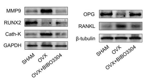
Application: WB    Species: rat    Sample: bone marrow

Figure 4. |Y1R antagonist treatment promoted bone formation and inhibit bone resorption in OVX rats. (A) The Alizarin Red S (yellow arrow head) and TRAP staining (red arrow head) of bone tissues in groups. (B) Immunohistochemical analysis of bone tissue among groups for MMP9 and RUNX2 (x 400). (C, D) Western blotting results of MMP, RUNX2, Cath-K, OPG and RANKL expression in bone marrow from rat femurs. The data are expressed as the means ± SD (n = 6 in each group). ***P<0.001; ****P<0.0001 vs. SHAM, and #P<0.05; ####P<0.0001 vs. OVX by one-way ANOVA and Tukey’s post hoc test

5). Hexokinase 2‐mediated glycolysis promotes receptor activator of NF‐κB ligand expression in Porphyromonas gingivalis lipopolysaccharide‐treated osteoblasts. JOURNAL OF PERIODONTOLOGY (PubMed: 34585393) [IF=4.3]

6). Role of microRNA-19b-3p on osteoporosis after experimental spinal cord injury in rats. ARCHIVES OF BIOCHEMISTRY AND BIOPHYSICS (PubMed: 33587904) [IF=3.9]

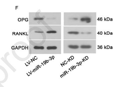
Application: WB    Species: rat    Sample: osteoblasts

Figure 2. MiR-19b-3p inhibits osteoblast differentiation. (A) RT-PCR analysis was used 443 to detect miRNA-19b-3p expression levels in BMSC-derived osteoblasts after 444 miR-19b-3p-overexpressed or knockdown by corresponding lentivirus. (B) ALP activity in 445 BMSC-derived osteoblasts after miRNA-19b-3p overexpression or knockdown by 446 corresponding lentivirus. (C) RT-PCR was used to detect EBF2 mRNA expression levels in BMSC-derived osteoblasts after miRNA-19b-3p overexpression or knockdown by 448 corresponding lentivirus. Western blot assay was used to detect (D and E) EBF2, (F-H) 449 RANKL and OPG protein expression levels in BMSC-derived osteoblasts after 450 miRNA-19b-3p overexpression or knockdown by corresponding lentivirus. (I) The ratio of 451 OPG to RANKL in BMSC-derived osteoblasts after miRNA-19b-3p overexpression or 452 knockdown by corresponding lentivirus (J) Alizarin red staining was used to detect cell 453 mineralization at day 21 after differentiation after miRNA-19b-3p overexpression or 454 knockdown at 100×. Data are analyzed using unpaired Student’s t-tests and presented as the 455 mean ± SD from three independent experiments. * p < 0.05; ** p < 0.01 vs. the indicated 456 group.

7). Nirogacestat suppresses RANKL-Induced osteoclast formation in vitro and attenuates LPS-Induced bone resorption in vivo. EXPERIMENTAL CELL RESEARCH (PubMed: 31211955) [IF=3.7]

Application: IHC    Species: mouse    Sample: BMMs

Fig. 7.| Histological analysis of the inhibitory effect of PF on LPS-induced bone resorption. (A–C) Representative images of HE and TRAP staining, showing the reduced osteolytic legion and TRAP-positive OCs in the PF-treated groups. (D–G) Representative images of IHC staining of RANKL, OPG, OCN and TNF-α. (H) TRAPpositive OCs number. (H) Quantitative analysis of the expression of RANKL, OPG and RANKL/OPG ratio. The data are presented as the mean ± SD (*p < 0.05,**p < 0.01, ***p < 0.001).

8). Rodent incisor and molar dental follicles show distinct characteristics in tooth eruption. ARCHIVES OF ORAL BIOLOGY (PubMed: 33845260) [IF=3.0]

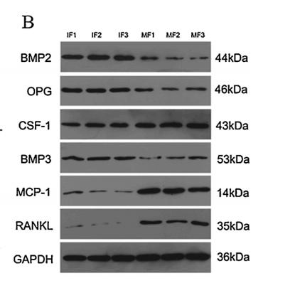
Application: WB    Species: Rat    Sample: Incisor dental follicle (IF) cells and molar dental follicle (MF) cells

Fig. 5. Differential expression patterns of tooth eruption-related genes and proteins between IF cells and MF cells. (A) Non-induced IF cells and MF cells showed different gene expression patterns; Data are mean ± SD of n = 3 replicates, one-way ANOVA, *p<0.05, **p<0.01, ***p<0.001, ****p<0.0001. (B) Non-induced and induced IF cells and MF cells showed different protein expression patterns. After induction with CLCCM and HERSCM, there were no significant changes of the expression patterns of tooth eruption-related proteins in IF cells and MF cells. (C) The grey value ratios of Western blotting results; Data are mean ± SD of n = 3 replicates, one-way ANOVA followed by Tukey post hoc test, *p<0.05, **p<0.01, ***p<0.001, ****p<0.0001. (IF1, MF1 represent the cells cultured by α-MEM; IF2, MF2 represent the cells cultured by α-MEM + CLCCM; IF3, MF3 represent the cells cultured by α-MEM+HERSCM).

9). Low frequency pulsed electromagnetic fields exposure alleviate the abnormal subchondral bone remodeling at the early stage of temporomandibular joint osteoarthritis. BMC Musculoskeletal Disorders (PubMed: 36384557) [IF=2.3]

10). Deletion of osteopontin in non-small cell lung cancer cells affects bone metabolism by regulating miR-34c/Notch1 axis: a clue to bone metastasis. European Journal of Histochemistry (PubMed: 37491944) [IF=2.0]

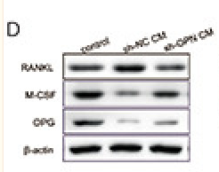
Application: WB    Species: Human    Sample: SaOS-2 cells

Figure 4.Deletion of osteopontin (OPN) promoted the differentiation and mineralization of osteoblasts. A) Osteocalcin-positive rate of SaOS-2 cells was detected by flow cytometry. B) The expression of osteocalcin in SaOS-2 cells was observed by immunofluorescence staining, and the fluorescence intensity was exhibited on the right. C) Alizarin Red S staining was performed on SaOS-2 cells to evaluate the mineralization. D) The protein expression of RANKL, M-CSF and OPG was detected by Western blot.

加载更多

限制条款

产品的规格、报价、验证数据请以官网为准,官网链接:www.affbiotech.com | www.affbiotech.cn(简体中文)| www.affbiotech.jp(日本語)

产品的数据信息为Affinity所有,未经授权不得收集Affinity官网数据或资料用于商业用途,对抄袭产品数据的行为我们将保留诉诸法律的权利。

产品相关数据会因产品批次、产品检测情况随时调整,如您已订购该产品,请以订购时随货说明书为准,否则请以官网内容为准,官网内容有改动时恕不另行通知。

Affinity保证所销售产品均经过严格质量检测。如您购买的商品在规定时间内出现问题需要售后时,请您在Affinity官方渠道提交售后申请。

产品仅供科学研究使用。不用于诊断和治疗。 

产品未经授权不得转售。

Affinity Biosciences将不会对在使用我们的产品时可能发生的专利侵权或其他侵权行为负责。Affinity Biosciences, Affinity Biosciences标志和所有其他商标所有权归Affinity Biosciences LTD.