, using CXCR4 Antibody at 1/1000 dilution.
5ug/NC membrane strip.
Exposure for 30s with Affinity™ ECL Kit(#KF8001).
Bands result from membrane strip incubation.")

 and mouse anti-beta tubulin Ab(T0023 1:200) for 1 hour at 37°C. An AlexaFluor594 conjugated goat anti-rabbit IgG(H+L) Ab(Red) and an AlexaFluor488 conjugated goat anti-mouse IgG(H+L) Ab(Green) were used as the secondary antibody.
The nuclear counter stain is DAPI(blue).")
 Photographs of the PASMCs migration through the polycarbonate membrane stained by 0.2% crystal violet in hypoxia and treated with increasing concentrations of quercetin for 24 h. (B) Quantification of the number of cells migrating through the polycarbonate membrane of average of 3 independent experiments. (C) Full-length blots of MMP-2, MMP-9, CXCR4, Integrin α1, β1, and α5 and GAPDH are presented. (D) Results were quantified by densitometry analysis of the bands form (C) and then normalization to GAPDH protein. *Po0.05, **Po0.01 compared with control; #Po0.05, ##Po0.01 compared with hypoxia and quercetin treated PASMCs.")
 The protein expression of c-Myc, vimentin, E-cadherin, HIF-1α, CXCR4, and SDF-1 in PANC-1 and SW1990 pancreatic cancer cells under different treatments was detected via western blot. (B) Quantification results of protein expressions of c-Myc, vimentin, E-cadherin, HIF-1α, CXCR4, and SDF-1 in PANC-1 pancreatic cancer cells. (C) Quantification results of protein expression of c-Myc, vimentin, E-cadherin, HIF-1α, CXCR4, and SDF-1 in SW1990 pancreatic cancer cells (* p < 0.05, ** p < 0.01 vs control, ▲ p < 0.05, ▲▲ p < 0.01 vs bufalin treatment group, n = 3).")
 Quantitative polymerase chain reaction (qPCR) data showing the levels of CTSS, SDF-1, CXCR4, IL-17, IL-18, MCP-1, ICAM-1, VCAM-1, TNF-α, p22phox, p47phox, p67phox, gp91phox, PGC1-α, and PPAR-γ mRNAs. (B) Representative immunoblotting images and quantitative data (C) for CTSS, SDF-1, CXCR4 in gastrocnemius muscles at Day 14 after stress (n = 4). (D and E) Activities of Na+-K+-ATPase and mitochondrial complex IV of the four groups of mice. Data are mean ± SEM, and p-values were determined by a one-way ANOVA followed by Bonferroni post hoc tests (C). **p")

产品描述
*The optimal dilutions should be determined by the end user. For optimal experimental results, antibody reuse is not recommended.
*Tips:
WB: 适用于变性蛋白样本的免疫印迹检测. IHC: 适用于组织样本的石蜡(IHC-p)或冰冻(IHC-f)切片样本的免疫组化/荧光检测. IF/ICC: 适用于细胞样本的荧光检测. ELISA(peptide): 适用于抗原肽的ELISA检测.
引用格式: Affinity Biosciences Cat# AF5279, RRID:AB_2837765.
展开/折叠
C-X-C chemokine receptor type 4; CD184; CD184 antigen; Chemokine (C X C motif) receptor 4; Chemokine CXC Motif Receptor 4; CXC-R4; CXCR-4; CXCR4; CXCR4_HUMAN; D2S201E; FB22; Fusin; HM89; HSY3RR; LAP 3; LAP3; LCR1; LESTR; Leukocyte derived seven transmembrane domain receptor; Leukocyte-derived seven transmembrane domain receptor; Lipopolysaccharide associated protein 3; Neuropeptide Y receptor Y3; NPY3R; NPYR; NPYRL; NPYY3; NPYY3R; Probable G protein coupled receptor lcr1 homolog; SDF 1 receptor; SDF-1 receptor; SEVEN-TRANSMEMBRANE-SEGMENT RECEPTOR; Stromal cell derived factor 1 receptor; Stromal cell-derived factor 1 receptor; WHIM; WHIMS;
抗原和靶标
A synthesized peptide derived from human CXCR4, corresponding to a region within C-terminal amino acids.
研究领域
· Cellular Processes > Transport and catabolism > Endocytosis. (View pathway)
· Environmental Information Processing > Signaling molecules and interaction > Cytokine-cytokine receptor interaction. (View pathway)
· Human Diseases > Cancers: Overview > Pathways in cancer. (View pathway)
· Organismal Systems > Immune system > Chemokine signaling pathway. (View pathway)
· Organismal Systems > Development > Axon guidance. (View pathway)
· Organismal Systems > Immune system > Leukocyte transendothelial migration. (View pathway)
· Organismal Systems > Immune system > Intestinal immune network for IgA production. (View pathway)
文献引用

Application: IHC Species: Mouse Sample:

Application: WB Species: Mouse Sample:

Application: IF/ICC Species: human Sample: breast cancers
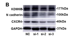
Application: WB Species: Human Sample: BMSCs

Application: WB Species: Mouse Sample: UCMSCs
限制条款
产品的规格、报价、验证数据请以官网为准,官网链接:www.affbiotech.com | www.affbiotech.cn(简体中文)| www.affbiotech.jp(日本語)产品的数据信息为Affinity所有,未经授权不得收集Affinity官网数据或资料用于商业用途,对抄袭产品数据的行为我们将保留诉诸法律的权利。
产品相关数据会因产品批次、产品检测情况随时调整,如您已订购该产品,请以订购时随货说明书为准,否则请以官网内容为准,官网内容有改动时恕不另行通知。
Affinity保证所销售产品均经过严格质量检测。如您购买的商品在规定时间内出现问题需要售后时,请您在Affinity官方渠道提交售后申请。产品仅供科学研究使用。不用于诊断和治疗。
产品未经授权不得转售。
Affinity Biosciences将不会对在使用我们的产品时可能发生的专利侵权或其他侵权行为负责。Affinity Biosciences, Affinity Biosciences标志和所有其他商标所有权归Affinity Biosciences LTD.