
, using PKM2 Antibody at 1/1000 dilution.
5ug/NC membrane strip.
Exposure for 30s with Affinity™ ECL Kit(#KF8001).
Bands result from membrane strip incubation.")


 and mouse anti-beta tubulin Ab(T0023 1:200) for 1 hour at 37°C. An AlexaFluor594 conjugated goat anti-rabbit IgG(H+L) Ab(Red) and an AlexaFluor488 conjugated goat anti-mouse IgG(H+L) Ab(Green) were used as the secondary antibody.
The nuclear counter stain is DAPI(blue).")
 promotes c-Myc protein degradation. Expression levels of c-Myc, HK2, PKM2, and LDHA were detected by Western blot (A) and qRT-PCR (B) analysis in MEG3 overexpression and knockdown colorectal cancer (CRC) cell lines. (C) MEG3 overexpression CRC cell lines were treated with 100 μg/ml of cycloheximide (CHX) and harvested at the indicated time points. c-Myc protein was detected by Western blot analysis, quantified by densitometry, and plotted against time to determine c-Myc stability. (D) CRC cells were transfected with pcDNA-c-Myc in combination with pcDNA-MEG3 in the presence of the HA-ubiquitin plasmid as indicated at the top. The cells were treated with MG132 (30 μM) for 6 h before harvesting, and the cell lysates were subjected to immunoprecipitation using anti-HA antibody. Ubiquitinated proteins were detected by Western blot with the anti-Flag antibody. (E) CRC cell lines that strongly express MEG3 were treated with 5 μM of MG132 for 12 h, and c-Myc protein was detected by Western blot. (F) Expression of FBXW7 was detected by Western blot in CRC cells that strongly and weakly expressed MEG3. (G) Level of FBXW7 was measured by Western blot in the pcDNA-MEG3 cells with MEG3 knockdown. Data are expressed as mean ± standard deviation from three independent experiments. *P < 0.05, **P < 0.01.")
 promotes c-Myc protein degradation. Expression levels of c-Myc, HK2, PKM2, and LDHA were detected by Western blot (A) and qRT-PCR (B) analysis in MEG3 overexpression and knockdown colorectal cancer (CRC) cell lines. (C) MEG3 overexpression CRC cell lines were treated with 100 μg/ml of cycloheximide (CHX) and harvested at the indicated time points. c-Myc protein was detected by Western blot analysis, quantified by densitometry, and plotted against time to determine c-Myc stability. (D) CRC cells were transfected with pcDNA-c-Myc in combination with pcDNA-MEG3 in the presence of the HA-ubiquitin plasmid as indicated at the top. The cells were treated with MG132 (30 μM) for 6 h before harvesting, and the cell lysates were subjected to immunoprecipitation using anti-HA antibody. Ubiquitinated proteins were detected by Western blot with the anti-Flag antibody. (E) CRC cell lines that strongly express MEG3 were treated with 5 μM of MG132 for 12 h, and c-Myc protein was detected by Western blot. (F) Expression of FBXW7 was detected by Western blot in CRC cells that strongly and weakly expressed MEG3. (G) Level of FBXW7 was measured by Western blot in the pcDNA-MEG3 cells with MEG3 knockdown. Data are expressed as mean ± standard deviation from three independent experiments. *P < 0.05, **P < 0.01.")
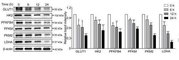
 The level of the consumption of glucose and the production of lactate was measured from the media of different groups. The fold change
between Cd-treated groups and non-treated control groups is represented using the Bar Chart (*P < .05 vs. cultivated 48 h non-treated control). (C) The relative
intracellular generation of ATP was measured (*P < .05 vs. Control). (D) Western blots of the relative aerobic glycolysis protein of GLUT1, HKII, PKM2 and LDHA in
both cells exposed to Cd for 0, 12, 24, 36 and 48 h. (E) Fold changes of them were normalized to the expression of β-actin. Each bar represents mean ± SD from three
independent experiments (*P < .05 vs. Control).")
 The intracellular activity levels of the metabolic enzymes HK2 and PKM2 were assessed by ELISA in Huh7, HepG2, and PLC/PRF/5 cells treated with 0.05 or 0.1 mg/mL ubenimex (**, P")
![PKM2 Antibody - Figure 5 BHD inhibited protein and mRNA expression of pyruvate kinase M2 (PKM2) and hypoxia-inducible factor-1 alpha (HIF-1α) as well as its target genes (GLUT1, PDK1, lactate dehydrogenase A [LDHA]) in aortas after high fat diet treatment.](http://img.affbiotech.cn/uploads/202409/5ce509e907ddfd09b9be49206226cfbb.png "Figure 5 BHD inhibited protein and mRNA expression of pyruvate kinase M2 (PKM2) and hypoxia-inducible factor-1 alpha (HIF-1α) as well as its target genes (GLUT1, PDK1, lactate dehydrogenase A [LDHA]) in aortas after high fat diet treatment. (a) Protein Levels of PKM2 were measured by Western blot analysis, and quantitative analysis was performed on the corresponding bands (n = 8–9). (b) Protein Levels of HIF-1α was measured by WB, and quantitative analysis was performed on the corresponding bands (n = 9). (c) The mRNA levels of GLUTA, (d) PDK1, and (e) LDHA in aortas (n = 6) were measured by qRT-PCR. Means ± standard error of mean. *P < 0.05, **P < 0.01, ***P < 0.001 versus Con; #P < 0.05, ##P < 0.01 versus Mod.")
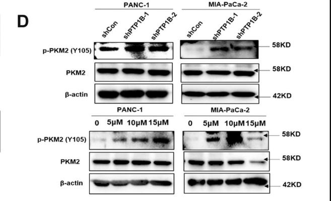
Western blot detected the expression levels of key autophagy-related proteins, including LC3B, P62 and Beclin1. GAPDH was defined as the internal control. (B)Quantitative statistics of protein expression levels of LC3B, P62 and Beclin1 after different treatments in BMSCs (n = 3). (C)Western blot detected the key protein expression levels of autophagic pathway, including p-mTOR, mTOR, p-ULK1, ULK1, PINK1 and Parkin1. GAPDH was defined as the internal control. (D)Quantitative statistics of protein expression levels of p-mTOR, mTOR, p-ULK1, ULK1, PINK1 and Parkin in different groups (n = 3). (E)Western blot detected the expression levels of PKM2 and p-PKM2 (Tyr105) in the three different groups. (F)Western blot detected the expression levels of p-AMPKα and total AMPKα after treatment of Shikonin in LVsh-PTP1B-transfected D-gal-induced Y-BMSCs. (G)Quantitative statistics of protein expression level of p-PKM2/PKM2 in the three different groups of D-gal-induced Y-BMSCs (n = 3). (H)Quantitative statistics of protein expression level of p-AMPKα/AMPKα after administration of Shikonin in LVsh-PTP1B-transfected D-gal-induced Y-BMSCs (n = 3). All data are shown as the mean ± SD. ****P ≤ 0.0001, ***P ≤ 0.001, **P < 0.01, *P < 0.05; NS, not significant (P > 0.05).")
.")

产品描述
*The optimal dilutions should be determined by the end user. For optimal experimental results, antibody reuse is not recommended.
*Tips:
WB: 适用于变性蛋白样本的免疫印迹检测. IHC: 适用于组织样本的石蜡(IHC-p)或冰冻(IHC-f)切片样本的免疫组化/荧光检测. IF/ICC: 适用于细胞样本的荧光检测. ELISA(peptide): 适用于抗原肽的ELISA检测.
引用格式: Affinity Biosciences Cat# AF5234, RRID:AB_2837720.
展开/折叠
CTHBP; Cytosolic thyroid hormone binding protein; Cytosolic thyroid hormone-binding protein; KPYM_HUMAN; MGC3932; OIP 3; OIP-3; OIP3; OPA interacting protein 3; Opa-interacting protein 3; p58; PK muscle type; PK, muscle type; PK2; PK3; PKM; PKM2; pykm; Pyruvate kinase 2/3; Pyruvate kinase 3; Pyruvate kinase isozymes M1/M2; Pyruvate kinase muscle; Pyruvate kinase muscle isozyme; pyruvate kinase PKM; Pyruvate kinase, muscle 2; TCB; THBP1; Thyroid hormone binding protein 1; Thyroid hormone binding protein cytosolic; Thyroid hormone-binding protein 1; Tumor M2 PK; Tumor M2-PK;
抗原和靶标
A synthesized peptide derived from human PKM2, corresponding to a region within the internal amino acids.
研究领域
· Human Diseases > Endocrine and metabolic diseases > Type II diabetes mellitus.
· Human Diseases > Infectious diseases: Viral > Human papillomavirus infection.
· Human Diseases > Cancers: Overview > Viral carcinogenesis.
· Human Diseases > Cancers: Overview > Central carbon metabolism in cancer. (View pathway)
· Metabolism > Carbohydrate metabolism > Glycolysis / Gluconeogenesis.
· Metabolism > Nucleotide metabolism > Purine metabolism.
· Metabolism > Carbohydrate metabolism > Pyruvate metabolism.
· Metabolism > Global and overview maps > Metabolic pathways.
· Metabolism > Global and overview maps > Carbon metabolism.
· Metabolism > Global and overview maps > Biosynthesis of amino acids.
· Organismal Systems > Endocrine system > Glucagon signaling pathway.
文献引用
Application: WB Species: human Sample: MIA PaCa-2 cells
Application: WB Species: Human Sample: pancreatic cancer tissue

Application: WB Species: Mouse Sample: RAW 264.7 cells

Application: WB Species: Human Sample: MCF-7 cells

Application: WB Species: human Sample: LC cells
限制条款
产品的规格、报价、验证数据请以官网为准,官网链接:www.affbiotech.com | www.affbiotech.cn(简体中文)| www.affbiotech.jp(日本語)产品的数据信息为Affinity所有,未经授权不得收集Affinity官网数据或资料用于商业用途,对抄袭产品数据的行为我们将保留诉诸法律的权利。
产品相关数据会因产品批次、产品检测情况随时调整,如您已订购该产品,请以订购时随货说明书为准,否则请以官网内容为准,官网内容有改动时恕不另行通知。
Affinity保证所销售产品均经过严格质量检测。如您购买的商品在规定时间内出现问题需要售后时,请您在Affinity官方渠道提交售后申请。产品仅供科学研究使用。不用于诊断和治疗。
产品未经授权不得转售。
Affinity Biosciences将不会对在使用我们的产品时可能发生的专利侵权或其他侵权行为负责。Affinity Biosciences, Affinity Biosciences标志和所有其他商标所有权归Affinity Biosciences LTD.