产品: CD83 抗体
货号: AF5233
描述: Rabbit polyclonal antibody to CD83
应用: WB IHC IF/ICC
文献验证: WB, IF/ICC
反应: Human, Mouse, Rat
预测: Pig
蛋白号: Q01151
RRID: AB_2837719

浏览相似产品>>

   规格 价格 库存
 50ul RMB¥ 1250 现货
 100ul RMB¥ 2300 现货
 200ul RMB¥ 3000 现货

货期: 当天发货

联系销售

产品描述

来源:
Rabbit
应用:
WB 1:500-1:2000, IF/ICC 1:100-1:500, IHC 1:50-1:200
*The optimal dilutions should be determined by the end user.
*Tips:

WB: 适用于变性蛋白样本的免疫印迹检测. IHC: 适用于组织样本的石蜡(IHC-p)或冰冻(IHC-f)切片样本的免疫组化/荧光检测. IF/ICC: 适用于细胞样本的荧光检测. ELISA(peptide): 适用于抗原肽的ELISA检测.

反应:
Human, Mouse, Rat
克隆:
Polyclonal
特异性:
CD83 Antibody detects endogenous levels of total CD83.
RRID:
AB_2837719
引用格式: Affinity Biosciences Cat# AF5233, RRID:AB_2837719.
偶联:
Unconjugated.
纯化:
The antiserum was purified by peptide affinity chromatography using SulfoLink™ Coupling Resin (Thermo Fisher Scientific).
保存:
Rabbit IgG in phosphate buffered saline , pH 7.4, 150mM NaCl, 0.02% sodium azide and 50% glycerol. Store at -20 °C. Stable for 12 months from date of receipt.
别名:

展开/折叠

B cell activation 45kDa cell surface glycoprotein Ig superfamily; B cell activation protein; B-cell activation protein; BL11; BL11 PEN; CD83; CD83 antigen (activated B lymphocytes, immunoglobulin superfamily); CD83 antigen; CD83 molecule; CD83_HUMAN; Cell surface glycoprotein; Cell surface protein HB15; HB15; hCD83;

抗原和靶标

免疫原:

A synthesized peptide derived from human CD83, corresponding to a region within the internal amino acids.

基因/基因ID:
描述:
May play a significant role in antigen presentation or the cellular interactions that follow lymphocyte activation.

文献引用

1). Targeted Covalent Nanodrugs Reinvigorate Antitumor Immunity and Kill Tumors via Improving Intratumoral Accumulation and Retention of Doxorubicin. ACS nano, 2024 (PubMed: 39760789) [IF=15.8]

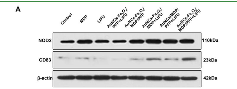
2). A Multifunctional Nanoparticle for Multimodal Imaging-Guided LIFU/Immuno-Synergistic Retinoblastoma Therapy. ACS Applied Materials & Interfaces, 2020 (PubMed: 31940169) [IF=8.3]

Application: WB    Species: Human    Sample: Y79 cells

Figure 12. (A,B) NOD2 and CD83 contents in Y79 tumors after different treatments. (C) IFN-γ and MCP-1 levels in Y79 tumors after different treatments. (D) H&E staining in major organs (heart, liver, spleen, lung, and kidney) of all groups after treatments. The scale bars are 50 μm. (n = 5, **p < 0.01).

3). Microglia Exhibit Distinct Heterogeneity Rather than M1/M2 Polarization within the Early Stage of Acute Ischemic Stroke. Aging and disease, 2023 (PubMed: 37199734) [IF=7.0]

Application: IF/ICC    Species: Mouse    Sample: Mic_M1L1 cells

Figure 4. Subpopulation analysis of M1-polarization-like microglia clusters. (A) Pseudotime plot (from monocle3) shows the differentiation trajectory between Mic_M1L1 and Mic_M1L2 cells. (B) Line plot of the fraction of cells in Mic_M1L1 and Mic_M1L2 subpopulations over time. (C) Expression of the ten genes with the highest Moran’s I score alongside the pseudotime trajectory. (D) Violin plot of the functional pathways revealed by gene set variation analysis (GSVA) in the Mic_M1L1 and Mic_M1L2 subpopulations. (E) Volcano plot displays the differentially expressed genes between Mic_M1L1 and Mic_M1L2 cells. Red dots represent the upregulated genes in the Mic_M1L1 cluster compared with the Mic_M1L2 cluster and blue dots represent the downregulated. (F) Relative activity of the highly expressed transcription factors (TF) in Mic_M1L2 cells compared with Mic_M1L1 cells. (G) Double immunofluorescence staining of Mic_M1L1 cells at each sampling time. Coronal brain sections are all stained with anti-Iba1(green, representing microglia), Nfkbiz (red, the marker of Mic_M1L1 cells), and DAPI (blue, representing cell nuclei) antibodies (N=3). The yellow bar represents 100μm. The arrows point to the cells simultaneously marked by Iba1 and Nfkbiz. (H) Double immunofluorescence staining of Mic_M1L2 cells at each sampling time. Coronal brain sections are all stained with anti-Iba1(green, representing microglia), Cd83 (red, the marker of Mic_M1L2 cells), and DAPI (blue, representing cell nuclei) antibodies (N=3). The yellow bar represents 100μm. The arrows point to the cells simultaneously marked by Iba1 and Cd83. (I) Bar plot of the ratio of Nfkbiz+Iba1+ cells (green plus red) compared with all DAPI+ cells in a 500μm2 area surrounding the infarction core (N=6). (J) Bar plot of the ratio of Cd83+Iba1+ cells (green plus red) compared with all DAPI+ cells in a 500μm2 area surrounding the infarction core (N=6).

4). Stromal and tumor immune microenvironment reprogramming through multifunctional cisplatin-based liposomes boosts the efficacy of anti-PD-1 immunotherapy in pancreatic cancer. Biomaterials science, 2023 (PubMed: 37921708) [IF=5.8]

5). Иммуногистохимическая характеристика биопсийных образцов десны в области беззубого альвеолярного края челюсти. Пародонтология, 2024

限制条款

产品的规格、报价、验证数据请以官网为准,官网链接:www.affbiotech.com | www.affbiotech.cn(简体中文)| www.affbiotech.jp(日本語)

产品的数据信息为Affinity所有,未经授权不得收集Affinity官网数据或资料用于商业用途,对抄袭产品数据的行为我们将保留诉诸法律的权利。

产品相关数据会因产品批次、产品检测情况随时调整,如您已订购该产品,请以订购时随货说明书为准,否则请以官网内容为准,官网内容有改动时恕不另行通知。

Affinity保证所销售产品均经过严格质量检测。如您购买的商品在规定时间内出现问题需要售后时,请您在Affinity官方渠道提交售后申请。

产品仅供科学研究使用。不用于诊断和治疗。 

产品未经授权不得转售。

Affinity Biosciences将不会对在使用我们的产品时可能发生的专利侵权或其他侵权行为负责。Affinity Biosciences, Affinity Biosciences标志和所有其他商标所有权归Affinity Biosciences LTD.