, using BMP2 Antibody at 1/1000 dilution.")


 and mouse anti-beta tubulin Ab(#T0023) for 1 hour at 37°C. An AlexaFluor594 conjugated goat anti-rabbit IgG Ab(Red) and an AlexaFluor488 conjugated goat anti-mouse IgG Ab(Green) were used as the secondary antibody.
The nuclear counter stain is DAPI (blue).")
 Non-induced IF cells and MF cells showed
different gene expression patterns; Data are mean ± SD of n = 3 replicates, one-way ANOVA, *p<0.05, **p<0.01, ***p<0.001, ****p<0.0001. (B) Non-induced
and induced IF cells and MF cells showed different protein expression patterns. After induction with CLCCM and HERSCM, there were no significant changes of
the expression patterns of tooth eruption-related proteins in IF cells and MF cells. (C) The grey value ratios of Western blotting results; Data are mean ± SD of n = 3
replicates, one-way ANOVA followed by Tukey post hoc test, *p<0.05, **p<0.01, ***p<0.001, ****p<0.0001. (IF1, MF1 represent the cells cultured by α-MEM;
IF2, MF2 represent the cells cultured by α-MEM + CLCCM; IF3, MF3 represent the cells cultured by α-MEM+HERSCM).")
‐thiazole‐4‐carboxylicacid methyl ester; TGF‐β, transforming growth factor‐β")
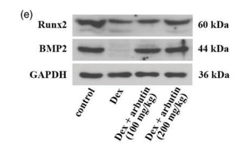
 lentivirus, respectively. 48 hours after transduction, cells were cultured in osteoblast induction medium. ALP staining was performed at days 7 and 14 following induction (a); corresponding ALP absorbance index was calculated (b). ARS staining of BMSCs was performed at days 14 and 21 following induction (c); corresponding ARS absorbance index was calculated (d). mRNA (e) and protein (f) levels of osteoblast-related genes Runx2, Bmp2, and OCN were detected by quantitative RT-PCR and western blot, respectively. β-Actin was used as an internal control. All data were expressed as means ± SD. ∗p < 0.05 and ∗∗p < 0.01.")

 hJBMMSCs. A. The ALP staining area was smaller and the color was lighter in DOP group than NC group on the 10th day (Mean ± SD, n = 6). B. The ALP activity was lower in DOP group than NC group after 7 days and 14 days of osteogenic induction (Mean ± SD, n = 6). C. After osteogenic induction, ARS (alizarin red S) staining on the 21st day showed that the number of mineralized nodules in DOP group was significantly less than that in NC group (Mean ± SD, n = 6). D. semiquantitative analysis showed that the OD (optical density) value of DOP group was significantly lower than that of NC group (Mean ± SD, n = 6). E. After osteogenic induction, the mRNA expressions of RUNX2, BMP2,ALP, OCN, SP7 and DLX5 in DOP group were significantly lower than those in NC group (Mean ± SD, n = 3). F. The protein expressions of RUNX2, BMP2, ALP and DLX5 in DOP group were significantly lower than those in NC group (Mean ± SD, n = 3). **P < 0.01,***P < 0.001.")
 promoted cell viability and osteogenic differentiation of rat bone marrow mesenchymal stem cells (BMSCs). (A) Morphological observation of primary cultured BMSCs at 1, 3, 5, and 7 days ( × 100, Scale bar = 100 μm). Then, BMSCs were cultured in the osteogenic induction and differentiation medium with/without IS (50, 100, and 200 mg/L) for 72 h. (B) IS (50, 100, and 200 mg/L) promoted cell viability of BMSCs, which was measured by the cell counting kit-8 analysis (n = 5). (C) IS advanced the levels of collagen type I (collagen I), alkaline phosphatase (ALP), and osteocalcin (OCN) of cultured supernatant, which was detected by the enzyme-linked immunosorbent assay (n = 6). (D) Alizarin red S (ARS) staining showed the area of ARS staining of BMSC with IS treatment was increased (n = 3). (E) They are characteristic photos of alizarin red S staining ( × 400, Scale bar = 300 μm). The expression levels of (F) bone morphogenetic protein-2 (BMP2), (G) smad1, (H) runt-related transcription factor 2 (RUNX2), (I) collagen I, (J) ALP in BMSCs were measured by Western blot (n = 3). IS promoted their expression. (K) The characteristic photos of Western blot. The experiment was repeated thrice, and all data were expressed with mean ± standard deviation. *P < 0.05, **P < 0.01, compared to the control (CN) group.")
 Molecular formula of Emo. (B, C) Immunoblot analysis of RUNX2, BMP2, and OCN. (D) Real-time PCR results of RUNX2, BMP2, and OCN. Data are expressed as mean ± standard deviation. ns, not significant")
, the immunohistochemical results of Piezo1, RUNX2, BMP2, CD31, HIF-a, and VEGF are presented. Additionally, (B–G) display the corresponding quantitative immunohistochemical results. In comparison to the model group, both the blank and WBVT groups exhibited significantly higher expression levels of Piezo1, RUNX2, BMP2, CD31, HIF-a, and VEGF. These differences were statistically significant (p < 0.05).")
 After transfection with BMP2-siRNA or pcDNA-BMP2, the protein expression of BMP2 was detected by WB to verify the transfection efficiency. (B) CCK8 was used to detect the viability of Nalm-6 cells after intervention or overexpression of BMP2. (C) The apoptosis of Nalm-6 cells was detected by flow cytometry. (D) Histogram of percentage of apoptotic cells. (E) The protein expressions of Bax, Bcl-2 and PCAN were detected by WB. (F) Histogram of gray level analysis of protein bands. *p < 0.05, **p < 0.01, ***p < 0.001.")

 IHC analysis of OCN in the femur (black arrows, n=5). (B) Western blotting analysis of ALP, BMP2 and Runx2 in tibias (n=3). Data are expressed as the mean ± SD. *P < 0.05, **P < 0.01 compared with the MOD group.")
 Histological analysis of the distal femur including HE staining, Masson staining, and immunohistochemistry. (B) The expression of ALP, BMP-2, RUNX-2, COL-1, and OCN in stained femurs were analyzed using Image J software.")
 After 14 days of osteogenic induction, the expression of Col1, Alp, Runx2, Bmp2, and Ocn mRNA was assessed via qRT-PCR. (B) The expression of Col1, Alp, Runx2, Bmp2, and Ocn was assessed via Western blotting analysis after 14 days of osteogenic induction. Quantification of protein expression is shown. Data are presented as the mean ± SD, n = 3 specimens/group")
 From top to bottom are the macroscopic view and microscopic view of Alizarin Red S-stained calcium nodules during osteogenic differentiation in h-BMSCs. (Scale bar = 10 μm). (B) The mRNA levels of typical osteogenic differentiation markers OCN, RUNX2 and BMP2 were measured by qRT-PCR at 0, 7, and 14 days. (C) The protein levels of typical osteogenic differentiation markers RUNX2 and BMP2 were detected by western-blotting at 0, 7, and 14 days. (D) Expression of hsa_circ_0001275/miR-422a during osteogenic differentiation of h-BMSCs at 0, 7, and 14 days. (E) Hsa_circ_0001275 was pictured under confocal microscopy by FISH in situ fluorescence hybridization technique. hsa_circ_0001275 appeared as red and DAPI staining as blue confirming that hsa_circ_0001275 was mainly located in the cytoplasm of h-BMSCs cells. *P < 0.05, **P < 0.01 and ****P < 0.0001. (For interpretation of the references to colour in this figure legend, the reader is referred to the web version of this article.)")
 The relative expression levels of miR-25-3p in ONFH-BMSCs and control BMSCs were examined by RT-qPCR analysis. (B) The BMSCs were transfected with miR-25-3p mimics or its control mimics NC, and the relative expression levels of miR-25-3p were determined by RT-qPCR analysis. (C) The BMSCs were treated with miR-25-3p inhibitor or its control inhibitor NC, and the relative expression levels of miR-25-3p were determined by RT-qPCR analysis. (D and E) The cell proliferation ability of BMSCs with corresponding treatment was revealed by the cell-cycle analysis, with the cell population of G1, G2, and S cell-cycle phase indicated. #p < 0.05 versus the corresponding NC group for comparing cell percentage in S phase; &p < 0.05 versus the corresponding NC group for comparing cell percentage in G1 phase; n = 3. (F and G) The protein expression levels of osteogenic marker genes in BMSCs of different groups were detected by western blot analysis. The quantitative analysis showed the protein expression levels of ALP, BMP2, RUNX2, and OCN were increased in the miRNA mimics group compared with the mimics NC group and were decreased in the miRNA inhibitor group compared with the inhibitor NC group. (H and I) The osteogenic differentiation ability of BMSCs at the 7th and 14th day after corresponding treatment was evaluated by alizarin red staining. Data were shown as mean ± SD. ∗∗p < 0.01, ∗∗∗p < 0.001, n = 3. Data between two groups were analyzed by Student's t test. Data among multiple groups were analyzed by one-way ANOVA test.")
 in the Control group, marked increase in expression (black arrows) in L-MTZ, increased expression (black arrows) in S-MTZ. BMP-2: expression findings between the groups, moderate expression (black arrow) in the Control group, increase in expression (black arrows) in L-MTZ, marked expression (black arrow) in S-MTZ. Runx2: moderate expression (black arrows) in the Control group, increase in expression (black arrows) in L-MTZ, marked expression (black arrows) in S-MTZ. ALP: moderate expression (black arrow) in the Control group, marked increase in expression (black arrows) in L-MTZ, moderate expression (black arrow) in S-MTZ. OCN: moderate expression (black arrow) in the control group, markedly increased expression (black arrow) in L-MTZ, and increased expression (black arrow) in S-MTZ. RANKL: Similar RANKL expressions (black arrows) in (A) Control group, (B) L-MTZ, and (C) S-MTZ, Streptavidin biotin peroxidase method, Scale bars = 50 μm Representative histopathological images among the groups. (A) Marked fibrous tissue, moderate new bone formation (white arrow with a black border), and residual graft materials (RG) in the Control group. (B) Decreased fibrous tissue, moderate residual graft material (RG), and marked new bone formation (white arrow with a black border) in L-MTZ. (C) Decreased fibrous tissue, moderately increased new bone formation (white arrow with a black border), and residual graft materials (RG) in S-MTZ. HE, Scale bars = 200 μm")
 ALP staining. (B) ARS staining. (C) Unstained MSMSCs transfected with shSLC7A2 at 21 days after osteogenic induction. Scale bars = 100 µm. (D) qPCR analysis of OCN, RUNX2, and BMP2 mRNA expression on D3, D7, D14, and D21 after osteogenic induction in shSLC7A2-transfected MSMSCs. Target mRNA levels were normalized to GAPDH. (E) WB analysis of OCN, RUNX2, and BMP2 protein expression at t D0, D7 and D14 after osteogenic induction in shSLC7A2-transfected MSMSCs. (F) Bar chart showing OCN, RUNX2, and BMP2 protein levels (normalized to GAPDH) quantified from WB analysis in shSLC7A2-transfected MSMSCs at D0, D7, and D14 after osteogenic induction. Results are representative of three independent experiments. Error bars indicate the standard error of the mean. * indicates P < 0.05 when comparing shSLC7A2-transfected MSMSCs at D3, D7, D14, and D21 with D0 within the same group. # P < 0.05 and ## P < 0.01 indicate statistically significant differences at the 0.05 and 0.01 levels, respectively,between the shSLC7A2 and shEV group at the same time point. Legend: shEV = short hairpin RNA empty vector; shSLC7A2 = Short hairpin RNA targeting solute carrier family 7 member 2; GAPDH = Glyceraldehyde 3-phosphate dehydrogenase; qPCR = quantitative PCR; WB = Western blotting; ALP: alkaline phosphatase; ARS:Alizarin Red S staining. OCN: Osteocalcin; RUNX2:Runt-related transcription factor2; BMP2: Bone Morphogenetic Protein 2.")

产品描述
*The optimal dilutions should be determined by the end user. For optimal experimental results, antibody reuse is not recommended.
*Tips:
WB: 适用于变性蛋白样本的免疫印迹检测. IHC: 适用于组织样本的石蜡(IHC-p)或冰冻(IHC-f)切片样本的免疫组化/荧光检测. IF/ICC: 适用于细胞样本的荧光检测. ELISA(peptide): 适用于抗原肽的ELISA检测.
引用格式: Affinity Biosciences Cat# AF5163, RRID:AB_2837649.
展开/折叠
BMP 2; BMP 2A; BMP2; BMP2A; Bone morphogenetic protein 2;
抗原和靶标
A synthesized peptide derived from human BMP2, corresponding to a region within the internal amino acids.
研究领域
· Environmental Information Processing > Signaling molecules and interaction > Cytokine-cytokine receptor interaction. (View pathway)
· Environmental Information Processing > Signal transduction > TGF-beta signaling pathway. (View pathway)
· Environmental Information Processing > Signal transduction > Hippo signaling pathway. (View pathway)
· Human Diseases > Cancers: Overview > Pathways in cancer. (View pathway)
· Human Diseases > Cancers: Specific types > Basal cell carcinoma. (View pathway)
文献引用

Application: WB Species: Rat Sample:

Application: IF/ICC Species: Rat Sample:

Application: WB Species: human Sample:

Application: WB Species: Rat Sample: rBMSCs

Application: WB Species: rat Sample: BMSCs

Application: IF/ICC Species: Rat Sample:
Application: WB Species: Human Sample: DPSCs

Application: WB Species: human Sample:
限制条款
产品的规格、报价、验证数据请以官网为准,官网链接:www.affbiotech.com | www.affbiotech.cn(简体中文)| www.affbiotech.jp(日本語)产品的数据信息为Affinity所有,未经授权不得收集Affinity官网数据或资料用于商业用途,对抄袭产品数据的行为我们将保留诉诸法律的权利。
产品相关数据会因产品批次、产品检测情况随时调整,如您已订购该产品,请以订购时随货说明书为准,否则请以官网内容为准,官网内容有改动时恕不另行通知。
Affinity保证所销售产品均经过严格质量检测。如您购买的商品在规定时间内出现问题需要售后时,请您在Affinity官方渠道提交售后申请。产品仅供科学研究使用。不用于诊断和治疗。
产品未经授权不得转售。
Affinity Biosciences将不会对在使用我们的产品时可能发生的专利侵权或其他侵权行为负责。Affinity Biosciences, Affinity Biosciences标志和所有其他商标所有权归Affinity Biosciences LTD.