产品: FAK 抗体
货号: AF6397
描述: Rabbit polyclonal antibody to FAK
应用: WB IHC IF/ICC
文献验证: WB
反应: Human, Mouse, Rat
预测: Pig, Horse, Sheep, Rabbit, Dog, Chicken, Xenopus
蛋白号: Q05397
RRID: AB_2835232

浏览相似产品>>

   规格 价格 库存
 50ul RMB¥ 1250 现货
 100ul RMB¥ 2300 现货
 200ul RMB¥ 3000 现货

货期: 当天发货

联系销售

产品描述

来源:
Rabbit
应用:
WB 1:500-1:2000, IHC 1:50-1:200, IF/ICC 1:100-1:500
*The optimal dilutions should be determined by the end user.
*Tips:

WB: 适用于变性蛋白样本的免疫印迹检测. IHC: 适用于组织样本的石蜡(IHC-p)或冰冻(IHC-f)切片样本的免疫组化/荧光检测. IF/ICC: 适用于细胞样本的荧光检测. ELISA(peptide): 适用于抗原肽的ELISA检测.

反应:
Human, Mouse, Rat
克隆:
Polyclonal
特异性:
FAK Antibody detects endogenous levels of total FAK.
RRID:
AB_2835232
引用格式: Affinity Biosciences Cat# AF6397, RRID:AB_2835232.
偶联:
Unconjugated.
纯化:
The antiserum was purified by peptide affinity chromatography using SulfoLink™ Coupling Resin (Thermo Fisher Scientific).
保存:
Rabbit IgG in phosphate buffered saline , pH 7.4, 150mM NaCl, 0.02% sodium azide and 50% glycerol. Store at -20 °C. Stable for 12 months from date of receipt.
别名:

展开/折叠

FADK 1; FADK; FAK related non kinase polypeptide; FAK1; FAK1_HUMAN; Focal adhesion kinase 1; Focal adhesion Kinase; Focal adhesion kinase isoform FAK Del33; Focal adhesion kinase related nonkinase; FRNK; p125FAK; pp125FAK; PPP1R71; Protein phosphatase 1 regulatory subunit 71; Protein tyrosine kinase 2; Protein-tyrosine kinase 2; Ptk2; PTK2 protein tyrosine kinase 2;

抗原和靶标

免疫原:

A synthesized peptide derived from human FAK, corresponding to a region within the internal amino acids.

基因/基因ID:
描述:
This gene encodes a cytoplasmic protein tyrosine kinase which is found concentrated in the focal adhesions that form between cells growing in the presence of extracellular matrix constituents.

研究领域

· Cellular Processes > Cellular community - eukaryotes > Focal adhesion.   (View pathway)

· Cellular Processes > Cell motility > Regulation of actin cytoskeleton.   (View pathway)

· Environmental Information Processing > Signal transduction > ErbB signaling pathway.   (View pathway)

· Environmental Information Processing > Signal transduction > PI3K-Akt signaling pathway.   (View pathway)

· Human Diseases > Drug resistance: Antineoplastic > Endocrine resistance.

· Human Diseases > Infectious diseases: Bacterial > Bacterial invasion of epithelial cells.

· Human Diseases > Infectious diseases: Parasitic > Amoebiasis.

· Human Diseases > Infectious diseases: Viral > Human papillomavirus infection.

· Human Diseases > Cancers: Overview > Pathways in cancer.   (View pathway)

· Human Diseases > Cancers: Overview > Transcriptional misregulation in cancer.

· Human Diseases > Cancers: Overview > Proteoglycans in cancer.

· Human Diseases > Cancers: Specific types > Small cell lung cancer.   (View pathway)

· Organismal Systems > Immune system > Chemokine signaling pathway.   (View pathway)

· Organismal Systems > Development > Axon guidance.   (View pathway)

· Organismal Systems > Immune system > Leukocyte transendothelial migration.   (View pathway)

文献引用

1). Anti-proliferation and anti-migration effects of Yishen Tongbi decoction in experimental rheumatoid arthritis by suppressing SLC3A2/integrin β3 signaling pathways. Phytomedicine, 2023 (PubMed: 36990010) [IF=6.7]

2). Catalpol ameliorates liver fibrosis via inhibiting aerobic glycolysis by EphA2/FAK/Src signaling pathway. Phytomedicine : international journal of phytotherapy and phytopharmacology, 2024 (PubMed: 39321687) [IF=6.7]

3). pH-responsive biomimetic zeolitic imidazolate framework-based nanoparticles for co-delivery of cetuximab and siRNA in synergistic therapy of laryngeal squamous cell carcinoma. Journal of pharmaceutical analysis, 2025 (PubMed: 40735032) [IF=6.1]

Application: WB    Species: Mouse    Sample: TU177 cells

Fig. 4. Evaluation of therapeutic efficiency and pH-responsive property of cetuximab (Cet)/focal adhesion kinase (FAK) small interfering RNA (siFAK)@zeolitic imidazolate framework-8 (ZIF-8)@TU177 cell membrane (TCM) in vitro. (A, B) The messenger RNA (mRNA) (A) and protein (B) levels of FAK in TU177 cells treated with free siFAK and siFAK-loaded nanocomplexes. (C) FAK mRNA levels of FAK in TU177 cells before and after three generations. (D–F) Viability (D), apoptosis (E), and live/dead status (F) of TU177 cells cultured with free Cet, free siFAK, Cet@ZIF-8@TCM, siFAK@ZIF-8@TCM, and Cet/siFAK@ZIF-8@TCM at pH 6.5 and 7.4. ∗P < 0.05 and ∗∗P < 0.01. ns: no significance. GAPDH: glyceraldehyde-3-phosphate dehydrogenase; PEA: phycoerythrin-area; UL: upper left; UR: upper right; LL: lower left; LR: lower right; FITC: fluorescein isothiocyanate.

4). Analysis of Expression of the ANG1, CaSR and FAK Proteins in Uterine Fibroids. International journal of molecular sciences, 2024 (PubMed: 39000274) [IF=5.6]

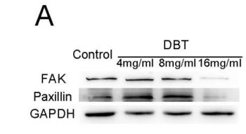
5). Danggui Buxue Tang promotes the adhesion and migration of bone marrow stromal cells via the focal adhesion pathway in vitro. JOURNAL OF ETHNOPHARMACOLOGY, 2019 (PubMed: 30445110) [IF=4.8]

Application: WB    Species: mouse    Sample: BMSCs

Figure 8 |Western blot analysis of key proteins in the focal adhesion pathway after DBT treatment for 6 days. (A, C) Representatives blot from 3 independent experiments. (B, D) Densitometric quantification. GAPDH was used as an internal control. Values are the mean ± SEM, where n = 3. (*p < 0.05 as compared to the control, LSD).

6). Phellinus linteus activates Treg cells via FAK to promote M2 macrophage polarization in hepatocellular carcinoma. Cancer immunology, immunotherapy : CII, 2024 (PubMed: 38240856) [IF=4.6]

Application: WB    Species: Mouse    Sample:

Fig. 3 FAK promotes the polarization of macrophages in the Treg-macrophage co-culture system through PI3K/AKT/JAK/STAT3 and p38/JNK/ERK signal pathway A, B The expression of the PI3K, p-PI3K, AKT, p-AKT, p-FAK, FAK, JAK2, STAT3, p-STAT3 and p38, p-p38/JNK, p-JNK/ERK1/2, p-ERK1/2 in Treg-macrophage co-culture system were observed by the western blot, n = 3 in each group. #P 

7). KRT7 promotes epithelial‑mesenchymal transition in ovarian cancer via the TGF‑β/Smad2/3 signaling pathway. ONCOLOGY REPORTS, 2021 (PubMed: 33416175) [IF=3.8]

Application: WB    Species: Human    Sample: HEY cells

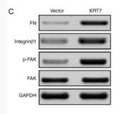
Figure 5. - KRT7 expression affects the integrin-β1-FAK signaling and TGF-β signaling pathways. (A and B) Expression of proliferation- and migration-associated genes (PCNA, MMP9 and TIMP-1) were evaluated using western blotting in HEY cells. (C and D) Western blotting of proteins involved in integrin-β1-FAK signaling pathway in the KRT7-overexpressing HEY cells. (E) Expression of MMPs after knockdown of KRT7 in OVCAR433 cells. (F and G) Expression of the TGF-β signaling pathway-related proteins was evaluated by western blotting in KRT7-overexpressing HEY cells and KRT7-knockdown OVCAR433 cells. All experiments were performed at least three times. Results are presented as the mean ± standard deviation. **P<0.01. FAK, focal adhesion kinase; PCNA, proliferating cell nuclear antigen; FN, fibronectin; TIMP-1, TIMP metallopeptidase inhibitor 1; p-, phosphorylated; MMP, matrix metalloproteinase; KRT7, keratin 7; sh, short hairpin RNA; NC, negative control.

8). FOXF1 inhibits invasion and metastasis of lung adenocarcinoma cells and enhances anti-tumor immunity via MFAP4/FAK signal axis. Scientific reports, 2024 (PubMed: 39271782) [IF=3.8]

Application: WB    Species: Human    Sample:

Fig. 6. FOXF1 can affect the activation of FAK signal pathway by regulating the expression of MFAP4. (a) The network diagram shows the signal pathways positively related to the high expression of FOXF1 and MFAP4. Green indicates that the activation of signaling pathways is significantly correlated with the expression of FOXF1, and the blue indicates that the activation of signaling pathways is significantly correlated with the expression of MFAP4. The network diagram displays the three signal pathways that are most relevant to both FOXF1 and MFAP4 (Pathways in Cancer, Focal Adhesion, Purine Metabolism). (b) GSEA shows that the high expression of FOXF1 and MFAP4 is significantly associated with Focal Adhesion signal pathway. (c) ssGSEA indicates that both high expression of FOXF1 and MFAP4 is significantly associated with Focal Adhesion signal pathway. (d) Protein expression of FAK and p-FAK (Tyr397) with FOXF1 and MFAP4 expression are detected by Western Blot. The network diagram was generated from “Cytoscape” software (version 3.10.2, https://cytoscape.org/) and the GSEA plots were generated from the “ggplot2” (version 3.5.0, https://github.com/tidyverse/ggplot2) package in R.

9). Niban apoptosis regulator 1 promotes gemcitabine resistance by activating the focal adhesion kinase signaling pathway in bladder cancer. Journal of Cancer, 2022 (PubMed: 35281857) [IF=3.3]

Application: WB    Species: Human    Sample: bladder cancer cells

Figure 7 High NIBAN1 expression correlates with highly enriched focal adhesion signaling in bladder cancer. A. Gene Set Enrichment Analysis of The Cancer Genome Atlas bladder cancer dataset revealed a highly enriched gene signature of focal adhesion pathway in NIBAN1-high bladder cancer. B. GEM-resistant cells T24GR exhibited highly activated FAK and its downstream SRC/AKT signaling compared to GEM-sensitive T24 and 5637 cells. β-Actin was used as a loading control. C. Differentially expressed genes in T24GR cells with or without NIBAN1 knockdown. D. Pathway enrichment analysis was conducted on downregulated genes (n = 173) using GO, KEGG, and Reactome Gene Sets annotations.

10). Inhibitory Impact Of Cinobufagin In Triple-Negative Breast Cancer Metastasis: Involvements Of Macrophage Reprogramming Through Upregulated MME and Inactivated FAK/STAT3 Signaling. Clinical breast cancer, 2024 (PubMed: 38378361) [IF=2.9]

加载更多

限制条款

产品的规格、报价、验证数据请以官网为准,官网链接:www.affbiotech.com | www.affbiotech.cn(简体中文)| www.affbiotech.jp(日本語)

产品的数据信息为Affinity所有,未经授权不得收集Affinity官网数据或资料用于商业用途,对抄袭产品数据的行为我们将保留诉诸法律的权利。

产品相关数据会因产品批次、产品检测情况随时调整,如您已订购该产品,请以订购时随货说明书为准,否则请以官网内容为准,官网内容有改动时恕不另行通知。

Affinity保证所销售产品均经过严格质量检测。如您购买的商品在规定时间内出现问题需要售后时,请您在Affinity官方渠道提交售后申请。

产品仅供科学研究使用。不用于诊断和治疗。 

产品未经授权不得转售。

Affinity Biosciences将不会对在使用我们的产品时可能发生的专利侵权或其他侵权行为负责。Affinity Biosciences, Affinity Biosciences标志和所有其他商标所有权归Affinity Biosciences LTD.