产品: SOX9 抗体
货号: AF6330
描述: Rabbit polyclonal antibody to SOX9
应用: WB IHC IF/ICC
文献验证: WB, IHC, IF/ICC
反应: Human, Mouse, Rat, Monkey
预测: Pig, Horse, Sheep, Rabbit, Dog, Chicken, Xenopus
蛋白号: P48436
RRID: AB_2835186

浏览相似产品>>

   规格 价格 库存
 50ul RMB¥ 1250 现货
 100ul RMB¥ 2300 现货
 200ul RMB¥ 3000 现货

货期: 当天发货

联系销售

产品描述

来源:
Rabbit
应用:
WB 1:500-1:2000, IHC 1:50-1:200, IF/ICC 1:100-1:500
*The optimal dilutions should be determined by the end user.
*Tips:

WB: 适用于变性蛋白样本的免疫印迹检测. IHC: 适用于组织样本的石蜡(IHC-p)或冰冻(IHC-f)切片样本的免疫组化/荧光检测. IF/ICC: 适用于细胞样本的荧光检测. ELISA(peptide): 适用于抗原肽的ELISA检测.

反应:
Human, Mouse, Rat, Monkey
克隆:
Polyclonal
特异性:
SOX9 Antibody detects endogenous levels of total SOX9.
RRID:
AB_2835186
引用格式: Affinity Biosciences Cat# AF6330, RRID:AB_2835186.
偶联:
Unconjugated.
纯化:
The antiserum was purified by peptide affinity chromatography using SulfoLink™ Coupling Resin (Thermo Fisher Scientific).
保存:
Rabbit IgG in phosphate buffered saline , pH 7.4, 150mM NaCl, 0.02% sodium azide and 50% glycerol. Store at -20 °C. Stable for 12 months from date of receipt.
别名:

展开/折叠

campomelic dysplasia autosomal sex reversal; CMD 1; CMD1; CMPD 1; CMPD1; SOX 9; Sox9; SOX9_HUMAN; SRA 1; SRA1; SRXX2; SRXY10; SRY (sex determining region Y) box 9 (campomelic dysplasia autosomal; SRY (sex determining region Y) box 9; SRY (sex determining region Y)-box 9; SRY (sex-determining region Y)-box 9 protein; SRY related HMG box gene 9; Transcription factor SOX 9; Transcription factor SOX-9; transcription factor SOX9;

抗原和靶标

免疫原:

A synthesized peptide derived from human SOX9, corresponding to a region within the internal amino acids.

基因/基因ID:
描述:
SOX9 Plays an important role in the normal skeletal development. May regulate the expression of other genes involved in chondrogenesis by acting as a transcription factor for these genes. Defects in SOX9 are the cause of campomelic dysplasia (CMD1).

研究领域

· Environmental Information Processing > Signal transduction > cAMP signaling pathway.   (View pathway)

文献引用

1). Targeting the immune privilege of tumor-initiating cells to enhance cancer immunotherapy. Cancer cell, 2024 (PubMed: 39515328) [IF=48.8]

2). Targeted Blockage of Pathological Extracellular Vesicles and Particles From Fibroblast-Like Synoviocytes for Osteoarthritis Relief: Proteomic Analysis and Cellular Effect. Journal of Extracellular Vesicles, 2025 [IF=14.5]

Application: IF/ICC    Species: Mouse    Sample:

FIGURE 5 Pathogenic FLS EVPs impair chondrogenic differentiation of mesenchymal stem cells. (A) Schematic diagram of EVP stimulation on mouse BMSCs isolated from the femoral bone marrow cavity of mice. (B) Internalization of EVPs by BMSCs. Scale bar = 50 µm. (C) Expression of chondrogenic differentiation-related genes in chondrogenesis-induced BMSCs after 7 days of treatment with different EVPs. (D) Representative Western blot images of COL10A1, the marker of chondrocyte hypertrophy. (E, F) Representative images of alcian blue staining and SA-β-gal staining in BMSCs after 7 days of stimulation with different EVPs. Scale bar = 1 mm and 200 µm separately. (G) Schematic diagram of section staining observation after inducing BMSCs to form chondrocyte pellets for 21 days while simultaneously stimulating with different EVPs. (H, I) Representative images of SOX9 fluorescence staining, safranin O (SO) staining, alcian blue (AB) staining and toluidine blue (TB) staining of BMSC-differentiated chondrocyte pellet sections. Scale bar = 50 µm. (J) Schematic diagram of EVP treatment on mouse ADSCs isolated from the iWAT of mice. (K) Internalization of EVPs by ADSCs. Scale bar = 50 µm. (L, M) Representative images of SA-β-gal staining and alcian blue staining of ADSCs after 7 days of chondrogenic induction and treatment with different EVPs. (N) Representative images of SOX9 staining in sections of chondrocyte pellets formed by ADSC after 21-day induction.

3). Plasma Membrane-Derived Biomimetic Apoptotic Nanovesicles Targeting Inflammation and Cartilage Degeneration for Osteoarthritis. Small methods, 2024 (PubMed: 39036830) [IF=10.7]

4). Lacc1-engineered extracellular vesicles reprogram mitochondrial metabolism to alleviate inflammation and cartilage degeneration in TMJ osteoarthritis. Journal of nanobiotechnology, 2025 (PubMed: 40186254) [IF=10.2]

5). HAMA-SBMA hydrogel with anti-inflammatory properties delivers cartilage organoids, boosting cartilage regeneration. Journal of nanobiotechnology, 2025 (PubMed: 40448111) [IF=10.2]

Application: IF/ICC    Species: Rat    Sample:

Fig. 4 HS suppresses inflammation and promotes chondrogenesis in CCO while enhancing integrin β1 expression. (A) Immunofluorescence staining for iNOS, Arg-1, CD86, and CD206 was performed after one day of co-culture of HS hydrogel with BMDM macrophages for M1 macrophage induction, utilizing DAPI (blue) for nuclei, specific staining (red) for iNOS, CD86 and specific staining (green) for CD206, Arg-1. (B) Quantitative analysis of Figure A. All three types of hydrogels can promote the polarization of macrophages toward the M2 phenotype. (C) SO, and AB staining after four days of co-culture of HS hydrogel with BMSCs for cartilage induction. HS600 can accumulate a greater amount of cartilage matrix. (D) Quantitative analysis of mRNA transcripts for COL2 and SOX9. The expression of cartilage genes is higher in the HS600 group. (E) Fluorescence staining for COL2 and SOX9, utilizing rhodamine phalloidin (red) for F-actin, DAPI (blue) for nuclei, and specific staining (green) for COL2 and SOX9. (F) Quantitative analysis of Figure E. The expression of cartilage-related proteins is higher in the HS600 group. (G) Fluorescence staining for β1, using rhodamine phalloidin (red) for F-actin, DAPI (blue) for nuclei, and specific staining (green) for β1. (H) Quantitative analysis of Figure G. The expression of integrin β1 protein is higher in the HS600 group. (I) Staining of F-actin (green) and cell nuclei (blue) after BMSCs were seeded on the hydrogel surface for 1 day. The morphology of BMSCs in the HS group can exhibit normal spreading. (J) Immunofluorescence staining of COL2, and β1 in CCO co-cultured with HS600. (K) Quantitative analysis of Figure J. After co-culturing HS600 with CCO, the expression of cartilage markers and the adhesion factor β1 are enhanced. All experiments were repeated three times. ns p > 0.05, *0.01 

6). Selenium nanozyme-crosslinked composite hydrogel for promoting cartilage regeneration in osteoarthritis via an integrated 'outside-in' and 'inside-out' strategy. Journal of colloid and interface science, 2025 (PubMed: 40252578) [IF=9.4]

7). A lithium-containing biomaterial promotes chondrogenic differentiation of induced pluripotent stem cells with reducing hypertrophy. Stem Cell Research & Therapy, 2020 (PubMed: 32085810) [IF=7.5]

Application: IF/ICC    Species: Human    Sample: iPSCs

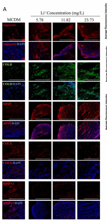
Fig. 7 The chondrogenic differentiation of iPSCs with different Li+ ions concentration in 14 days. a Immunofluorescence of chondrocytes specified proteins (COL II, Aggrecan, and SOX9) and hypertrophic specified protein (COL X and MMP13). b The average fluorescence intensity of COL II, Aggrecan, SOX9, MMP13, and COL X proteins in each group, n = 5. c The relative genes expression of chondrogenic differentiation cultured with Li+ ions, n = 3. d The relative genes expression of hypertrophic differentiation cultured with Li+ ions, n = 3. Data presented as mean ± SEM. Scale bar = 200 μm. CTR: MCDM without any Li+ ions. *p < 0.05, **p < 0.01, ***p < 0.001

8). Vitamin K2 ameliorates osteoarthritis by suppressing ferroptosis and extracellular matrix degradation through activation GPX4's dual functions. Biomedicine & pharmacotherapy = Biomedecine & pharmacotherapie, 2024 (PubMed: 38759289) [IF=6.9]

Application: WB    Species: Rat    Sample:

Fig. 8. GPX4 inhibitor RSL3 substantially attenuates VK2 protection of chondrocytes. (A-I) WB results for GPX4, NFκB, pMAPK/MAPK, Aggrecan, CollagenⅡ, SOX9, MMP3, MMP13 and Tubulin; (J) Quantitive analysis of relative Glutathione Peroxidase Activity; (K-L) GSH contents and ratio of GSH/GSSG; (M) Quantitative results of MDA content assay.

9). Combination of curcumin and catalase protects against chondrocyte injury and knee osteoarthritis progression by suppressing oxidative stress. Biomedicine & pharmacotherapy = Biomedecine & pharmacotherapie, 2023 (PubMed: 37879214) [IF=6.9]

10). TCF12 regulates the TGF-β/Smad2/3 signaling pathway to accelerate the progression of osteoarthritis by targeting CXCR4. Journal of orthopaedic translation, 2023 (PubMed: 38235367) [IF=5.9]

加载更多

限制条款

产品的规格、报价、验证数据请以官网为准,官网链接:www.affbiotech.com | www.affbiotech.cn(简体中文)| www.affbiotech.jp(日本語)

产品的数据信息为Affinity所有,未经授权不得收集Affinity官网数据或资料用于商业用途,对抄袭产品数据的行为我们将保留诉诸法律的权利。

产品相关数据会因产品批次、产品检测情况随时调整,如您已订购该产品,请以订购时随货说明书为准,否则请以官网内容为准,官网内容有改动时恕不另行通知。

Affinity保证所销售产品均经过严格质量检测。如您购买的商品在规定时间内出现问题需要售后时,请您在Affinity官方渠道提交售后申请。

产品仅供科学研究使用。不用于诊断和治疗。 

产品未经授权不得转售。

Affinity Biosciences将不会对在使用我们的产品时可能发生的专利侵权或其他侵权行为负责。Affinity Biosciences, Affinity Biosciences标志和所有其他商标所有权归Affinity Biosciences LTD.