


affects tumor functions related to vasculogenic mimicry (VM) formation in hepatocellular carcinoma cells..e Western blot analysis of the expression levels of VE–Cad, vascular endothelial growth factor receptor 1 (VEGFR1), and VEGFR2 influenced by overexpressing TP and adding dT in glucose-free cultured PLC-PRF-5 cells. The ratio of densitometry value to the corresponding glyceraldehyde 3-phosphate dehydrogenase (GAPDH) value was used to indicate the relative protein expression. NG means “No Glucose” (mean ± SD; n = 3 in triplicate; **P < 0.01)")
 on hepatocellular carcinoma (HCC) growth,metastasis, and vasculogenic mimicry (VM) formation in the xenograft model. d Analysis of Twist1, TP, VE–Cad, vascular endothelial growth factor receptor1 (VEGFR1), and VEGFR2 expression levels in xenograft tumors. Images were taken at 400 magnification.")
 or MCC950 (10µM), or NLRP3 siRNA. Western blots showed the effects of acarbose on the protein expression of ZO-1 and VE-Cadherin (A, B).")
 to develop a rat model of T2DM that were treated with acarbose (30mg/kg/d) and MCC950(3mg/kg/d) for 5 weeks. Representative immunofluorescent images and summarized data showed the role of acarbose and MCC950 on the expression of ZO-1 and VE-Cadherin in the arterial endothelium (A-B).")
 and VE‐Cadherin
(b) in mice defect implanted the
LV‐NC‐iPSC‐MSCs/HA scaffold or
LV‐Sema3A/HIF1α‐iPSC‐MSCs/HA
scaffold at 8 weeks after surgery (scale
bar = 100 μm). HA, hydroxyapatite; HIF1α,
hypoxia‐inducible factor‐1α; iPSC, induced
pluripotent stem cell; LV, lentivirus; MSC,
mesenchymal stem or stromal cell; RUNX2,
runt‐related transcription factor 2;
Sema3A, semaphorin 3A")
 VE-cad, p-VE-cad, and Occludin protein levels in HUVECs were determined (n=5).")
 VE-cad, p-VE-cad, and Occludin protein levels in HUVECs were determined (n=5). (C) Immunofluorescence localization and relative expression of VE-cad in HUVECs. All photographs were taken at 40× magnification.")
 (N= 3 indicates independent experiments for RT-qPCR and N=2 for immunoblotting with six mice/group(12 femora and 12 tibias). *p<0.05 **p<0.01, and ***p<0.001")

 (N= 3 indicates independent experiments for RT-qPCR and N=2 for immunoblotting with six mice/group (12 femora and 12 tibias). *p<0.05 **p<0.01, and ***p<0.001")
, anti-CD144 (VE-cadherin), anti-CD106 (ICAM-1), and anti-VEGFR2—the cell shows the expression of these endothelial cell-specific molecules. b The mean fluorescence intensity for endothelial cells particular marker in the graph was relatively quantified by ImageJ, which shows the order of the endothelial cell-specific markers (PECAM-1> VE-cadherin > ICAM-1 > VEGFR2, N = 3 experimental repeats with three mice/group (6 femora and 6 tibias), scale bar 5μm, ×20 magnification")
-cadherin protein expression level is markedly increased in mouse aortic endothelial cells (MAECs) cultured in high glucose (HG) for 7 days and in thoracic aorta endothelial cells of streptozotocin (STZ)-induced mice with diabetes with no change in VE-cadherin expression level; HG exposure promotes intercellular permeability in MAECs. Representative western blot analysis images (A) and summary data (B) showing p-VE-cadherin and VE-cadherin expression levels in MAECs cultured in normal glucose (NG) or HG medium. (C, E) MAECs were cultured in HG medium for 7 days and then fixed and incubated with anti-p-VE-cadherin antibody (red), anti-VE-cadherin antibody (red) and 4',6-diamidino-2-phenylindole (DAPI) (blue; cell nuclei) and then imaged with a confocal microscope. Representative confocal microscopy images and the final merged images are shown (C, E). (D, F) Fluorescence intensity profiles and summary data for anti-p-VE-cadherin antibody and anti-VE-cadherin antibody in the regions delineated by the corresponding yellow line shown in (C) and (E). (G) Representative images of thoracic aorta endothelium immunostaining (brown; eg, red arrowheads) for expression levels of p-VE-cadherin and VE-cadherin in a mouse model of diabetes mellitus (DM) and control mice. Magnification, ×200. (H) Quantification of p-VE-cadherin and VE-cadherin expression levels in the thoracic aorta endothelium of a mouse model of diabetes and control mice. IOD, integrated optical density. (I) HG-induced transendothelial electrical resistance (TER) was examined in vitro after MAECs were cultured in HG medium for 7 days. (J) FD-20 permeability was tested in a monolayer of aortic endothelial cells using a transwell permeability assay. OD, optical density; values, means±SEM (n=4–8 samples). *P<0.05, ***p<0.001 compared with NG-cultured cells or control groups.")
-cadherin protein expression level is markedly increased in mouse aortic endothelial cells (MAECs) cultured in high glucose (HG) for 7 days and in thoracic aorta endothelial cells of streptozotocin (STZ)-induced mice with diabetes with no change in VE-cadherin expression level; HG exposure promotes intercellular permeability in MAECs. Representative western blot analysis images (A) and summary data (B) showing p-VE-cadherin and VE-cadherin expression levels in MAECs cultured in normal glucose (NG) or HG medium. (C, E) MAECs were cultured in HG medium for 7 days and then fixed and incubated with anti-p-VE-cadherin antibody (red), anti-VE-cadherin antibody (red) and 4',6-diamidino-2-phenylindole (DAPI) (blue; cell nuclei) and then imaged with a confocal microscope. Representative confocal microscopy images and the final merged images are shown (C, E). (D, F) Fluorescence intensity profiles and summary data for anti-p-VE-cadherin antibody and anti-VE-cadherin antibody in the regions delineated by the corresponding yellow line shown in (C) and (E). (G) Representative images of thoracic aorta endothelium immunostaining (brown; eg, red arrowheads) for expression levels of p-VE-cadherin and VE-cadherin in a mouse model of diabetes mellitus (DM) and control mice. Magnification, ×200. (H) Quantification of p-VE-cadherin and VE-cadherin expression levels in the thoracic aorta endothelium of a mouse model of diabetes and control mice. IOD, integrated optical density. (I) HG-induced transendothelial electrical resistance (TER) was examined in vitro after MAECs were cultured in HG medium for 7 days. (J) FD-20 permeability was tested in a monolayer of aortic endothelial cells using a transwell permeability assay. OD, optical density; values, means±SEM (n=4–8 samples). *P<0.05, ***p<0.001 compared with NG-cultured cells or control groups.")
-cadherin protein expression level is markedly increased in mouse aortic endothelial cells (MAECs) cultured in high glucose (HG) for 7 days and in thoracic aorta endothelial cells of streptozotocin (STZ)-induced mice with diabetes with no change in VE-cadherin expression level; HG exposure promotes intercellular permeability in MAECs. Representative western blot analysis images (A) and summary data (B) showing p-VE-cadherin and VE-cadherin expression levels in MAECs cultured in normal glucose (NG) or HG medium. (C, E) MAECs were cultured in HG medium for 7 days and then fixed and incubated with anti-p-VE-cadherin antibody (red), anti-VE-cadherin antibody (red) and 4',6-diamidino-2-phenylindole (DAPI) (blue; cell nuclei) and then imaged with a confocal microscope. Representative confocal microscopy images and the final merged images are shown (C, E). (D, F) Fluorescence intensity profiles and summary data for anti-p-VE-cadherin antibody and anti-VE-cadherin antibody in the regions delineated by the corresponding yellow line shown in (C) and (E). (G) Representative images of thoracic aorta endothelium immunostaining (brown; eg, red arrowheads) for expression levels of p-VE-cadherin and VE-cadherin in a mouse model of diabetes mellitus (DM) and control mice. Magnification, ×200. (H) Quantification of p-VE-cadherin and VE-cadherin expression levels in the thoracic aorta endothelium of a mouse model of diabetes and control mice. IOD, integrated optical density. (I) HG-induced transendothelial electrical resistance (TER) was examined in vitro after MAECs were cultured in HG medium for 7 days. (J) FD-20 permeability was tested in a monolayer of aortic endothelial cells using a transwell permeability assay. OD, optical density; values, means±SEM (n=4–8 samples). *P<0.05, ***p<0.001 compared with NG-cultured cells or control groups.")
 OCM-1 and C918 cells were treated for 24 h with ART. Western blotting showed that ART suppressed the protein expression levels of Wnt5a, p-CaMKII, HIF-1α, VE-cadherin, EphA2, VEGFA, VEGFR2, and PDGFR in OCM-1 and C918 cells. GAPDH was used as an internal control. The results are represented as the mean ± SEM from three independent samples. *P < 0.05, **P < 0.01, ***P < 0.001.")
 VE-cadherin, (E and J) α-SMA, (F and K) desmin, and (G and L) FSP-1. Data are shown as the mean ± SD (n = 8). ∗∗p < 0.01, ∗∗∗p < 0.001. EndMT, endothelial–mesenchymal transition; TFP12, tissue factor pathway inhibitor 2.")

 α-SMA-positive microvessels in each group were evaluated by immunofluorescence (scale bar: 50 µm). (B) Statistical chart of the number of α-SMA marked microvessels in each group. (C) IHC for CD34-positive microvessels in each group (original magnification ×200; scale bar, 50 µm). (D) The statistical chart describes the CD34-positive vessel density. (E) The H&E staining shows the microvessels and subcutaneous histology in each group (original magnification ×100; scale bar, 100 μm). (F) The statistical chart describes the percentage of microvascular density (MVD). (G, I) IHC for Cadherin 5 and VEGF in the multiterritory perforator flap in each group (original magnification ×200; scale bar, 50 µm). (H, J) Statistical chart of the Cadherin 5 and VEGF expression levels estimated by IHC. (K) Western blotting of angiogenesis markers, Cadherin 5, VEGF, and MMP9 in the control, DATS, and DATS+3MA groups. All the gel electrophoresis experiments were carried out under the same experimental conditions. (L) Statistical chart of the quantification of Cadherin 5, VEGF, and MMP9 expressions in the perforator flaps. *p < 0.05, vs. control group; #p < 0.05, vs. DATS group. The obtained data were expressed as means ± SEM, n = 6 for every group. DATS, diallyl trisulfide; IHC, immunohistochemistry.")
 α-SMA-positive microvessels in each group were evaluated by immunofluorescence (scale bar: 50 µm). (B) Statistical chart of the number of α-SMA marked microvessels in each group. (C) IHC for CD34-positive microvessels in each group (original magnification ×200; scale bar, 50 µm). (D) The statistical chart describes the CD34-positive vessel density. (E) The H&E staining shows the microvessels and subcutaneous histology in each group (original magnification ×100; scale bar, 100 μm). (F) The statistical chart describes the percentage of microvascular density (MVD). (G, I) IHC for Cadherin 5 and VEGF in the multiterritory perforator flap in each group (original magnification ×200; scale bar, 50 µm). (H, J) Statistical chart of the Cadherin 5 and VEGF expression levels estimated by IHC. (K) Western blotting of angiogenesis markers, Cadherin 5, VEGF, and MMP9 in the control, DATS, and DATS+3MA groups. All the gel electrophoresis experiments were carried out under the same experimental conditions. (L) Statistical chart of the quantification of Cadherin 5, VEGF, and MMP9 expressions in the perforator flaps. *p < 0.05, vs. control group; #p < 0.05, vs. DATS group. The obtained data were expressed as means ± SEM, n = 6 for every group. DATS, diallyl trisulfide; IHC, immunohistochemistry.")
, Occludin (B), Claudin-5 (C), and VE-Cadherin (D) were selected as the markers reflecting the integrity of the vascular endothelium. CD31 was applied explicitly for labeling the microvessels. Scale bar indicates 50 μm. (E) The qPCR analysis of ZO-1, Occludin, Claudin-5, and VE-Cadherin transcription in hBMECs treated by multiple dosages of IL-17A (0, 1, 5, 10, and 20 ng/mL). GAPDH was used as the internal reference. Data were presented as mean ± SD from three independent experiments. (F) Western blot analysis of ZO-1, Occludin, Claudin-5, and VE-Cadherin in hBMECs in response to multiple dosages of IL-17A. β-Actin was used as the loading control.")
 Representative western blot images and (B) summary data showing VE-cadherin expression level in HCAECs cultured in NG or HG medium. (C) Representative western blot images and (D) summary data showing IGFBP3 siRNA knockdown of VE-cadherin level. (E) High glucose-induced TER was examined via transendothelial electrical resistance in vitro after HCAECs were cultured in HG medium for 7 days. (F) FD20 permeability was tested in monolayer aortic endothelial cells through transwell permeability assay. (G, H) Summary data showing TER and FD20 permeability in HCAECs cultured in HG or NG medium for 7 days in the presence or absence of the SOCE agonist ATP (100 µM) or the SOCE inhibitor BTP2 (10 µM). The migration of human colonic cells was investigated using the wound-repair ratio. (I) Representative photos showing cell migration of HCAECs cultured in HG or NG medium for 7 days in the presence or absence of ATP (100 µM) or BTP2 (10 µM), and the ratios of cell migration are shown in (J). (K) Representative photos showing cell migration of HCAECs transfected with IGFBP3 siRNA and cultured in HG medium for 7 days, and the ratios of cell migration are shown in (L). β-tubulin or GAPDH was used as loading controls. Values represent mean±SEM (n=4–5 samples). *P<0.05, **P<0.01, ***P<0.001 compared with NG-cultured cells or control groups. FD20, fluorescein isothiocyanate (FITC)-labeled dextran 20 kDa; GAPDH, glyceraldehyde 3-phosphate dehydrogenase; HCAECs, human coronary artery endothelial cells; HG, high glucose; IGFBP3, insulin-like growth factor binding protein 3; NG, normal glucose; OD, optical density; siRNA, small interfering RNA; SOCE, store-operated Ca2+ entry; TER, transendothelial electrical resistance; VE-cadherin, vascular endothelial cadherin.")
 and claudin-5 (C) were detected by immunofluorescence staining, in which VE-cadherin and claudin-5 were labeled with green fluorescence, while the nuclear was stained by DAPI with blue fluorescence. The mean values ± SD was shown for each bar. * (P < 0.05) or ** (P < 0.01) or *** (P < 0.001) represents significance, ns represents no significance. Original magnification: × 20. BLM: Bleomycin, WYHZTL: Wenyang Huazhuo Tongluo formula")
 Image of Masson staining. Magnification of microscopic image: 100 ×. (B) Image of VE-CAD (VE-CAD with brown tint), (C) Piezo1 (Piezo1 with brown color), and (D) YAP1 (YAP1 with brown color) immunochemistry staining in the dermis. Statistics of the area of VE-CAD (E), YAP1 (F), and Piezo1 (G) per area in immunochemistry staining. The expression of YAP1 after eight weeks of UVR was the least. When normal tissue progressed to AK, it exceeded normal skin. When it moved into cSCC, the positive area increased rapidly. Piezo1 represented the opposite trend compared with YAP1. (H) The trend lines of YAP1, LVs, and Piezo1 value. (I)Statistics of collagen thickness. (J) Statistics of the grade of collagen disorganization. The amount of collagen decreased. The collagen bundle thickened and disorderly arranged in a mass accumulation. Magnification of microscopic image: 100 ×. (K) The potential lymphatic-centered immune microenvironment markers in different skin stages induced by UVR.*, p < 0.05 and **, p")
: negative staining of SOX17 in ESCC (A), positive staining of SOX17 in the nucleus of normal esophageal tissue (B), positive staining of SOX4 in the nucleus of ESCC (C), negative staining of SOX4 in the normal esophageal tissue (D), positive staining of VE-cadherin in the cell membrane and plasma of ESCC (E), negative staining of VE-cadherin in the normal esophageal tissue (F), VM in ESCC (G, red arrow), positive staining of normal blood vessels with CD34 (H, black arrow).")
. Quantitative western blot data (C, D). (a indicates the control group; b indicates the control siRNA group; and c indicates the VE-cadherin siRNA group). *P")
–(c) Graphical representation of SOX17, Cyclin D1, and VE-cadherin protein expression, respectively (∗P < 0.05); (d) Western blot analysis of SOX17, Cyclin D1, and VE-cadherin protein levels in ESCC tissues and nontumor tissues (T1 and T2 correspond to ESCC tissues; N1 and N2 correspond to normal mucosal tissues); and (e)–(g) Graphical representation of SOX17, Cyclin D1, and VE-cadherin mRNA expression, respectively (∗P < 0.05 vs. adjacent normal tissues).")
: negative staining of SOX17 in ESCC (a), positive staining of SOX17 in the nucleus of normal esophageal tissue (b), positive staining of cyclin D1 in the nucleus of ESCC (c), negative staining of Cyclin D1 in the normal esophageal tissue (d), positive staining of VE-cadherin in the cell membrane and plasma of ESCC (e), negative staining of VE-cadherin in the normal esophageal tissue (f), VM in ESCC (g) (red arrow), and positive staining of normal blood vessels with CD34 (h) (black arrow).")
. B. Western blotting analysis of the expression of VM-associated biomarkers after FGFR1 blockade. Representative images of the wound healing assays of SGC-7901 (C) and HGC-27 (D) cells after FGFR1 blockade (100 × magnification). E shows the fold changes in migration. Transwell assays showing the migration (F) and invasion (G) of GC cells after FGFR1 blockade (200 × magnification). The number of invading cells showing migration and invasion are shown in (H) and (I). WT: Wild Type; *p < 0.05, **p < 0.01.")
 Representative immunohistochemistry images of VEGF-A, VEGF-R1, and VEGF-R2 and immunofluorescence images of VE-Cadherin (green) in the retina (magnification, ×40). (B-E) Quantification of the expression of VEGF-A (B), VEGF-R1 (C), VEGF-R2 (D) and Ve-Cadherin (E). (F) Western blotting detection of VEGF-A, VEGF-R1, and VEGF-R2. (G-I) Bar charts indicate VEGF-A/GAPDH (G), VEGF-R1/GAPDH (H) and VEGF-R2/GAPDH (I). nsp > 0.05, *p ≤ 0.05, **p ≤ 0.01, ***p ≤ 0.001.")
 VM formation ability was assessed in all of three types cells. (B and C ) The efficacy of PD-L1 knockdown using siRNA was evaluated through Western blotting and q-PCR. (D) The correlation between angiogenesis and PD-L1 levels was analyzed using the TCGA-Lung Adenocarcinoma (TCGA-LUAD) dataset, which comprised 465 samples. (E and F) VM-related genes’ expression in mRNA and protein levels was assessed through Western blotting and q-PCR analyses. (G) The number of VM structures decreased considerably following PD-L1 siRNA transfection")
. B Histopathological scores of the small intestine were determined from the H&E staining images (n = 6). C Occludin and D ZO-1 mRNA expression in the small intestine (n = 3). E Representative Western blot bands of VE-cadherin, Integrin-β1, Talin, and PV-1. F Quantitative analysis of the Western blot band densities of VE-cadherin, Integrin-β1, Talin, and PV-1 (n = 3). G Spearman analysis of the relationships among intestinal barrier proteins (ZO-1, Occludin, VE-cadherin, Integrin-β1, Talin, and PV-1) and HIFs (HIF-1α and HIF-2α) presented as a heatmap. The data are presented as the mean ± SEM. #P")
产品描述
*The optimal dilutions should be determined by the end user. For optimal experimental results, antibody reuse is not recommended.
*Tips:
WB: 适用于变性蛋白样本的免疫印迹检测. IHC: 适用于组织样本的石蜡(IHC-p)或冰冻(IHC-f)切片样本的免疫组化/荧光检测. IF/ICC: 适用于细胞样本的荧光检测. ELISA(peptide): 适用于抗原肽的ELISA检测.
引用格式: Affinity Biosciences Cat# AF6265, RRID:AB_2835123.
展开/折叠
7B 4; 7B4; 7B4 antigen; CADH5_HUMAN; Cadherin 5; Cadherin 5 type 2; Cadherin 5, type 2 (vascular endothelium); Cadherin 5, type 2, VE cadherin (vascular epithelium); cadherin, vascular endothelial; cadherin, vascular endothelial, 1; Cadherin-5; Cadherin5; CD 144; CD144; CD144 antigen; CDH 5; CDH5; CDH5 protein; Endothelial specific cadherin; FLJ17376; OTTHUMP00000174777; Vascular endothelial cadherin; Vascular epithelium cadherin; VE Cad; VE-cadherin; VEC;
抗原和靶标
A synthesized peptide derived from human VE-Cadherin, corresponding to a region within C-terminal amino acids.
研究领域
· Environmental Information Processing > Signaling molecules and interaction > Cell adhesion molecules (CAMs). (View pathway)
· Organismal Systems > Immune system > Leukocyte transendothelial migration. (View pathway)
文献引用

Application: WB Species: human Sample: U87 and U251 cells

Application: WB Species: Mouse Sample:

Application: WB Species: human Sample:

Application: WB Species: Mouse Sample:
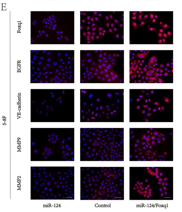
Application: IF/ICC Species: mouse Sample: 5–8F cells
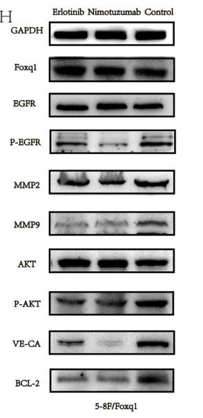
Application: WB Species: mouse Sample: 5–8F cells
限制条款
产品的规格、报价、验证数据请以官网为准,官网链接:www.affbiotech.com | www.affbiotech.cn(简体中文)| www.affbiotech.jp(日本語)产品的数据信息为Affinity所有,未经授权不得收集Affinity官网数据或资料用于商业用途,对抄袭产品数据的行为我们将保留诉诸法律的权利。
产品相关数据会因产品批次、产品检测情况随时调整,如您已订购该产品,请以订购时随货说明书为准,否则请以官网内容为准,官网内容有改动时恕不另行通知。
Affinity保证所销售产品均经过严格质量检测。如您购买的商品在规定时间内出现问题需要售后时,请您在Affinity官方渠道提交售后申请。产品仅供科学研究使用。不用于诊断和治疗。
产品未经授权不得转售。
Affinity Biosciences将不会对在使用我们的产品时可能发生的专利侵权或其他侵权行为负责。Affinity Biosciences, Affinity Biosciences标志和所有其他商标所有权归Affinity Biosciences LTD.