产品: PKC-pan 抗体
货号: AF6197
描述: Rabbit polyclonal antibody to PKC-pan
应用: WB IHC IF/ICC
文献验证: WB, IF/ICC
反应: Human, Mouse, Rat
预测: Pig, Bovine, Horse, Sheep, Rabbit, Dog, Chicken, Xenopus
蛋白号: P17252 | P05771 | P05129 | P24723
RRID: AB_2835078

浏览相似产品>>

   规格 价格 库存
 50ul RMB¥ 1250 现货
 100ul RMB¥ 2300 现货
 200ul RMB¥ 3000 现货

货期: 当天发货

联系销售

产品描述

来源:
Rabbit
应用:
WB 1:500-1:2000, IHC 1:50-1:200, IF/ICC 1:100-1:500
*The optimal dilutions should be determined by the end user.
*Tips:

WB: 适用于变性蛋白样本的免疫印迹检测. IHC: 适用于组织样本的石蜡(IHC-p)或冰冻(IHC-f)切片样本的免疫组化/荧光检测. IF/ICC: 适用于细胞样本的荧光检测. ELISA(peptide): 适用于抗原肽的ELISA检测.

反应:
Human, Mouse, Rat
克隆:
Polyclonal
特异性:
PKC-pan Antibody detects endogenous levels of total PKC-pan.
RRID:
AB_2835078
引用格式: Affinity Biosciences Cat# AF6197, RRID:AB_2835078.
偶联:
Unconjugated.
纯化:
The antiserum was purified by peptide affinity chromatography using SulfoLink™ Coupling Resin (Thermo Fisher Scientific).
保存:
Rabbit IgG in phosphate buffered saline , pH 7.4, 150mM NaCl, 0.02% sodium azide and 50% glycerol. Store at -20 °C. Stable for 12 months from date of receipt.
别名:

展开/折叠

AAG6; Aging associated gene 6; aPKC; KPCA_HUMAN; PKC alpha; PKC-A; PKC-alpha; PKCA; PRKACA; PRKCA; Protein Kinase C alpha; Protein kinase C alpha type; KPCG_HUMAN; MGC57564; OTTHUMP00000067291; PKC-gamma; PKCC; PKCG; PRKCG; Protein kinase C gamma; Protein kinase C gamma polypeptide; Protein kinase C gamma type; Protein kinase C, gamma; SCA 14; SCA14;

抗原和靶标

免疫原:

A synthesized peptide derived from human PKC-pan, corresponding to a region within the internal amino acids.

基因/基因ID:
描述:
Protein Kinase C (PKC) isoforms are serine/threonine kinases involved in signal transduction pathways that govern a wide range of physiological processes including differentiation, proliferation, gene expression, brain function, membrane transport and the organization of cytoskeletal and extracellular matrix proteins.

研究领域

· Cellular Processes > Cellular community - eukaryotes > Focal adhesion.   (View pathway)

· Cellular Processes > Cellular community - eukaryotes > Gap junction.   (View pathway)

· Environmental Information Processing > Signal transduction > MAPK signaling pathway.   (View pathway)

· Environmental Information Processing > Signal transduction > ErbB signaling pathway.   (View pathway)

· Environmental Information Processing > Signal transduction > Ras signaling pathway.   (View pathway)

· Environmental Information Processing > Signal transduction > Rap1 signaling pathway.   (View pathway)

· Environmental Information Processing > Signal transduction > Calcium signaling pathway.   (View pathway)

· Environmental Information Processing > Signal transduction > NF-kappa B signaling pathway.   (View pathway)

· Environmental Information Processing > Signal transduction > HIF-1 signaling pathway.   (View pathway)

· Environmental Information Processing > Signal transduction > Phosphatidylinositol signaling system.

· Environmental Information Processing > Signal transduction > Sphingolipid signaling pathway.   (View pathway)

· Environmental Information Processing > Signal transduction > Phospholipase D signaling pathway.   (View pathway)

· Environmental Information Processing > Signal transduction > mTOR signaling pathway.   (View pathway)

· Environmental Information Processing > Signal transduction > PI3K-Akt signaling pathway.   (View pathway)

· Environmental Information Processing > Signal transduction > Wnt signaling pathway.   (View pathway)

· Human Diseases > Drug resistance: Antineoplastic > EGFR tyrosine kinase inhibitor resistance.

· Human Diseases > Endocrine and metabolic diseases > Insulin resistance.

· Human Diseases > Substance dependence > Amphetamine addiction.

· Human Diseases > Substance dependence > Morphine addiction.

· Human Diseases > Infectious diseases: Bacterial > Vibrio cholerae infection.

· Human Diseases > Infectious diseases: Bacterial > Pathogenic Escherichia coli infection.

· Human Diseases > Infectious diseases: Parasitic > Leishmaniasis.

· Human Diseases > Infectious diseases: Parasitic > African trypanosomiasis.

· Human Diseases > Infectious diseases: Parasitic > Amoebiasis.

· Human Diseases > Infectious diseases: Viral > Hepatitis B.

· Human Diseases > Infectious diseases: Viral > Influenza A.

· Human Diseases > Cancers: Overview > Pathways in cancer.   (View pathway)

· Human Diseases > Cancers: Overview > Proteoglycans in cancer.

· Human Diseases > Cancers: Overview > MicroRNAs in cancer.

· Human Diseases > Cancers: Specific types > Glioma.   (View pathway)

· Human Diseases > Cancers: Specific types > Non-small cell lung cancer.   (View pathway)

· Human Diseases > Cancers: Specific types > Hepatocellular carcinoma.   (View pathway)

· Human Diseases > Cancers: Overview > Choline metabolism in cancer.   (View pathway)

· Organismal Systems > Immune system > Chemokine signaling pathway.   (View pathway)

· Organismal Systems > Circulatory system > Adrenergic signaling in cardiomyocytes.   (View pathway)

· Organismal Systems > Circulatory system > Vascular smooth muscle contraction.   (View pathway)

· Organismal Systems > Development > Axon guidance.   (View pathway)

· Organismal Systems > Immune system > Natural killer cell mediated cytotoxicity.   (View pathway)

· Organismal Systems > Immune system > B cell receptor signaling pathway.   (View pathway)

· Organismal Systems > Immune system > Fc epsilon RI signaling pathway.   (View pathway)

· Organismal Systems > Immune system > Fc gamma R-mediated phagocytosis.   (View pathway)

· Organismal Systems > Immune system > Leukocyte transendothelial migration.   (View pathway)

· Organismal Systems > Environmental adaptation > Circadian entrainment.

· Organismal Systems > Nervous system > Long-term potentiation.

· Organismal Systems > Nervous system > Retrograde endocannabinoid signaling.   (View pathway)

· Organismal Systems > Nervous system > Glutamatergic synapse.

· Organismal Systems > Nervous system > Cholinergic synapse.

· Organismal Systems > Nervous system > Serotonergic synapse.

· Organismal Systems > Nervous system > GABAergic synapse.

· Organismal Systems > Nervous system > Dopaminergic synapse.

· Organismal Systems > Nervous system > Long-term depression.

· Organismal Systems > Sensory system > Inflammatory mediator regulation of TRP channels.   (View pathway)

· Organismal Systems > Endocrine system > Insulin secretion.   (View pathway)

· Organismal Systems > Endocrine system > Melanogenesis.

· Organismal Systems > Endocrine system > Thyroid hormone synthesis.

· Organismal Systems > Endocrine system > Thyroid hormone signaling pathway.   (View pathway)

· Organismal Systems > Endocrine system > Oxytocin signaling pathway.

· Organismal Systems > Endocrine system > Aldosterone synthesis and secretion.

· Organismal Systems > Endocrine system > Relaxin signaling pathway.

· Organismal Systems > Excretory system > Aldosterone-regulated sodium reabsorption.

· Organismal Systems > Excretory system > Endocrine and other factor-regulated calcium reabsorption.

· Organismal Systems > Digestive system > Salivary secretion.

· Organismal Systems > Digestive system > Gastric acid secretion.

· Organismal Systems > Digestive system > Pancreatic secretion.

· Organismal Systems > Digestive system > Carbohydrate digestion and absorption.

文献引用

1). Integrative Single-Cell Transcriptomics and Epigenomics Mapping of the Fetal Retina Developmental Dynamics. Advanced science (Weinheim, Baden-Wurttemberg, Germany), 2023 (PubMed: 37017569) [IF=15.1]

Application: IF/ICC    Species: human    Sample:

Figure 1 A single-cell regulatory atlas of the developing human retina. A) Schematic of experimental design. B) Retinal structure schematic. C) Immunofluorescence analysis of primary human retinal tissue validating the expression of cell-type-specific markers, including NRL (rods); RGR (MGCs); CALIBINDIN (HCs); PKC (BCs); CALRETININ (HCs, ACs, and RGCs); and BRN3A (RGCs). Nuclei are counterstained with DAPI. Scale bar, 50 µm. D) UMAP embedding of the retina scRNA-seq dataset, with individual cells colored by age and annotated cell types. E) Violin plot showing relative expression of transcripts with high specificity for individual cell types ordered by cell type. F) UMAP embedding of the retina scATAC-seq dataset, with individual cells colored by age and annotated cell types. G) Coverage plots and gene score of known marker genes for cell types. Gene color representing the direction of gene translation (red: right; blue: left). H) UMAP embedding of the developmental trajectories of RPCs, MGCs, and NPCs colored by pseudo-time. Red arrows show two developmental directions from RPCs. I) UMAP embedding of the developmental trajectories of RPCs and RPC-derived cells from scRNA-seq colored by pseudo-time. White arrows show three neuronal lineages. Abbreviations: PCW, post-conception weeks; RGCs, retinal ganglion cells; HCs, horizontal cells; ACs, amacrine cells; BCs, bipolar cells; MGCs, Müller glial cells; GCL, ganglion cell layer; ONL, outer nuclear layer; OPL, outer plexiform layer; INL, inner nuclear layer; IPL, inner plexiform layer.

2). Opsin 5 is a key regulator of ultraviolet radiation induced melanogenesis in human epidermal melanocytes. British Journal of Dermatology, 2021 (PubMed: 33400324) [IF=11.0]

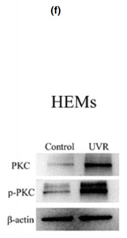
Application: WB    Species: Human    Sample: human epidermal melanocytes(HEMs)

Figure 4 Ultraviolet radiation (UVR) activates the Ca2+ /protein kinase C (PKC)/microphthalmia-associated transcription factor (MITF) signalling pathway through opsin 5 (OPN5) and upregulates expression of tyrosinase (TYR), tyrosinase-related protein 1 (TRP1) and TRP2. (a, b) Images of representative human epidermal melanocytes (HEMs) and PIG1 human melanocyte cells loaded with the Ca2+ indicator Fluo-3 and irradiated with UVR. Scale bar = 20 µm. HEMs and PIG1 calcium fluxes were quantified by flow cytometry. Data are shown as mean  SEM of three independent experiments. Statistical significance was determined by one-way ANOVA followed by Tukey’s test. **P < 0 01 ***P < 0 001. (c, d) Cells were transfected with (c) short interfering (si)RNA against OPN5, or (d) lentivirus overexpressing OPN5 (H-OPN5), then irradiated with or without UVR and lysed after 48 h. Lysates were analysed by Western blotting using the indicated antibodies. (e, f) Cells were irradiated with and without UVR, and mitogen-activated protein kinase (MAPK) and PKC protein and phosphorylated (p-) protein levels were determined by Western blot analysis. (g) Cells were transfected with short interfering RNA (RNAi) against OPN5 irradiated with UVR and lysed after 48 h. Lysates were analysed by Western blotting using the indicated antibodies. (h, i) After inhibiting the expression of MITF with ML329, cells were irradiated with or without UVR, and lysed 48 h later to observe PKC protein expression. Beta-actin was used as a loading control. CAMKII, calmodulin-dependent protein kinase II.

3). SiNiSan ameliorates depression-like behavior in rats by enhancing synaptic plasticity via the CaSR-PKC-ERK signaling pathway. BIOMEDICINE & PHARMACOTHERAPY, 2020 (PubMed: 31958763) [IF=6.9]

Application: WB    Species: rat    Sample: HIP

Fig. 7. |Effect of SNS on the PKC/ERK1/2 signal pathway in the HIP and PFC of stressed rats. (A): Representative immunoblots for PKC, ERK1/2, p-ERK1/2, and tubulin in the HIP region; Levels of PKC and ratio of p-ERK1/2/ERK1/2 in the HIP of rats in each group

4). FGFR2 Mutation p.Cys342Arg Enhances Mitochondrial Metabolism-Mediated Osteogenesis via FGF/FGFR-AMPK-Erk1/2 Axis in Crouzon Syndrome. Cells, 2022 (PubMed: 36231091) [IF=6.0]

Application: WB    Species: Mice    Sample: MC3T3-E1 cells

Figure 2 Constitutive activation of FGFR2 (p.Cys342Arg) enhanced osteogenesis via Erk1/2 MAPK signaling pathway: (A) Schematic representation of the relative linear location in which the FGFR2 mutation is identified as illustrated by the large red arrow shown in the context of the protein structure. (B) CCK-8 assay was carried out to assess cell proliferation. Proliferation of MC3T3-E1 cells was more active in the MT group. (C) Relative expressions of osteogenic marker measured by qPCR. The expressions of Alp, ColIα2, Runx2, Opn and Ocn mRNA in differentiated MC3T3-e1 were remarkably increased in the MT group. Western blot analysis showed that the level of ALP, COLI and RUNX2 were also increased in the MT group. (D) ALP staining, alizarin red staining and quantitative tests. ALP staining showed increased crystal violet-staining cells in the MT group compared with the WT group. Quantitative experiment demonstrated that ALP activity is more active in the MT group. Alizarin red staining and quantitative test showed there were more mineralized nodules and mineral content in the MT group than in the WT group. (E) Western blot analysis demonstrated that the levels of p-FGFR2 and p-Erk1/2 were increased in the MT group. There was no significant change in the expression of key proteins in other downstream pathways. The western blot results of FGF/FGFR2-Erk1/2 was circled by the red frame. p values were significant at * p < 0.05, ** p < 0.01, *** p < 0.001 and **** p < 0.0001.

5). Inhibition of Hypoxia-Inducible Factor Prolyl-Hydroxylase Modulates Platelet Function. THROMBOSIS AND HAEMOSTASIS, 2022 (PubMed: 35477177) [IF=5.0]

6). Proteomics analysis of tumor exosomes reveals vital pathways of Jinfukang inhibiting circulating tumor cells metastasis in lung cancer. JOURNAL OF ETHNOPHARMACOLOGY, 2020 (PubMed: 32240782) [IF=4.8]

Application: WB    Species: human    Sample: CTC-TJH-01 cells

Fig. 6. |Jinfukang inhibits the migration of CTC cells through EGF signaling pathway. (A) CTC-TJH-01 cells were treated with Jinfukang (700 μg/mL) for 24 h. Protein level of EGF, p-mTOR, p-STAT1, STAT3, p-STAT3, PRKCA, p-PRKCA, Akt1/2/3 and p-Akt were analyzed by Western blot analysis.

7). In vitro evaluation of the neuroprotective effect of oligo-porphyran from Porphyra yezoensis in PC12 cells. JOURNAL OF APPLIED PHYCOLOGY, 2019 [IF=2.8]

8). ErbB4 mediates amyloid β‐induced neurotoxicity through JNK/tau pathway activation: implications for Alzheimer's disease. Journal of Comparative Neurology, 2021 (PubMed: 34212389) [IF=2.3]

Application: WB    Species: Human    Sample: SH-SY5Y cells

FIGURE 7 The effect of ErbB4 deletion on molecules downstream of ErbB4 in Aβ-treated SH-SY5Y cells. (a) qPCR was performed to detect the effect of ErbB4 siRNA on Src mRNA expression. Src mRNA expression was reduced in Aβ-treated SH-SY5Y cells after ErbB4 knockdown by siRNA. *p < .05. Differences were assessed by one-way ANOVA with Newman–Keuls post hoc test; values represent mean ± SEM. β-actin was used as control. (b) Western blotting was performed to detect the effect of ErbB4 siRNA on (pan) PKC phosphorylation and protein expression. (c-f) qPCR was performed to detect the effect of ErbB4 siRNA on Fyn (c), CDK5 (d), mTOR (e), and Abl (f) mRNA, respectively

限制条款

产品的规格、报价、验证数据请以官网为准,官网链接:www.affbiotech.com | www.affbiotech.cn(简体中文)| www.affbiotech.jp(日本語)

产品的数据信息为Affinity所有,未经授权不得收集Affinity官网数据或资料用于商业用途,对抄袭产品数据的行为我们将保留诉诸法律的权利。

产品相关数据会因产品批次、产品检测情况随时调整,如您已订购该产品,请以订购时随货说明书为准,否则请以官网内容为准,官网内容有改动时恕不另行通知。

Affinity保证所销售产品均经过严格质量检测。如您购买的商品在规定时间内出现问题需要售后时,请您在Affinity官方渠道提交售后申请。

产品仅供科学研究使用。不用于诊断和治疗。 

产品未经授权不得转售。

Affinity Biosciences将不会对在使用我们的产品时可能发生的专利侵权或其他侵权行为负责。Affinity Biosciences, Affinity Biosciences标志和所有其他商标所有权归Affinity Biosciences LTD.