产品: CDK1/CDC2 抗体
货号: AF6108
描述: Rabbit polyclonal antibody to CDK1/CDC2
应用: WB IF/ICC
文献验证: WB
反应: Human, Mouse, Rat
预测: Pig, Zebrafish, Bovine, Horse, Rabbit, Dog, Chicken, Xenopus
蛋白号: P06493
RRID: AB_2834995

浏览相似产品>>

   规格 价格 库存
 50ul RMB¥ 1250 现货
 100ul RMB¥ 2300 现货
 200ul RMB¥ 3000 现货

货期: 当天发货

联系销售

产品描述

来源:
Rabbit
应用:
WB 1:500-1:2000, IF/ICC 1:100-1:500
*The optimal dilutions should be determined by the end user.
*Tips:

WB: 适用于变性蛋白样本的免疫印迹检测. IHC: 适用于组织样本的石蜡(IHC-p)或冰冻(IHC-f)切片样本的免疫组化/荧光检测. IF/ICC: 适用于细胞样本的荧光检测. ELISA(peptide): 适用于抗原肽的ELISA检测.

反应:
Human, Mouse, Rat
克隆:
Polyclonal
特异性:
CDK1/CDC2 Antibody detects endogenous levels of total CDK1/CDC2.
RRID:
AB_2834995
引用格式: Affinity Biosciences Cat# AF6108, RRID:AB_2834995.
偶联:
Unconjugated.
纯化:
The antiserum was purified by peptide affinity chromatography using SulfoLink™ Coupling Resin (Thermo Fisher Scientific).
保存:
Rabbit IgG in phosphate buffered saline , pH 7.4, 150mM NaCl, 0.02% sodium azide and 50% glycerol. Store at -20 °C. Stable for 12 months from date of receipt.
别名:

展开/折叠

Cdc 2; Cdc2; CDC28A; CDK 1; CDK1; CDK1_HUMAN; CDKN1; CELL CYCLE CONTROLLER CDC2; Cell division control protein 2; Cell division control protein 2 homolog; Cell division cycle 2 G1 to S and G2 to M; Cell division protein kinase 1; Cell Divsion Cycle 2 Protein; Cyclin Dependent Kinase 1; Cyclin-dependent kinase 1; DKFZp686L20222; MGC111195; p34 Cdk1; p34 protein kinase; P34CDC2;

抗原和靶标

免疫原:

A synthesized peptide derived from human CDK1/CDC2, corresponding to a region within N-terminal amino acids.

基因/基因ID:
描述:
The protein encoded by this gene is a member of the Ser/Thr protein kinase family. This protein is a catalytic subunit of the highly conserved protein kinase complex known as M-phase promoting factor (MPF), which is essential for G1/S and G2/M phase transitions of eukaryotic cell cycle. Mitotic cyclins stably associate with this protein and function as regulatory subunits.

研究领域

· Cellular Processes > Cell growth and death > Cell cycle.   (View pathway)

· Cellular Processes > Cell growth and death > Oocyte meiosis.   (View pathway)

· Cellular Processes > Cell growth and death > p53 signaling pathway.   (View pathway)

· Cellular Processes > Cell growth and death > Cellular senescence.   (View pathway)

· Cellular Processes > Cellular community - eukaryotes > Gap junction.   (View pathway)

· Human Diseases > Infectious diseases: Viral > Herpes simplex infection.

· Human Diseases > Infectious diseases: Viral > Epstein-Barr virus infection.

· Human Diseases > Cancers: Overview > Viral carcinogenesis.

· Organismal Systems > Endocrine system > Progesterone-mediated oocyte maturation.

文献引用

1). Secalonic acid D induces cell apoptosis in both sensitive and ABCG2-overexpressing multidrug resistant cancer cells through upregulating c-Jun expression. Acta Pharmaceutica Sinica B, 2019 (PubMed: 31193763) [IF=14.7]

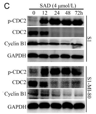
Application: WB    Species: human    Sample: S1 and S1-MI-80 cells

Figure 2 | Effect of SAD on cell cycle and apoptosis. (A) The cell cycle analysis was determined by PI staining and flow cytometry cell quest software. S1 and S1-MI-80 cells were treated with 4 μmol/L SAD for 12, 24, 48, and 72 h, respectively. The content of G2/M phase was increased in a time-dependent pattern. (B) Histograms of cell cycle distribution in non-treated and treated S1 and S1-MI-80 cells. (C) S1 and S1-MI-80 cells were treated with SAD (4 μmol/L) for four different time points. Western blot analysis was used to detect the levels of CDC2, p-CDC2 and cyclin B1 protein after SAD treatment.

2). Silica nanoparticles cause spermatogenesis dysfunction in mice via inducing cell cycle arrest and apoptosis. ECOTOXICOLOGY AND ENVIRONMENTAL SAFETY, 2022 (PubMed: 35051769) [IF=6.2]

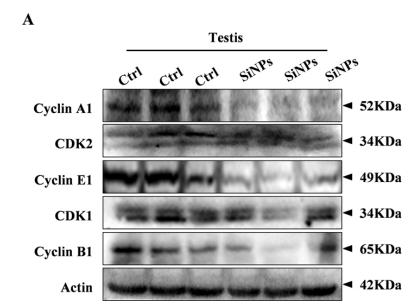
Application: WB    Species: mouse    Sample: testis

Fig. 5. |SiNPs affected the protein expressions of cell cycle regulators. (A) The protein expressions of cyclin A1, CDK2, cyclin E1, CDK1, and cyclin B1 were significantly decreased in the testis after exposure to SiNPs.

3). CENPF interaction with PLA2G4A promotes glioma growth by modulating mTORC1 and NF-κB pathways. Cancer cell international, 2025 (PubMed: 40025532) [IF=5.8]

4). Cyclovirobuxine D inhibits growth and progression of non‑small cell lung cancer cells by suppressing the KIF11‑CDC25C‑CDK1‑CyclinB1 G2/M phase transition regulatory network and the NFκB/JNK signaling pathway. International Journal of Oncology, 2023 (PubMed: 36929198) [IF=4.5]

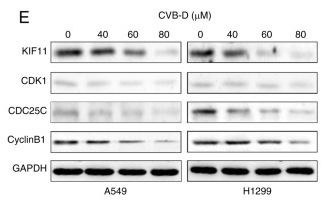
Application: WB    Species: Human    Sample: NSCLC cells

Figure 6 KIF11, KIF11-CDK1-CDC25C-cyclinB1 oncogenic network and NF-kB/JNK signaling pathway are the potential CVB-D therapeutic targets in the NSCLC cells. (A) Venn diagram showed the overlap number of potential CVB-D target genes in NSCLC cells by intersecting four LUAD datasets. (B) Protein-protein interaction network analysis revealed interaction between the 10 overlapping DEGs. The disconnected nodes are hidden. (C) The interactive heatmap from GEPIA 2 database showed expression of the 6 DEGs in LUAD and normal lung tissues. KIF11 is the most differentially expressed gene between LUAD and normal lung tissues. (D) Venn diagram revealed 2 common genes (KIF11 and CDK1) based on intersection between the 10 overlapping DEGs and the 100 predicted CVB-D target genes extracted from the Swisstarget prediction database. (E) Western blot analysis identified the expression of KIF11, CDK1, CDC25C and cyclinB1 proteins in the control and CVB-D-treated NSCLC cells. (F and G) Immunohistochemical assay results showed the expression levels and localization of the KIF11 protein in the control and CVB-D-treated A549 xenograft tumor tissues. (H) Western blot assay results revealed the KIF11 protein levels in the si-NC and si-KIF11-transfected NSCLC cells. (I) Western blot analysis demonstrated the expression levels of the oncogenic signaling network proteins (KIF11, CDK1, CDC25C and CyclinB1) and the NF-κB/JNK signaling pathway proteins (p65, p-p65, JNK and p-JNK) in the si-NC- and si-KIF11-transfected NSCLC cells. **P

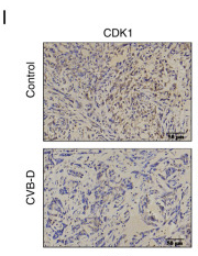
Application: IHC    Species: Mouse    Sample:

Figure 7 CVB-D inhibits in vivo progression of A549 xenografts tumors. (A) Experimental design for tumor xenograft experiments of NSCLC in nude mice to evaluate the in vivo therapeutic effects of CVB-D. Specific time points for CVB-D treatment and experimental analysis are indicated. (B) Representative images of the NSCLC xenograft tumor models with or without CVB-D treatment. (C) Representative images of the NSCLC xenograft tumor tissues from control and CVB-D-treated mice. (D and E) NSCLC xenograft tumor weights and volumes in the control and CVB-D treatment groups of nude mice. (F) Representative H&E and TUNEL staining images of the A549 xenograft tumor sections in the control and CVB-D treatment groups of nude mice (magnification, ×200; scale bar, 50 µm). (G) The proportion of apoptotic cells in the A549 xenograft tumors derived from the control and CVB-D-treatment groups of nude mice based on TUNEL staining. (H) The body weights of the xenograft tumor model mice in the control and CVB-D treatment groups. (I and J) Immunohistochemical assay data revealed the expression levels and distribution of CDK1, CDC25C, cyclinB1, Ki67, Bcl-2 and N-cadherin proteins in the A549 xenograft tumors derived from the control and CVB-D treatment groups (magnification, ×200; scale bar, 50 µm). *P

限制条款

产品的规格、报价、验证数据请以官网为准,官网链接:www.affbiotech.com | www.affbiotech.cn(简体中文)| www.affbiotech.jp(日本語)

产品的数据信息为Affinity所有,未经授权不得收集Affinity官网数据或资料用于商业用途,对抄袭产品数据的行为我们将保留诉诸法律的权利。

产品相关数据会因产品批次、产品检测情况随时调整,如您已订购该产品,请以订购时随货说明书为准,否则请以官网内容为准,官网内容有改动时恕不另行通知。

Affinity保证所销售产品均经过严格质量检测。如您购买的商品在规定时间内出现问题需要售后时,请您在Affinity官方渠道提交售后申请。

产品仅供科学研究使用。不用于诊断和治疗。 

产品未经授权不得转售。

Affinity Biosciences将不会对在使用我们的产品时可能发生的专利侵权或其他侵权行为负责。Affinity Biosciences, Affinity Biosciences标志和所有其他商标所有权归Affinity Biosciences LTD.