产品: 磷酸化 CDK5 (Tyr239) 抗体
货号: AF3628
描述: Rabbit polyclonal antibody to Phospho-CDK5 (Tyr239)
应用: IHC
文献验证: IHC
反应: Human, Mouse, Rat
蛋白号: Q00535
RRID: AB_2846942

浏览相似产品>>

   规格 价格 库存
 100ul RMB¥ 2800 现货
 200ul RMB¥ 3800 现货

货期: 当天发货

联系销售

产品描述

来源:
Rabbit
应用:
IHC 1:50-1:200
*The optimal dilutions should be determined by the end user.
*Tips:

WB: 适用于变性蛋白样本的免疫印迹检测. IHC: 适用于组织样本的石蜡(IHC-p)或冰冻(IHC-f)切片样本的免疫组化/荧光检测. IF/ICC: 适用于细胞样本的荧光检测. ELISA(peptide): 适用于抗原肽的ELISA检测.

反应:
Human, Mouse, Rat
克隆:
Polyclonal
特异性:
Phospho-CDK5 (Tyr239) Antibody detects endogenous levels of CDK5 only when phosphorylated at Tyr239.
RRID:
AB_2846942
引用格式: Affinity Biosciences Cat# AF3628, RRID:AB_2846942.
偶联:
Unconjugated.
纯化:
The antibody is from purified rabbit serum by affinity purification via sequential chromatography on phospho-peptide and non-phospho-peptide affinity columns.
保存:
Rabbit IgG in phosphate buffered saline , pH 7.4, 150mM NaCl, 0.02% sodium azide and 50% glycerol. Store at -20 °C. Stable for 12 months from date of receipt.
别名:

展开/折叠

Cdk 5; Cdk5; CDK5_HUMAN; Cell division protein kinase 5; Crk6; Cyclin dependent kinase 5; Cyclin-dependent kinase 5; Protein kinase CDK5 splicing; PSSALRE; Serine threonine protein kinase PSSALRE; Serine/threonine-protein kinase PSSALRE; Tau protein kinase II catalytic subunit; TPKII catalytic subunit;

抗原和靶标

免疫原:

A synthesized peptide derived from human CDK5 around the phosphorylation site of Tyr239.

基因/基因ID:

研究领域

· Human Diseases > Neurodegenerative diseases > Alzheimer's disease.

· Human Diseases > Substance dependence > Cocaine addiction.

· Organismal Systems > Development > Axon guidance.   (View pathway)

文献引用

1). Glucagon Enhances Chemotherapy Efficacy By Inhibition of Tumor Vessels in Colorectal Cancer. Advanced science (Weinheim, Baden-Wurttemberg, Germany), 2024 (PubMed: 38072640) [IF=15.1]

2). Osteopontin deficiency promotes cartilaginous endplate degeneration by enhancing the NF-κB signaling to recruit macrophages and activate the NLRP3 inflammasome. Bone research, 2024 (PubMed: 39242551) [IF=14.3]

3). Cyclin G2 in macrophages triggers CTL-mediated antitumor immunity and antiangiogenesis via interferon-gamma. Journal of Experimental & Clinical Cancer Research, 2022 (PubMed: 36566226) [IF=11.3]

Application: IHC    Species: Mouse    Sample:

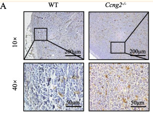
Fig. 3 Cyclin G2 in macrophages suppresses tumors by inhibiting tumor blood vessels after IFN-γ treatment. A CD31 immunohistochemical staining of the LLC tumors isolated from mice in the WT and Ccng2−/− groups, representative images are shown. 10× Scale bar = 200 μm; 40× Scale bar = 50 μm. B Graph showing the number of blood vessels in each field (n = 5). Data were analyzed using the unpaired Student’s t-test. Data are presented as the mean ± SEM. C, D Tube formation of SVEC4–10 cells treated with conditioned medium from BMDMs isolated from WT and Ccng2−/− C57BL/6 mice. Scale bar = 500 μm (representing 3 independent experiments). E Western blot showing successful knockdown of cyclin G2 in THP-1 cells. GAPDH was used as a loading control. F RT-qPCR was used to measure CCNG2 mRNA expression levels in THP-1 stable cell lines (Nonsense, shcyclin G2#1, and shcyclin G2#2). β-actin was used as an internal control. G Western blotting demonstrated successful cyclin G2 overexpression in THP-1 cells. β-tubulin was used as a loading control. H Measurement of CCNG2 mRNA expression levels in THP-1 stable cell lines (Vector and Flag-cyclin G2) by RT-qPCR. β-actin was used as an internal control (representing 2 independent experiments). I Tube formation by HUVECs treated with conditioned medium from THP-1 stable cell lines (Nonsense, shcyclin G2#1, and shcyclin G2#2) cells. Scale bar = 200 μm (representing 3 independent experiments). J Tube formation by HUVECs treated with conditioned medium from THP-1 stable cell lines (Vector and Flag-cyclin G2) cells. Scale bar = 200 μm (representing 3 independent experiments). Data in D, F, H, I, and J were analyzed with the unpaired Student’s t-test. Data are presented as the mean ± SD. *p 

4). Inhibition of neutrophil extracellular trap formation attenuates NLRP1-dependent neuronal pyroptosis via STING/IRE1α pathway after traumatic brain injury in mice. Frontiers in Immunology, 2023 (PubMed: 37143681) [IF=5.7]

Application: IHC    Species: Mouse    Sample: brain tissue

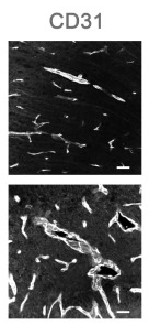
Figure 2 Neutrophils infiltration and NETs formation in mice brain tissue. (A) Representative immunofluorescence staining of Ly6G-positive neutrophils (red) and H3cit -positive(green) and CD31-labeled blood vessels (white) in the mice brain tissue sections post-CCT 3 days. Nuclei were stained with DAPI (blue). (B) Representative Western blot bands of MPO, PAD4, H3Cit, and H3 and statistical analysis expression level of MPO (C), PAD4 (D), and H3Cit (E) in mice brain tissue (n=6). Data are represented as mean ± SD. *P < 0.05, **P < 0.01.

5). Generation and in vivo characterization of a novel high-affinity human antibody targeting carcinoembryonic antigen. mAbs, 2023 (PubMed: 37243574) [IF=5.6]

6). CCN1 Is a Therapeutic Target for Reperfused Ischemic Brain Injury. Translational stroke research, 2024 (PubMed: 39028413) [IF=3.8]

7). Analysis of the expression, function and signaling of glycogen phosphorylase isoforms in hepatocellular carcinoma. Oncology Letters, 2022 (PubMed: 35761940) [IF=2.5]

Application: IF/ICC    Species: Human    Sample: HCC cell

Figure 6. Inhibition of PYGB suppresses HCC growth. (A) HCC cell viability analysis of CP91149 treatment. (B) HCC cell viability analysis of CP91149 treatment combined with sorafenib. (C) Tumor growth curve of HCC under vehicle (n=6), CP91149 (n=6), sorafenib (n=6), or combination treatment (n=6). (D) HCC tumor weight. (E) HCC tumor pictures. (F) H&E (scale bar, 200 µm) staining of tumor tissues and immunohistochemical analysis of Ki67 (scale bar, 100 µm) and CD31 (scale bar, 50 µm). (G) Quantification of Ki67 positive cells. (H) Quantification of blood vessel density. *P

8). Increased expression of osteopontin in subchondral bone promotes bone turnover and remodeling, and accelerates the progression of OA in a mouse model. Aging (Albany NY), 2022 (PubMed: 34982732) [IF=0.4]

限制条款

产品的规格、报价、验证数据请以官网为准,官网链接:www.affbiotech.com | www.affbiotech.cn(简体中文)| www.affbiotech.jp(日本語)

产品的数据信息为Affinity所有,未经授权不得收集Affinity官网数据或资料用于商业用途,对抄袭产品数据的行为我们将保留诉诸法律的权利。

产品相关数据会因产品批次、产品检测情况随时调整,如您已订购该产品,请以订购时随货说明书为准,否则请以官网内容为准,官网内容有改动时恕不另行通知。

Affinity保证所销售产品均经过严格质量检测。如您购买的商品在规定时间内出现问题需要售后时,请您在Affinity官方渠道提交售后申请。

产品仅供科学研究使用。不用于诊断和治疗。 

产品未经授权不得转售。

Affinity Biosciences将不会对在使用我们的产品时可能发生的专利侵权或其他侵权行为负责。Affinity Biosciences, Affinity Biosciences标志和所有其他商标所有权归Affinity Biosciences LTD.