, using ICAM1 Antibody at 1/1000 dilution.
5ug/NC membrane strip.
Exposure for 5s with Affinity™ ECL Kit(#KF8003).
Bands result from membrane strip incubation.")
, using ICAM1 Antibody at 1/1000 dilution.")


 and mouse anti-beta tubulin Ab(T0023 1:200) for 1 hour at 37°C. An AlexaFluor594 conjugated goat anti-rabbit IgG(H+L) Ab(Red) and an AlexaFluor488 conjugated goat anti-mouse IgG(H+L) Ab(Green) were used as the secondary antibody.
The nuclear counter stain is DAPI(blue).")

 The expressions of TNF-α, IL-6, IL-1β, and
ICAM-1 were evaluated by Western blotting at 3 days post-SCI in each group (n = 6). (B) Quantification of TNF-α, IL-6, IL-1β, and ICAM-1
expressions (n = 6, all the data are expressed as means ± SD, two-way ANOVA followed by Tukey's post hoc test was applied). (C–F)
Immunofluorescence staining was used to detect the level of TNF-α, IL-6, IL-1β, and ICAM-1 from each group (n = 6, scale bar = 50 µm). (G)
Statistical analysis of immunofluorescence staining for positive expression of TNF-α, IL-6, IL-1β, and ICAM-1 in nerve cells from each group
(n = 6, all the data are expressed as means ± SD, two-way ANOVA followed by Tukey's post hoc test was applied). * means p < 0.05; **means
p < 0.01; and *** means p < 0.001")
 was
analyzed by western blot in the KPNA2-overexpressing (A, B) or KPNA2-silenced cells (F, G). The nuclear translocation of p65 (nucleus) was detected by immunofluorescence assay in the KPNA2-overexpressing (C, D) or KPNA2-silenced cells (H, I). The NF-κB binding activity was analyzed by EMSA in the KPNA2-
overexpressing (E) or KPNA2-silenced cells (J). Parental, blank control group; NC, negative control group; OV-KPNA2, KPNA2 overexpressed group; siRNA1-KPNA2,
KPNA2-1 silencing group; siRNA2-KPNA2, KPNA2-2 silencing group. The results were obtained in three independent experiments. Mean values were compared by
One-way ANOVA. (**p < 0.01, ***p < 0.001, ****p < 0.001).")

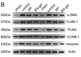
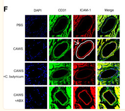
. The upper panel presents immunohistochemistry results, and the lower panel shows the quantified relative expression levels.*P<0.05 vs. control; #P<0.05 vs. model; ^P<0.05 vs. model + DEX. DEX, dexmedetomidine; 4‑PBA, 4‑phenylbutyric acid; ICAM‑1, intercellular adhesion molecule‑1; VCAM‑1, vascular adhesion molecule‑1.")

. The upper panel presents immunohistochemistry results, and the lower panel shows the quantified relative expression levels. *P<0.05 vs. control; #P<0.05 vs. model; ^P<0.05 vs. model + DEX. DEX, dexmedetomidine; 4-PBA, 4-phenylbutyric acid; ICAM-1, intercellular adhesion molecule-1; VCAM-1, vascular adhesion molecule-1.")
 The mRNA levels of ICAM-1 and VCAM-1 were measured by quantitative real-time PCR. (C-J) The level of p-MAPK/MAPK, p-ERK/ERK, ICAM, VCAM-1, Aβ1–42, and p-Tau/Tau were measured by Western blot. The data are expressed as mean ± standard deviation, n = 3. **p")
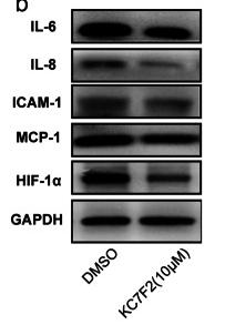
 Representative immunoblot and relative quantification of TLR4 and MyD88 in RAW264.7 cells. (D,E) Representative immunofluorescence images of TLR4-positive and MyD88-positive in RAW264.7 cells. Scale bars: 25 μm. (F–H) Representative immunoblot and relative quantification of p-IκBα (S32/S36), IκBα, p-p65 (S536), and p65 in RAW264.7 cells. (I) Representative immunofluorescence images of NF-κB p65 in RAW264.7 cells. Scale bars: 100 μm. (J–N) Representative immunoblot and relative quantification of CD68, MCP-1, ICAM1, and VCAM1 in RAW264.7 cells. (O–R) mRNA levels of Ccl2, Ccl3, Ccl4, and Cxcl10 in RAW264.7 cells. Data are presented as mean ± SEM (n = 3). * p < 0.05, ** p < 0.01, *** p < 0.001, and **** p < 0.0001 vs. the Con group. # p < 0.05, ## p < 0.01, ### p < 0.001, and #### p < 0.0001 vs. the LPS group.")
 AGS and (B) HGC-27 cells. Western blotting analysis the protein expression levels of VCAM1, ICAM1, PTGS2, IL6 and CCL2 (C) AGS and (D) HGC-27 cells. In order to save and recycle antibodies and save time, membranes were cut in horizontal strips at molecular weight ranges for target proteins and the information was shown in the Supplementary Fig. 1 and Fig. 2. Differences between two groups were assessed by Students t test, and differences among three groups were assessed by one-way analysis of variance (ANOVA).")

产品描述
*The optimal dilutions should be determined by the end user. For optimal experimental results, antibody reuse is not recommended.
*Tips:
WB: 适用于变性蛋白样本的免疫印迹检测. IHC: 适用于组织样本的石蜡(IHC-p)或冰冻(IHC-f)切片样本的免疫组化/荧光检测. IF/ICC: 适用于细胞样本的荧光检测. ELISA(peptide): 适用于抗原肽的ELISA检测.
引用格式: Affinity Biosciences Cat# AF6088, RRID:AB_2834982.
展开/折叠
Antigen identified by monoclonal antibody BB2; BB 2; BB2; CD 54; CD_antigen=CD54; CD54; Cell surface glycoprotein P3.58; Human rhinovirus receptor; ICAM 1; ICAM-1; ICAM1; ICAM1_HUMAN; intercellular adhesion molecule 1 (CD54), human rhinovirus receptor; Intercellular adhesion molecule 1; Major group rhinovirus receptor; MALA 2; MALA2; MyD 10; MyD10; P3.58; Surface antigen of activated B cells, BB2;
抗原和靶标
A synthesized peptide derived from human ICAM1, corresponding to a region within C-terminal amino acids.
研究领域
· Environmental Information Processing > Signal transduction > NF-kappa B signaling pathway. (View pathway)
· Environmental Information Processing > Signaling molecules and interaction > Cell adhesion molecules (CAMs). (View pathway)
· Environmental Information Processing > Signal transduction > TNF signaling pathway. (View pathway)
· Human Diseases > Infectious diseases: Parasitic > African trypanosomiasis.
· Human Diseases > Infectious diseases: Parasitic > Malaria.
· Human Diseases > Infectious diseases: Bacterial > Staphylococcus aureus infection.
· Human Diseases > Infectious diseases: Viral > Influenza A.
· Human Diseases > Infectious diseases: Viral > HTLV-I infection.
· Human Diseases > Infectious diseases: Viral > Epstein-Barr virus infection.
· Human Diseases > Immune diseases > Rheumatoid arthritis.
· Human Diseases > Cardiovascular diseases > Viral myocarditis.
· Organismal Systems > Immune system > Natural killer cell mediated cytotoxicity. (View pathway)
· Organismal Systems > Immune system > Leukocyte transendothelial migration. (View pathway)
文献引用
Application: WB Species: Rat Sample:

Application: IF/ICC Species: Rat Sample:

Application: WB Species: human Sample: HUVECs
Application: WB Species: Human Sample: HUVECs
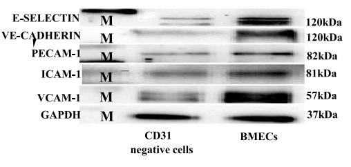
Application: WB Species: mouse Sample: primary bone marrow endothelial cells

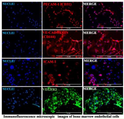
Application: IF/ICC Species: mouse Sample: primary bone marrow endothelial cells
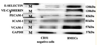
Application: WB Species: Mice Sample: bMECs
Application: IF/ICC Species: Mice Sample: BMECs
Application: IF/ICC Species: Mouse Sample:
限制条款
产品的规格、报价、验证数据请以官网为准,官网链接:www.affbiotech.com | www.affbiotech.cn(简体中文)| www.affbiotech.jp(日本語)产品的数据信息为Affinity所有,未经授权不得收集Affinity官网数据或资料用于商业用途,对抄袭产品数据的行为我们将保留诉诸法律的权利。
产品相关数据会因产品批次、产品检测情况随时调整,如您已订购该产品,请以订购时随货说明书为准,否则请以官网内容为准,官网内容有改动时恕不另行通知。
Affinity保证所销售产品均经过严格质量检测。如您购买的商品在规定时间内出现问题需要售后时,请您在Affinity官方渠道提交售后申请。产品仅供科学研究使用。不用于诊断和治疗。
产品未经授权不得转售。
Affinity Biosciences将不会对在使用我们的产品时可能发生的专利侵权或其他侵权行为负责。Affinity Biosciences, Affinity Biosciences标志和所有其他商标所有权归Affinity Biosciences LTD.