产品: CD20 抗体
货号: DF13319
描述: Rabbit polyclonal antibody to CD20
应用: WB IHC IF/ICC
文献验证: WB, IHC, IF/ICC
反应: Human
预测: Pig, Bovine, Sheep, Dog
分子量: 33kDa; 33kD(Calculated).
蛋白号: P11836
RRID: AB_2846338

浏览相似产品>>

   规格 价格 库存
 100ul RMB¥ 2300 现货
 200ul RMB¥ 3000 现货

货期: 当天发货

联系销售

产品描述

来源:
Rabbit
应用:
IF/ICC 1:100-1:500, WB 1:500-1:2000, IHC 1:50-1:200
*The optimal dilutions should be determined by the end user.
*Tips:

WB: 适用于变性蛋白样本的免疫印迹检测. IHC: 适用于组织样本的石蜡(IHC-p)或冰冻(IHC-f)切片样本的免疫组化/荧光检测. IF/ICC: 适用于细胞样本的荧光检测. ELISA(peptide): 适用于抗原肽的ELISA检测.

反应:
Human
预测:
Pig(88%), Bovine(%), Sheep(%), Dog(%)
克隆:
Polyclonal
特异性:
CD20 Antibody detects endogenous levels of total CD20.
RRID:
AB_2846338
引用格式: Affinity Biosciences Cat# DF13319, RRID:AB_2846338.
偶联:
Unconjugated.
纯化:
The antiserum was purified by peptide affinity chromatography using SulfoLink™ Coupling Resin (Thermo Fisher Scientific).
保存:
Rabbit IgG in phosphate buffered saline , pH 7.4, 150mM NaCl, 0.02% sodium azide and 50% glycerol. Store at -20 °C. Stable for 12 months from date of receipt.
别名:

展开/折叠

APY; ATOPY; B lymphocyte antigen CD20; B Lymphocyte Cell Surface Antigen B1; B-lymphocyte antigen CD20; B-lymphocyte cell-surface antigen B1; B-lymphocyte surface antigen B1; B1; Bp 35; Bp35; CD 20; CD20; CD20 antigen; CD20 receptor; CD20_HUMAN; CVID5; Fc epsilon receptor I beta chain; Fc Fragment of IgE high affinity I receptor for beta polypeptide; FCER1B; High affinity immunoglobulin epsilon receptor subunit beta; IgE Fc receptor subunit beta; IGEL; IGER; IGHER; LEU 16; LEU16; leukocyte surface antigen Leu 16; Leukocyte surface antigen Leu-16; Ly44; Membrane spanning 4 domains A1; Membrane spanning 4 domains subfamily A member 2; Membrane-spanning 4-domains subfamily A member 1; MGC3969; MS4A1; MS4A2; S7;

抗原和靶标

免疫原:
Uniprot:
基因/基因ID:
表达:
P11836 CD20_HUMAN:

Expressed on B-cells.

序列:
MTTPRNSVNGTFPAEPMKGPIAMQSGPKPLFRRMSSLVGPTQSFFMRESKTLGAVQIMNGLFHIALGGLLMIPAGIYAPICVTVWYPLWGGIMYIISGSLLAATEKNSRKCLVKGKMIMNSLSLFAAISGMILSIMDILNIKISHFLKMESLNFIRAHTPYINIYNCEPANPSEKNSPSTQYCYSIQSLFLGILSVMLIFAFFQELVIAGIVENEWKRTCSRPKSNIVLLSAEEKKEQTIEIKEEVVGLTETSSQPKNEEDIEIIPIQEEEEEETETNFPEPPQDQESSPIENDSSP

种属预测

种属预测:

score>80的预测可信度较高,可尝试用于WB检测。*预测模型主要基于免疫原序列比对,结果仅作参考,不作为质保凭据。

Species
Results
Score
Pig
88
Bovine
88
Sheep
88
Dog
88
Rabbit
75
Horse
0
Xenopus
0
Zebrafish
0
Chicken
0
Model Confidence:
High(score>80) Medium(80>score>50) Low(score<50) No confidence

研究背景

功能:

B-lymphocyte-specific membrane protein that plays a role in the regulation of cellular calcium influx necessary for the development, differentiation, and activation of B-lymphocytes. Functions as a store-operated calcium (SOC) channel component promoting calcium influx after activation by the B-cell receptor/BCR.

翻译修饰:

Phosphorylated on serines and threonines in resting B-cells. Protein kinase C/PKC can use CD20 as substrate.

细胞定位:

Cell membrane>Multi-pass membrane protein. Cell membrane>Lipid-anchor.
Note: Constitutively associated with membrane rafts.

Extracellular region or secreted Cytosol Plasma membrane Cytoskeleton Lysosome Endosome Peroxisome ER Golgi apparatus Nucleus Mitochondrion Manual annotation Automatic computational assertionSubcellular location
组织特异性:

Expressed on B-cells.

亚基结构:

Forms homotetramers. Interacts with the heavy and light chains of cell surface IgM, the antigen-binding components of the BCR.

蛋白家族:

Belongs to the MS4A family.

研究领域

· Organismal Systems > Immune system > Hematopoietic cell lineage.   (View pathway)

文献引用

1). Dupilumab improve acquired reactive perforating collagenosis characterized by type 2 inflammation. Frontiers in Immunology, 2023 (PubMed: 37638036) [IF=5.7]

Application: IHC    Species: Human    Sample:

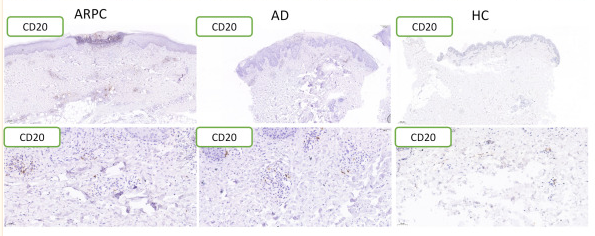
Figure 3 Representative images and data reporting hematoxylin and eosin staining, CD3, CD20, GATA3 and T-bet staining of APRC, AD and HC samples. Magnification: ×40, ×200, AD, atopic dermatitis, ARPC, acquired reactive perforating collagenosis, HC, healthy control, HE, hematoxylin and eosin.

2). Identification and validation of immune and diagnostic biomarkers for interstitial cystitis/painful bladder syndrome by integrating bioinformatics and machine-learning. Frontiers in immunology, 2025 (PubMed: 39917301) [IF=5.7]

3). Pyruvate dehydrogenase kinase 4‐mediated metabolic reprogramming is involved in rituximab resistance in diffuse large B‐cell lymphoma by affecting the expression of MS4A1/CD20. CANCER SCIENCE, 2021 (PubMed: 34252986) [IF=4.5]

Application: IF/ICC    Species: Human    Sample: DLBCL cells

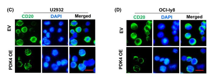
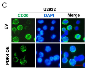
FIGURE 4 Pyruvate dehydrogenase kinase 4 (PDK4) has a negative regulatory effect on MS4A1/CD20 expression in diffuse large B- cell lymphoma cells. A, B, Real- time quantitative PCR analysis of PDK4 and MS4A1 mRNA expression in U2932 and OCI- Ly8 cells transfected with PDK4 overexpressing (PDK4 OE) plasmid or empty vector (EV). C, D, CD20 (green) molecules in the plasma membrane showed a reduction after PDK4 overexpression in both U2932 and OCI- Ly8 cells. Scale bar, 15 μm. E, F, Western blot analysis of PDK4 and CD20 protein levels in U2932 and OCI- Ly8 cells with PDK4 shRNA interference or transfected with PDK4 OE plasmid or EV. *P < .05, **P < .01, ***P < .001

Application: WB    Species: Human    Sample: DLBCL cells

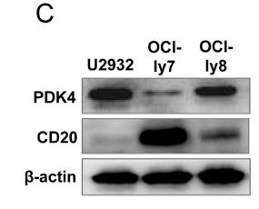
FIGURE 2 High pyruvate dehydrogenase kinase 4 (PDK4) is associated with rituximab (RTX) resistance and low MS4A1/CD20 in diffuse large B- cell lymphoma (DLBCL) cells. A, B, Real- time quantitative PCR analysis of PDK4 and MS4A1 mRNA expression in DLBCL cell lines U2932, OCI- ly7, and OCI- ly8. C, Western blot analysis of PDK4 and CD20 protein levels in DLBCL cell lines U2932, OCI- ly7, and OCI- ly8. D, E, Annexin V- phycoerythrin (PE)/7- AAD double staining analysis of the three DLBCL cell lines treated with RTX (50 μg/mL). F, G, Mitochondrial membrane potential of DLBCL cells following treatment with RTX for 48 hours and stained with JC- 1 probe. Representative pictures of JC- 1 staining are shown. Scale bar, 25 μm. ***P < .001

4). Tumor necrosis factor-α-inducible protein 8-like protein 3 (TIPE3): a novel prognostic factor in colorectal cancer. BMC cancer, 2023 (PubMed: 36755222) [IF=3.8]

Application: IHC    Species: human    Sample:

Fig. 3 Images of immunohistochemical staining immune cells in CRC tissue (200×): A, B Immunohistochemical staining of CD8+ T cells: A High expression; B Low expression; C, D Immunohistochemical staining of CD20+ B cells: C High expression; D Low expression; E, F Immunohistochemical staining of CD66b+ neutrophils: E High expression; F Low expression. Scale bar = 100 μm

5). PDK4-Mediated Metabolic Reprogramming Promotes Rituximab Resistance in Diffuse Large B-Cell Lymphoma Via Negative Regulation of MS4A1/CD20. Research Square, 2021

Application: WB    Species: Human    Sample: DLBCL cells

Figure 2. High PDK4 is associated with rituximab (RTX) resistance and low MS4A1/CD20 in DLBCL cells. A and B, qRT-PCR analysis of PDK4 and MS4A1 mRNA expression in DLBCL cell lines U2932, OCI-ly7 and OCI-ly8. C, western blotting analysis of PDK4 and CD20 protein levels in DLBCL cell lines U2932, OCI-ly7 and OCI-ly8. D and E, annexin V-FITC/PI double staining analysis of the three DLBCL cell lines treated with rituximab (50 μg/ml).

Application: IF/ICC    Species: Human    Sample: DLBCL cells

Figure 2. High PDK4 is associated with rituximab (RTX) resistance and low MS4A1/CD20 in DLBCL cells. A and B, qRT-PCR analysis of PDK4 and MS4A1 mRNA expression in DLBCL cell lines U2932, OCI-ly7 and OCI-ly8. C, western blotting analysis of PDK4 and CD20 protein levels in DLBCL cell lines U2932, OCI-ly7 and OCI-ly8. D and E, annexin V-FITC/PI double staining analysis of the three DLBCL cell lines treated with rituximab (50 μg/ml).

限制条款

产品的规格、报价、验证数据请以官网为准,官网链接:www.affbiotech.com | www.affbiotech.cn(简体中文)| www.affbiotech.jp(日本語)

产品的数据信息为Affinity所有,未经授权不得收集Affinity官网数据或资料用于商业用途,对抄袭产品数据的行为我们将保留诉诸法律的权利。

产品相关数据会因产品批次、产品检测情况随时调整,如您已订购该产品,请以订购时随货说明书为准,否则请以官网内容为准,官网内容有改动时恕不另行通知。

Affinity保证所销售产品均经过严格质量检测。如您购买的商品在规定时间内出现问题需要售后时,请您在Affinity官方渠道提交售后申请。

产品仅供科学研究使用。不用于诊断和治疗。 

产品未经授权不得转售。

Affinity Biosciences将不会对在使用我们的产品时可能发生的专利侵权或其他侵权行为负责。Affinity Biosciences, Affinity Biosciences标志和所有其他商标所有权归Affinity Biosciences LTD.