产品: PDLIM3 抗体
货号: DF12525
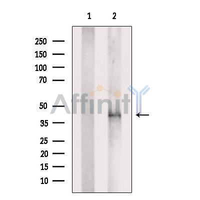
描述: Rabbit polyclonal antibody to PDLIM3
应用: WB IHC IF/ICC
文献验证: WB, IHC
反应: Human, Mouse, Rat
预测: Dog
蛋白号: Q53GG5
RRID: AB_2845487

浏览相似产品>>

   规格 价格 库存
 50ul RMB¥ 1250 现货
 100ul RMB¥ 2300 现货
 200ul RMB¥ 3000 现货

货期: 当天发货

联系销售

产品描述

来源:
Rabbit
应用:
WB 1:500-1:2000, IHC 1:50-1:200, IF/ICC 1:100-1:500
*The optimal dilutions should be determined by the end user.
*Tips:

WB: 适用于变性蛋白样本的免疫印迹检测. IHC: 适用于组织样本的石蜡(IHC-p)或冰冻(IHC-f)切片样本的免疫组化/荧光检测. IF/ICC: 适用于细胞样本的荧光检测. ELISA(peptide): 适用于抗原肽的ELISA检测.

反应:
Human, Mouse, Rat
克隆:
Polyclonal
特异性:
ALP Antibody detects endogenous levels of total ALP.
RRID:
AB_2845487
引用格式: Affinity Biosciences Cat# DF12525, RRID:AB_2845487.
偶联:
Unconjugated.
纯化:
The antiserum was purified by peptide affinity chromatography using SulfoLink™ Coupling Resin (Thermo Fisher Scientific).
保存:
Rabbit IgG in phosphate buffered saline , pH 7.4, 150mM NaCl, 0.02% sodium azide and 50% glycerol. Store at -20 °C. Stable for 12 months from date of receipt.
别名:

展开/折叠

Actinin associated LIM protein; Actinin-associated LIM protein; Alpha actinin 2 associated LIM protein; Alpha-actinin-2-associated LIM protein; DKFZp686L0362; Enigma homolog; PDLI3_HUMAN; PDLIM 3; PDLIM3; PDZ and LIM domain 3; PDZ and LIM domain protein 3; ALP;PDLI3;

抗原和靶标

免疫原:

A synthesized peptide derived from human ALP, corresponding to a region within the internal amino acids.

基因/基因ID:

文献引用

1). Osteoblastic and anti-osteoclastic activities of strontium-substituted silicocarnotite ceramics: In vitro and in vivo studies. Bioactive Materials, 2020 (PubMed: 32280833) [IF=18.9]

Application: IHC    Species: Mice    Sample: bone and osteoid tissues

Fig. 9 (A) Masson staining: the region between surrounding bone and scaffold (40x & 100x); (B) TRAP staining: TRAP positive cells (black arrows); (C) IHC staining of RUNX-2; (D) IHC staining of ALP; (E) Quantitative analysis for TRAP staining and IHC staining. * Compared with CPS group; # compared with Sr-5 group.

2). Fusion peptide engineered “statically-versatile” titanium implant simultaneously enhancing anti-infection, vascularization and osseointegration. Biomaterials, 2021 (PubMed: 33069134) [IF=12.8]

3). On-demand storage and release of antimicrobial peptides using Pandora's box-like nanotubes gated with a bacterial infection-responsive polymer. Theranostics, 2020 (PubMed: 31903109) [IF=12.4]

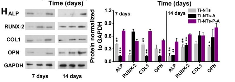
Application: WB    Species: Human    Sample: Human bone mesenchymal stem cells(hBMSCs)

Figure 5. In vitro biocompatibilities and osteogenic activities of the substrates. (A) CCK-8 assay of hBMSCs on the indicated surfaces after 1, 3 and 7 days of culture. * denotes p < 0.05 and ** denotes p < 0.01 compared to Ti-NTs, # denotes p < 0.05 compared to the corresponding control group without peptides. Error bars denote the standard deviations over quadruplicate measurements with separately implants. (B) Confocal fluorescence microscopy images of hBMSCs stained with vinculin, F-actin and DAPI after being cultured for 24 h. Scale bar, 50 μm. (C-F) qRT-PCR assay of osteogenic gene expression of (C) ALP, (D) RUNX-2, (E) COL1 and (F) OPN of hBMSCs after 7 and 14 days of culture. * denotes p < 0.05, ** denotes p < 0.01 and *** denotes p < 0.001 compared to Ti-NTs-P-A; # denotes p < 0.05, & denotes p < 0.01 and $ denotes p < 0.001 compared to the corresponding control group without peptides. All error bars denote the standard deviations over quadruplicate measurements with separately implants. (G) Immunofluorescence staining of hBMSCs cultured on Ti-NTs-P-A for 7 days (ALP and RUNX-2) and 14 days (OPN). The images were obtained by confocal fluorescence microscopy. Scale bar, 50 μm. (H) Western blotting of hBMSCs cultured on the substrates for 7 and 14 days. At each time point, left lane was Ti-NTs, middle lane was Ti-NTs-A and right lane was Ti-NTs-P-A. * denotes p < 0.05, ** denotes p < 0.01 and *** denotes p < 0.001 compared to Ti-NTs-P-A. Error bars denote the standard deviations over triplicate measurements with separately Western blotting results.

4). ZIF-8 Modified Multifunctional Bone-Adhesive Hydrogels Promoting Angiogenesis and Osteogenesis for Bone Regeneration. ACS Applied Materials & Interfaces, 2020 (PubMed: 32814397) [IF=8.3]

5). Insulin promotes the bone formation capability of human dental pulp stem cells through attenuating the IIS/PI3K/AKT/mTOR pathway axis. Stem cell research & therapy, 2024 (PubMed: 39075596) [IF=7.5]

Application: WB    Species: Human    Sample: DPSCs

Fig. 3 10− 6 M insulin promotes the osteogenic differentiation of human DPSCs. A 10− 6 M insulin up-regulated the mRNA levels of COL-1, ALP, OCN, and RUNX2 in DPSCs at day 3 and day 7 (n = 3). B 10− 6 M insulin promoted the protein expressions of COL-1, ALP, OCN, and RUNX2 in DPSCs at day 7 (n = 3). Representative western blotting (left) and quantification analysis (right). Full-length blots/gels are presented in Supplementary Fig. 1. C 10− 6 M insulin increased the secretion of extracellular bone metabolism and biochemical markers in DPSCs at day 1–7 and day 7–14 (n = 6). D 10− 6 M insulin enhanced alkaline phosphatase staining of DPSCs at day 21 (scale bars: 100 μm). E 10− 6 M insulin enhanced Alizarin red staining of DPSCs at day 21 (scale bars: 100 μm). F 10− 6 M insulin promoted the mineralized matrix formation of DPSCs at day 21 (n = 6). Data are expressed as the mean ± SD. *P 

6). MagT1 regulated the odontogenic differentiation of BMMSCs induced byTGC-CM via ERK signaling pathway. Stem Cell Research & Therapy, 2019 (PubMed: 30704530) [IF=7.5]

Application: WB    Species: rat    Sample: BMMSCs

Fig. 4 |Inactivation of ERK/MAPK signaling pathway inhibited the odontogenic differentiation of BMMSCs induced by TGC-CM. a ERK inhibitor U0126 significantly inhibited the mineralization and b reduced ALP, DSP, and DMP-1 protein levels of BMMSCs during odontogenic differentiation induced by TGC-CM for 7 days. Scale bar = 100 μm

7). miR-155-5p/Bmal1 Modulates the Senescence and Osteogenic Differentiation of Mouse BMSCs through the Hippo Signaling Pathway. Stem cell reviews and reports, 2024 (PubMed: 38150082) [IF=4.5]

8). Activation of osteoblast ferroptosis via the METTL3/ASK1‐p38 signaling pathway in high glucose and high fat (HGHF)‐induced diabetic bone loss. FASEB JOURNAL, 2022 (PubMed: 35104016) [IF=4.4]

9). Easily attainable and low immunogenic stem cells from exfoliated deciduous teeth enhanced the in vivo bone regeneration ability of gelatin/bioactive glass microsphere composite scaffolds. Frontiers in bioengineering and biotechnology, 2022 (PubMed: 36568292) [IF=4.3]

Application: IHC    Species: human    Sample:

FIGURE 6. Immunohistochemical staining of ALP, COL1, OCN, and RUNX2 after implantation for 4 and 8 weeks.

10). VX-765 ameliorates CKD VSMC calcification by regulating STAT3 activation. European Journal of Pharmacology, 2023 (PubMed: 36858340) [IF=4.2]

加载更多

限制条款

产品的规格、报价、验证数据请以官网为准,官网链接:www.affbiotech.com | www.affbiotech.cn(简体中文)| www.affbiotech.jp(日本語)

产品的数据信息为Affinity所有,未经授权不得收集Affinity官网数据或资料用于商业用途,对抄袭产品数据的行为我们将保留诉诸法律的权利。

产品相关数据会因产品批次、产品检测情况随时调整,如您已订购该产品,请以订购时随货说明书为准,否则请以官网内容为准,官网内容有改动时恕不另行通知。

Affinity保证所销售产品均经过严格质量检测。如您购买的商品在规定时间内出现问题需要售后时,请您在Affinity官方渠道提交售后申请。

产品仅供科学研究使用。不用于诊断和治疗。 

产品未经授权不得转售。

Affinity Biosciences将不会对在使用我们的产品时可能发生的专利侵权或其他侵权行为负责。Affinity Biosciences, Affinity Biosciences标志和所有其他商标所有权归Affinity Biosciences LTD.