产品: Cleaved-Caspase 1 (Ala317), p10 抗体
货号: AF4022
描述: Rabbit polyclonal antibody to Cleaved-Caspase 1 (Ala317), p10
应用: WB IHC IF/ICC
文献验证: WB, IHC, IF/ICC
反应: Human, Mouse, Rat
预测: Pig, Bovine, Horse, Rabbit, Dog
蛋白ID: P29466
RRID: AB_2845464

浏览相似产品>>

   规格 价格 库存
 50ul RMB¥ 1250 现货
 100ul RMB¥ 2300 现货

货期: 当天发货

联系销售

产品描述

来源:
Rabbit
应用:
WB 1:500-1:2000, IHC 1:50-1:200, IF/ICC 1:100-1:500
*The optimal dilutions should be determined by the end user. For optimal experimental results, antibody reuse is not recommended.
*Tips:

WB: 适用于变性蛋白样本的免疫印迹检测. IHC: 适用于组织样本的石蜡(IHC-p)或冰冻(IHC-f)切片样本的免疫组化/荧光检测. IF/ICC: 适用于细胞样本的荧光检测. ELISA(peptide): 适用于抗原肽的ELISA检测.

反应:
Human, Mouse, Rat
克隆:
Polyclonal
特异性:
Cleaved-Caspase-1 (Ala317,p10) Antibody detects endogenous levels of fragment of activated Caspase-1 resulting from cleavage adjacent to Ala317.
RRID:
AB_2845464
引用格式: Affinity Biosciences Cat# AF4022, RRID:AB_2845464.
偶联:
Unconjugated.
纯化:
The antiserum was purified by peptide affinity chromatography using SulfoLink™ Coupling Resin (Thermo Fisher Scientific).
保存:
Rabbit IgG in phosphate buffered saline , pH 7.4, 150mM NaCl, 0.02% sodium azide and 50% glycerol. Store at -20 °C. Stable for 12 months from date of receipt.
别名:

展开/折叠

CASP-1; CASP1; CASP1_HUMAN; Caspase 1; Caspase-1 subunit p10; ICE; IL-1 beta-converting enzyme; IL-1BC; IL1 beta converting enzyme; IL1B convertase; Interleukin 1 beta convertase; Interleukin 1B converting enzyme; Interleukin-1 beta convertase; Interleukin-1 beta-converting enzyme; p45;

抗原和靶标

免疫原:

The antiserum was produced against synthesized peptide derived from human Caspase 1.

基因/基因ID:

研究领域

· Cellular Processes > Cell growth and death > Necroptosis.   (View pathway)

· Human Diseases > Neurodegenerative diseases > Amyotrophic lateral sclerosis (ALS).

· Human Diseases > Infectious diseases: Bacterial > Salmonella infection.

· Human Diseases > Infectious diseases: Bacterial > Pertussis.

· Human Diseases > Infectious diseases: Bacterial > Legionellosis.

· Human Diseases > Infectious diseases: Viral > Influenza A.

· Organismal Systems > Immune system > NOD-like receptor signaling pathway.   (View pathway)

· Organismal Systems > Immune system > Cytosolic DNA-sensing pathway.   (View pathway)

文献引用
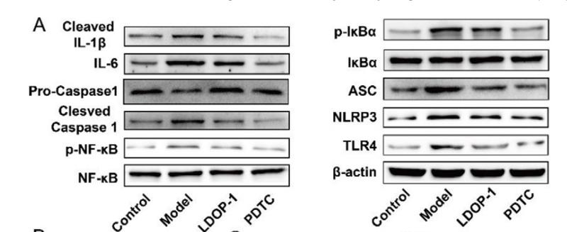
1). An artificial metabzyme for tumour-cell-specific metabolic therapy. Nature nanotechnology, 2024 (PubMed: 39103450) [IF=38.1]

2). NAD+-Boosters Improve Mitochondria Quality Control In Parkinson's Disease Models Via Mitochondrial UPR. Advanced science (Weinheim, Baden-Wurttemberg, Germany), 2025 (PubMed: 40685704) [IF=15.1]

3). p66Shc Contributes to Liver Fibrosis through the Regulation of Mitochondrial Reactive Oxygen Species. Theranostics, 2019 (PubMed: 30867846) [IF=12.4]

Application: WB    Species: mouse    Sample: Liver

Figure 2.| p66Shc silencing attenuates liver fibrosis in mice. p66Shc silencing was induced via lentivirus delivered to C57BL/6 mice exposed to CCl4 (2 ml/kg). (A) Liver p66Shc mRNA expression, n=6. (B) Liver p66Shc, SOD2, UCP1, Col1a1, α-SMA protein, n=3. (C) H2O2 content, n=8. (D) SOD activity, n=8. (E) Cytochrome c expression in the cytoplasm and mitochondria, n=3. (F) H&E and Masson staining. Scale bar, 200 μm. (G) Ishak score of Masson staining. (H) Serum ALT and AST levels, n=8. (I) Liver NLRP3 inflammasome protein expression, n=3.

4). Inflammation-driven biomimetic nano-polyphenol drug delivery system alleviates severe acute pancreatitis by inhibiting macrophage PANoptosis and pancreatic enzymes oversecretion. Journal of advanced research, 2025 (PubMed: 40210149) [IF=11.4]

Application: WB    Species: Mouse    Sample: BMDMs

Fig. 4. PTX formulations inhibited macrophage PANoptosis. (A) Gene clustering analysis of PTX, FePTX, FePTX@CM-treated BMDMs. (B-C) GSEA analysis of transcriptomics results of BMDMs after LPS intervention. (D-E) Inflammatory-related pathway analysis in transcriptomics results of BMDMs after different PTX formulation interventions. (F) Heatmap of transcriptional profiling of inflammatory factors in BMDMs after PTX formulation interventions. (G) Results of qPCR sequencing (n = 4). (H) Cell viability of BMDMs with different treatments (n = 4). (I) PI staining images of BMDMs after LPS and FePTX@CM NPs treatment and its analysis (n = 4). Scale bar = 100 μm. (J) WB detection of the PANoptosis biomarkers in BMDMs upon treatment with FePTX@CM NPs. (K) TEM images of BMDMs after different treatments. Scale bar = 5 μm. (L) Co-IP analysis of PANoptosomes biomarkers in RAW264.7 after various interventions (n = 4). *P < 0.05, **P < 0.01, ***P < 0.001, N.s. = no significance. Data = mean ± SD.

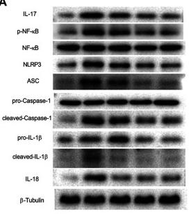
5). Water-extracted Lonicera japonica polysaccharide attenuates allergic rhinitis by regulating NLRP3-IL-17 signaling axis. Carbohydrate Polymers, 2022 (PubMed: 36184153) [IF=10.7]

6). An elastase-responsive "plug-and-play" quercetin-loaded engineered extracellular vesicle platform anchored with anti-PRDX1 antibody for severe acute pancreatitis therapy. Journal of controlled release : official journal of the Controlled Release Society, 2026 (PubMed: 41621758) [IF=10.5]

7). AIM2 Inflammasome Mediates Hallmark Neuropathological Alterations and Cognitive Impairment in a Mouse Model of Vascular Dementia. Molecular Psychiatry, 2020 (PubMed: 33299135) [IF=9.6]

8). NLRP3-GABA signaling pathway contributes to the pathogenesis of impulsive-like behaviors and cognitive deficits in aged mice. Journal of neuroinflammation, 2023 (PubMed: 37434240) [IF=9.3]

Application: IF/ICC    Species: Mouse    Sample: hippocampus

Fig. 6 Cognitive impairment may be related to pyroptosis-associated inflammatory responses in astrocytes. A Representative photomicrographs of NLRP3-, IL-18-, IL-1β- and cleaved caspase-1-positive cells in the CA1 of the hippocampus at 12 h and 10 days after surgical exposure. Scale bar = 50 μm. B Image of CA1 in hippocampus. C–J Co-stained area of NLRP3-, IL-18-, IL-1β-, cleaved caspase-1- and GFAP-positive cells in the CA1 of the hippocampus. K Representative western blot of NLRP3, IL-1β, IL-18 and cleaved caspase-1 at 12 h after surgical exposure. L–O The optical density values of NLRP3, IL-1β, IL-18, and cleaved caspase-1 at 12 h after surgical exposure. P Representative western blot of NLRP3, IL-1β, IL-18 and cleaved caspase-1 at 10 days after surgical exposure. Q–T The optical density values of NLRP3, IL-1β, IL-18 and cleaved caspase-1 at 10 days after surgical exposure. Data are presented as the mean ± SD (n = 6 mice/group or n = 3 mice/group). Data were analyzed by one-way ANOVA with Tukey’s multiple comparison test or Kruskal–Wallis and Dunn’s multiple comparison test. ***P 

9). Mesenchymal stem cells protect against TBI-induced pyroptosis in vivo and in vitro through TSG-6. Cell Communication and Signaling, 2022 (PubMed: 35982465) [IF=8.4]

10). JC124 confers multimodal neuroprotection in epilepsy by suppressing NLRP3 inflammasome activation: evidence from animal and human neuronal models. Cell communication and signaling : CCS, 2025 (PubMed: 40629339) [IF=8.4]

Application: WB    Species: mouse    Sample:

Fig. 3 From: JC124 confers multimodal neuroprotection in epilepsy by suppressing NLRP3 inflammasome activation: evidence from animal and human neuronal models JC124 treatment inhibits NLRP3 inflammasome activation in KA-induced epileptic mice. A Representative WB bands. In KA-induced epileptic mice, treatment with JC124 significantly reduced the relative intensities of (B) NLRP3/GAPDH, (C) ASC/GAPDH, (D) pro-Caspase-1/GAPDH, and (E) Caspase-1 p10/GAPDH in the hippocampus (n = 3). F–H Representative immunohistochemical images of NLRP3 (brown), ASC (brown), and Caspase-1 p10 (brown) in the hippocampal CA1 region. Scale bar, 50 μm. The black arrows indicate typical positively stained cells. KA-induced epileptic mice treated with JC124 showed a significant decrease in the mean optical densities of the (I) NLRP3, (J) ASC, and (K) Caspase-1 p10 (n = 5). L-N Representative immunofluorescence images of NLRP3 (green)/GFAP (red), NLRP3 (green)/Iba1 (red), NLRP3 (green)/NeuN (red), ASC (green)/GFAP (red), ASC (green)/Iba1 (red), ASC (green)/NeuN (red), Caspase-1 p10 (green)/GFAP (red), Caspase-1 p10 (green)/Iba1 (red), and Caspase-1 p10 (green)/NeuN (red) in the hippocampal CA1 region (n = 3). Scale bar, 50 μm. The white arrows indicate typical co-labeled cells. Data are represented as mean ± S.E.M. *p < 0.05, **p < 0.01, ***p < 0.001

Application: IF/ICC    Species: mouse    Sample:

Fig. 3 From: JC124 confers multimodal neuroprotection in epilepsy by suppressing NLRP3 inflammasome activation: evidence from animal and human neuronal models JC124 treatment inhibits NLRP3 inflammasome activation in KA-induced epileptic mice. A Representative WB bands. In KA-induced epileptic mice, treatment with JC124 significantly reduced the relative intensities of (B) NLRP3/GAPDH, (C) ASC/GAPDH, (D) pro-Caspase-1/GAPDH, and (E) Caspase-1 p10/GAPDH in the hippocampus (n = 3). F–H Representative immunohistochemical images of NLRP3 (brown), ASC (brown), and Caspase-1 p10 (brown) in the hippocampal CA1 region. Scale bar, 50 μm. The black arrows indicate typical positively stained cells. KA-induced epileptic mice treated with JC124 showed a significant decrease in the mean optical densities of the (I) NLRP3, (J) ASC, and (K) Caspase-1 p10 (n = 5). L-N Representative immunofluorescence images of NLRP3 (green)/GFAP (red), NLRP3 (green)/Iba1 (red), NLRP3 (green)/NeuN (red), ASC (green)/GFAP (red), ASC (green)/Iba1 (red), ASC (green)/NeuN (red), Caspase-1 p10 (green)/GFAP (red), Caspase-1 p10 (green)/Iba1 (red), and Caspase-1 p10 (green)/NeuN (red) in the hippocampal CA1 region (n = 3). Scale bar, 50 μm. The white arrows indicate typical co-labeled cells. Data are represented as mean ± S.E.M. *p < 0.05, **p < 0.01, ***p < 0.001

加载更多

限制条款

产品的规格、报价、验证数据请以官网为准,官网链接:www.affbiotech.com | www.affbiotech.cn(简体中文)| www.affbiotech.jp(日本語)

产品的数据信息为Affinity所有,未经授权不得收集Affinity官网数据或资料用于商业用途,对抄袭产品数据的行为我们将保留诉诸法律的权利。

产品相关数据会因产品批次、产品检测情况随时调整,如您已订购该产品,请以订购时随货说明书为准,否则请以官网内容为准,官网内容有改动时恕不另行通知。

Affinity保证所销售产品均经过严格质量检测。如您购买的商品在规定时间内出现问题需要售后时,请您在Affinity官方渠道提交售后申请。

产品仅供科学研究使用。不用于诊断和治疗。 

产品未经授权不得转售。

Affinity Biosciences将不会对在使用我们的产品时可能发生的专利侵权或其他侵权行为负责。Affinity Biosciences, Affinity Biosciences标志和所有其他商标所有权归Affinity Biosciences LTD.