产品: PDGFB 抗体
货号: AF0240
描述: Rabbit polyclonal antibody to PDGFB
应用: WB IHC IF/ICC
反应: Human, Mouse, Rat
预测: Rabbit
分子量: 27kDa,22kDa; 27kD(Calculated).
蛋白号: P01127
RRID: AB_2833415

浏览相似产品>>

   规格 价格 库存
 50ul RMB¥ 1250 现货
 100ul RMB¥ 2300 现货
 200ul RMB¥ 3000 现货

货期: 当天发货

联系销售

产品描述

来源:
Rabbit
应用:
WB 1:500-1:3000, IF/ICC 1:100-1:500, IHC 1:50-1:200
*The optimal dilutions should be determined by the end user.
*Tips:

WB: 适用于变性蛋白样本的免疫印迹检测. IHC: 适用于组织样本的石蜡(IHC-p)或冰冻(IHC-f)切片样本的免疫组化/荧光检测. IF/ICC: 适用于细胞样本的荧光检测. ELISA(peptide): 适用于抗原肽的ELISA检测.

反应:
Human,Mouse,Rat
预测:
Rabbit(85%)
克隆:
Polyclonal
特异性:
PDGFB Antibody detects endogenous levels of total PDGFB.
RRID:
AB_2833415
引用格式: Affinity Biosciences Cat# AF0240, RRID:AB_2833415.
偶联:
Unconjugated.
纯化:
The antiserum was purified by peptide affinity chromatography using SulfoLink™ Coupling Resin (Thermo Fisher Scientific).
保存:
Rabbit IgG in phosphate buffered saline , pH 7.4, 150mM NaCl, 0.02% sodium azide and 50% glycerol. Store at -20 °C. Stable for 12 months from date of receipt.
别名:

展开/折叠

Becaplermin; PDGF 2; PDGF B chain; PDGF subunit B; PDGF-2; PDGF2; Pdgfb; PDGFB_HUMAN; Platelet derived growth factor 2; Platelet-derived growth factor B chain; Platelet-derived growth factor beta polypeptide; Platelet-derived growth factor subunit B; Proto-oncogene c-Sis; SIS; SSV;

抗原和靶标

免疫原:
Uniprot:
基因/基因ID:
表达:
P01127 PDGFB_HUMAN:

Expressed at high levels in the heart, brain (sustantia nigra), placenta and fetal kidney. Expressed at moderate levels in the brain (hippocampus), skeletal muscle, kidney and lung.

描述:
PDGFB Platelet-derived growth factor is a potent mitogen for cells of mesenchymal origin. Binding of this growth factor to its affinity receptor elicits a variety of cellular responses. It is released by platelets upon wounding and plays an important role in stimulating adjacent cells to grow and thereby heals the wound.
序列:
MNRCWALFLSLCCYLRLVSAEGDPIPEELYEMLSDHSIRSFDDLQRLLHGDPGEEDGAELDLNMTRSHSGGELESLARGRRSLGSLTIAEPAMIAECKTRTEVFEISRRLIDRTNANFLVWPPCVEVQRCSGCCNNRNVQCRPTQVQLRPVQVRKIEIVRKKPIFKKATVTLEDHLACKCETVAAARPVTRSPGGSQEQRAKTPQTRVTIRTVRVRRPPKGKHRKFKHTHDKTALKETLGA

种属预测

种属预测:

score>80的预测可信度较高,可尝试用于WB检测。*预测模型主要基于免疫原序列比对,结果仅作参考,不作为质保凭据。

Species
Results
Score
Rabbit
85
Horse
77
Bovine
77
Sheep
77
Dog
69
Pig
0
Xenopus
0
Zebrafish
0
Chicken
0
Model Confidence:
High(score>80) Medium(80>score>50) Low(score<50) No confidence

翻译修饰 - P01127 作为底物

Site PTM Type Enzyme
T190 O-Glycosylation
S196 Phosphorylation

研究背景

功能:

Growth factor that plays an essential role in the regulation of embryonic development, cell proliferation, cell migration, survival and chemotaxis. Potent mitogen for cells of mesenchymal origin. Required for normal proliferation and recruitment of pericytes and vascular smooth muscle cells in the central nervous system, skin, lung, heart and placenta. Required for normal blood vessel development, and for normal development of kidney glomeruli. Plays an important role in wound healing. Signaling is modulated by the formation of heterodimers with PDGFA (By similarity).

细胞定位:

Secreted.
Note: Released by platelets upon wounding.

Extracellular region or secreted Cytosol Plasma membrane Cytoskeleton Lysosome Endosome Peroxisome ER Golgi apparatus Nucleus Mitochondrion Manual annotation Automatic computational assertionSubcellular location
组织特异性:

Expressed at high levels in the heart, brain (sustantia nigra), placenta and fetal kidney. Expressed at moderate levels in the brain (hippocampus), skeletal muscle, kidney and lung.

亚基结构:

Antiparallel homodimer; disulfide-linked. Antiparallel heterodimer with PDGFA; disulfide-linked. The PDGFB homodimer interacts with PDGFRA and PDGFRB homodimers, and with heterodimers formed by PDGFRA and PDGFRB. The heterodimer composed of PDGFA and PDGFB interacts with PDGFRB homodimers, and with heterodimers formed by PDGFRA and PDGFRB. Interacts with XLKD1 (By similarity). Interacts with LRP1. Interacts with SORL1 (via the N-terminal ectodomain).

蛋白家族:

Belongs to the PDGF/VEGF growth factor family.

研究领域

· Cellular Processes > Cellular community - eukaryotes > Focal adhesion.   (View pathway)

· Cellular Processes > Cellular community - eukaryotes > Gap junction.   (View pathway)

· Cellular Processes > Cell motility > Regulation of actin cytoskeleton.   (View pathway)

· Environmental Information Processing > Signal transduction > MAPK signaling pathway.   (View pathway)

· Environmental Information Processing > Signal transduction > Ras signaling pathway.   (View pathway)

· Environmental Information Processing > Signal transduction > Rap1 signaling pathway.   (View pathway)

· Environmental Information Processing > Signaling molecules and interaction > Cytokine-cytokine receptor interaction.   (View pathway)

· Environmental Information Processing > Signal transduction > Phospholipase D signaling pathway.   (View pathway)

· Environmental Information Processing > Signal transduction > PI3K-Akt signaling pathway.   (View pathway)

· Environmental Information Processing > Signal transduction > Jak-STAT signaling pathway.   (View pathway)

· Human Diseases > Drug resistance: Antineoplastic > EGFR tyrosine kinase inhibitor resistance.

· Human Diseases > Infectious diseases: Viral > HTLV-I infection.

· Human Diseases > Cancers: Overview > Pathways in cancer.   (View pathway)

· Human Diseases > Cancers: Overview > MicroRNAs in cancer.

· Human Diseases > Cancers: Specific types > Renal cell carcinoma.   (View pathway)

· Human Diseases > Cancers: Specific types > Glioma.   (View pathway)

· Human Diseases > Cancers: Specific types > Prostate cancer.   (View pathway)

· Human Diseases > Cancers: Specific types > Melanoma.   (View pathway)

· Human Diseases > Cancers: Overview > Choline metabolism in cancer.   (View pathway)

文献引用

1). Soft apoptotic-cell-inspired nanoparticles persistently bind to macrophage membranes and promote anti-inflammatory and pro-healing effects. Acta Biomaterialia (PubMed: 34245890) [IF=9.7]

Application: IHC    Species: Rat    Sample: Raw 264.7 cells

Fig. 6. Soft d -PSLs evidently promote chronic wound healing in STZ induced diabetic rats. (a) Representative images of the wound closure process at days 0, 1, 3, 7, and 14. (b) Percentage of remaining wound area expressed as the ratio of remaining wound area to the initial wound area. (c) Representative images of H&E staining (Scale bars = 50 μm) and IHC staining (Scale bars = 20 μm). (d-g) Quantification of CD86, CD206, CD31 and PDGF-BB positive area in IHC images. The proportion of the positive area of each field was quantified using Image J software. n = 6 in each group. ns, no significant difference. ∗∗ P < 0.01; ∗∗∗ P < 0.001.

2). Mechanical stretching can modify the papillary dermis pattern and papillary fibroblast characteristics during skin regeneration. Journal of Investigative Dermatology (PubMed: 35181299) [IF=6.5]

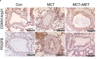
3). Metformin inhibits pulmonary artery smooth muscle cell proliferation by upregulating p21 via NONRATT015587. 2. International Journal of Molecular Medicine (PubMed: 35147202) [IF=5.4]

Application: IHC    Species: Rat    Sample: pulmonary artery tissue

Figure 1 Expression of NONRATT015587.2 and p21 in MCT-induced pulmonary hypertension rats. (A) Assessment of RVSP in MCT-induced PAH rats, controls and following MCT+MET treatment. (B) Changes in the RV/(LV+S) ratio. (C) NONRATT015587.2 gene expression was calculated in each group. (D) The expression of CDKN1A/p21 and PDGFB in pulmonary artery tissue was analyzed by immunohistochemistry staining (scale bars, 100 µm). (E) p21 protein expression was determined via western blotting and subsequently quantified. (F) Fluorescence in situ hybridization was performed to analyze the subcellular location of NONRATT015587.2. In this experiment, 18s was used as the cytoplasmic reference gene and u6 was used as the nuclear reference gene. DAPI was used for nuclear staining (scale bars, 50 µm). Data represent mean ± standard deviation (SD). *P<0.05 and **P<0.01 as indicated (n= ≥3). Con, control; MCT, monocrotaline; MET, metformin; PDGFB, platelet-derived growth factor subunit B; RV/(LV+S), right ventricle/left ventricle plus septum; RVSP, right ventricular systolic pressure.

4). Rnf-213 Knockout Induces Pericyte Reduction and Blood-Brain Barrier Impairment in Mouse. Molecular Neurobiology (PubMed: 37438553) [IF=5.1]

5). Lipoxin A4 inhibited the activation of hepatic stellate cells -T6 cells by modulating profibrotic cytokines and NF-κB signaling pathway. PROSTAGLANDINS & OTHER LIPID MEDIATORS (PubMed: 31698141) [IF=2.9]

Application: WB    Species: mouse    Sample: HSC-T6 cells

Figure 6.| LXA4 inhibited LPS-activated NF-κB signal pathway in HSC-T6 cells. (a-c) The protein levels of NF-κB P65, P-P65, and I-κBα in HSC-T6 cells were measured by western blot and optical density analysis was shown (n=6). **P < 0.01 compared with control group, ##P < 0.01 compared with LPS group, ΔP < 0.05 and ΔΔP < 0.01 compared with LPS + LXA4 group.

限制条款

产品的规格、报价、验证数据请以官网为准,官网链接:www.affbiotech.com | www.affbiotech.cn(简体中文)| www.affbiotech.jp(日本語)

产品的数据信息为Affinity所有,未经授权不得收集Affinity官网数据或资料用于商业用途,对抄袭产品数据的行为我们将保留诉诸法律的权利。

产品相关数据会因产品批次、产品检测情况随时调整,如您已订购该产品,请以订购时随货说明书为准,否则请以官网内容为准,官网内容有改动时恕不另行通知。

Affinity保证所销售产品均经过严格质量检测。如您购买的商品在规定时间内出现问题需要售后时,请您在Affinity官方渠道提交售后申请。

产品仅供科学研究使用。不用于诊断和治疗。 

产品未经授权不得转售。

Affinity Biosciences将不会对在使用我们的产品时可能发生的专利侵权或其他侵权行为负责。Affinity Biosciences, Affinity Biosciences标志和所有其他商标所有权归Affinity Biosciences LTD.