 Antibody at 1/1000 dilution.
5ug/NC membrane strip.
Exposure for 4min with Affinity™ ECL Kit(#KF8003).
Bands result from membrane strip incubation.")

 Expression of proliferation- and migration-associated genes (PCNA, MMP9 and TIMP-1) were evaluated using western blotting in HEY cells. (C and D) Western blotting of proteins involved in integrin-β1-FAK signaling pathway in the KRT7-overexpressing HEY cells. (E) Expression of MMPs after knockdown of KRT7 in OVCAR433 cells. (F and G) Expression of the TGF-β signaling pathway-related proteins was evaluated by western blotting in KRT7-overexpressing HEY cells and KRT7-knockdown OVCAR433 cells. All experiments were performed at least three times. Results are presented as the mean ± standard deviation. **P<0.01. FAK, focal adhesion kinase; PCNA, proliferating cell nuclear antigen; FN, fibronectin; TIMP-1, TIMP metallopeptidase inhibitor 1; p-, phosphorylated; MMP, matrix metalloproteinase; KRT7, keratin 7; sh, short hairpin RNA; NC, negative control.")
 The expression of TGF-β, α-sma, Smad2/3, pSmad2/3, Col-1and β-actin in mouse skin.")
 Representative western blots and quantitative analysis of (B) TGF‑β1/β‑actin, (C) p‑Smad2/3/Smad2/3 and (D) p‑ERK1/2/ERK1/2 levels in rats.")
 Western blots and densi-
tometry data showing the effects of CX-5461 on
phosphorylation of Smad2/3 and p38 in serum +
TGF-β-stimulated cells. Stim, stimulation with
serum + TGF-β. (B) Western blots for the steady
state levels of ribosomal proteins RpS6 and RpL10a
were performed using purified ribosome samples.
The total protein in the supernatant was used as
input control. The quantitative data were expressed
as mean ± S.E.M. *P < 0.05, one-way ANOVA (n =
3). NS, no significance.")
 Expression levels of TGFb1, SMAD2/3, and quantification. (d‒g) Expression levels of TGFb2, TGFb3, pSMAD2/3 (Thr8), and quantification. (h‒k) Expression levels of Akt and pAkt and quantification. n ¼ 3 biological replicates. Data are represented as mean SD; one-way ANOVA with Fisher’s posthoc test. *P < 0.05 versus control and #P < 0.05 versus FB-Exo. Akt, protein kinase B; ESC-Exo, epidermal stem cellederived exosome; FB, fibroblast; FB-Exo, fibroblast-derived exosome; pAkt, phosphorylated protein kinase B; pSMAD, phosphorylated SMAD.")
発色: TGFβR1、BMPR1A。AP(赤)発色: pSmad2/3、pSmad1/5/9。黒矢頭: 陽性共局在細 胞を示した。Scale bar: 50µm。B: TGFβR1 / pSmad2/3陽性共局在細胞比率の比較結果。C: BMPR1A/pSmad1/5/9 陽性共局在細胞比率の比較結果。D: pSmad1/5/9 / pSmad2/3比率の比較結果。B-D:すべてのデータは平均値 ±SDで示した。")
 and finasteride (10 mg/kg; 10–100 μM) on the expression of TGFBR2, TGF-β1, p-Smad2/3, Smad2/3, N-cadherin, E-cadherin and vimentin in prostate tissues at 14 days (A) and WPMY-1 cells at 24 h (B). (C-D) Data are presented as mean ± SEM. (*p < 0.05 compared with control, #p < 0.05 compared with TP or testosterone group).")
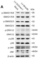
 or LV-Bmp8a cells (c, d). Protein expression levels were quantified using ImageJ software and normalized to the amount of total protein (n = 3). e, f Representative Oil Red O staining photographs of LV-bmp8a and LV-Bmp8a 3T3-L1 cells were induced to adipogenic in the presence of DMH1 or TP0427736 HCL, dimethylsulfoxide (DMSO) as a vehicle and subjected to OD492 quantifications (n = 3). Scale bar = 20 µm. g Schematic diagram of BMP8 mediated signal transduction. BMP8 can activate Smad1/5/8 signal transduction through the receptor complex formed by type I receptor ALK2, ALK3, or ALK6 and type II receptor ACVR2A or BMPR2. Meanwhile, BMP8 can also activate Smad2/3 signal transduction through the receptor complex formed by type I receptors ALK4 or ALK5 and type II receptors ACVR2A, ACVR2B, or TGFBR2. h Non-expression of mouse Alk6 gene in 3T3-L1 cells (n = 3). i The qPCR quantification of the type I receptor (Alk2, Alk3, Alk4, Alk5, Alk7) and type II receptor (Acvr2a, Acvr2b, Bmpr2, Tgrβr2) transcripts expressed in 3T3-L1 cells (n = 3). j, k Quantification of the activity of BRE-driven luciferase reporters with pCMV-bmp8a (j) or pCMV-Bmp8a (k) cotransfected with pCMV-Alk2, pCMV-Alk3, pCMV-Bmpr2, pCMV-Acrv2a, respectively (n = 3). Renilla luciferase was used as the internal control. l, m Quantification of the activity of CAGA-driven luciferase reporters with pCMV-bmp8a (l) or pCMV-Bmp8a (m) cotransfected with pCMV-Alk2, pCMV-Alk3, pCMV-Bmpr2, and pCMV-Acrv2a, respectively (n = 3). Renilla luciferase was used as the internal control. Data were representative of at least three independent experiments. Data were analyzed by One-way ANOVA and presented as mean ± SD")
 transfection for 18 hours and further treatment with or without TGFβ2 for 48 hours. (A, B) Western blot analysis and densitometry quantification showed protein levels of p-smad2 (phosphorylation of smad2) and p-smad3 (phosphorylation of smad3) in the siNC, siNC+T2, siLSS, and siLSS+T2 groups. (C) Immunofluorescent staining of p-smad2/3 (red) and smad2/3 (red) merged with DAPI (blue) in siNC, siNC+T2, siLSS, and siLSS+T2 groups. (D, E) Western blot analysis and densitometry quantification showed protein levels of p-smad2 and p-smad3 (phosphorylation of Smad2/3) in oeNC, oeNC+T2, oeLSS, and oeLSS+T2 groups. (F) Immunofluorescent staining of p-smad2/3 (red) and smad2/3 (red) merged with DAPI (blue) in oeNC, oeNC+T2, oeLSS, and oeLSS+T2 groups. Representative images of three replicated experiments were presented. All scale bars, 100 µm. ns, not significant. *P < 0.05; **P < 0.01; ***P < 0.001. Data are shown as mean ± SD (n = 3).")
 transfection for 18 hours and further treatment with or without TGFβ2 for 48 hours. (A, B) Western blot analysis and densitometry quantification showed protein levels of p-smad2 (phosphorylation of smad2) and p-smad3 (phosphorylation of smad3) in the siNC, siNC+T2, siLSS, and siLSS+T2 groups. (C) Immunofluorescent staining of p-smad2/3 (red) and smad2/3 (red) merged with DAPI (blue) in siNC, siNC+T2, siLSS, and siLSS+T2 groups. (D, E) Western blot analysis and densitometry quantification showed protein levels of p-smad2 and p-smad3 (phosphorylation of Smad2/3) in oeNC, oeNC+T2, oeLSS, and oeLSS+T2 groups. (F) Immunofluorescent staining of p-smad2/3 (red) and smad2/3 (red) merged with DAPI (blue) in oeNC, oeNC+T2, oeLSS, and oeLSS+T2 groups. Representative images of three replicated experiments were presented. All scale bars, 100 µm. ns, not significant. *P < 0.05; **P < 0.01; ***P < 0.001. Data are shown as mean ± SD (n = 3).")
, M2-conditioned medium only and verteporfin (1.5 μmol/L, 3 h) followed by M2-conditioned medium treatment (M2/VTP) for 36 h. Protein levels of collagen I, fibronectin, YAP, TAZ and components of TGF-β1/smad signalling were determined by western blotting. (A) Representative images of western blot images. (B) Analysis of relative protein expression. GAPDH or β-tubulin was used as a loading control, as appropriate. *P")
 Representative images of Periodic Acid Schiff staining (PAS, ×400, bar: 20 μm). (B) Representative images of Masson's trichrome staining (Masson, ×400, bar: 20 μm). (C) Statistics of mesangial area. (D) Statistics of mesangial density. (E) Statistical result of Masson. (F) Representative WB images of TGF-β1, Smad2/3, and p-Smad2/3. (G) Statistical results of TGF-β1. (H) Statistical results of p-Smad2/3 to Smad2/3. Data are expressed as mean ± SD, n = 4. **P < 0.01 versus Control; #P < 0.05, ##P < 0.01 versus Model.")
 Protein levels of Smad2/3, p-Smad2/3, Smad4, Vimentin, α-SMA, and E-cadherin in type II AECs co-cultured with Tregs supernatant were detected by Western blot. *P<0.05, **P<0.01, ***P<0.001, n = 3.")
 Primary fibroblasts were treated with IL-20 (200 ng/ml) for 48 h. Bromodeoxyuridine (BrdU) incorporation was measured using flow cytometry. Flow cytometry plots show side scatter (SSC) vs. BrdU fluorescence for three individual samples: Non-BrdU (left), Untreated control (middle), and IL-20 (200 ng/ml) treatment (right). (b) Quantification of BrdU+ cell percentages. Data are presented as the mean ± SEM (*P < 0.05, ****P < 0.0001). The data shown are representative of three independent experiments with similar results. (c) Primary fibroblasts were treated with IL-20 (200 ng/ml) for 10 h. Gene expression of the fibrotic marker α-SMA was analyzed using RT-qPCR with specific primers. Ctrl: untreated control. Data are presented as the mean ± SEM (**P < 0.01). The data shown are representative of three independent experiments with similar results. (d) Primary fibroblasts were incubated with IL-20 (200 ng/ml) for the indicated times. Cell lysates were collected and analyzed by immunoblotting with a specific antibody against α-SMA, p-Smad2/3, Smad2/3, p-STAT3, and STAT3. Tubulin was used as a loading control. (e) Primary fibroblasts were incubated with IL-20 (200 ng/ml), or IL-20 (200 ng/ml) plus 7E (2 μg/ml) for 5 days. Cell lysates were collected and analyzed by immunoblotting with a specific antibody against α-SMA and p-Smad2/3. Tubulin was used as a loading control.")
 Western blotting analysis of TGF-β1 (B), Smad2, Smad3, P-smad2/3 (C), Smad7 (D), c-Jun (E), and GAPDH expression in liver tissue. (F) Immunohistochemistry staining of P-smad2/3 in the liver tissues (magnification, 10× and 20×). (G) Quantification of areas of positive staining using ImageJ software. The proportion of cells stained brown (positive expression) to the entire field of view was analyzed. In the experiment, we randomly selected more than 3 repetitions of different fields of view. Data are expressed as mean ± standard deviation. * P < 0.05 and *** P < 0.001 versus the control group; ## P < 0.01 and ### P < 0.001 versus the CCl4 group; ns: no significance.")
 Western blotting analysis of TGF-β1 (B), Smad2, Smad3, P-smad2/3 (C), Smad7 (D), c-Jun (E), and GAPDH expression in liver tissue. (F) Immunohistochemistry staining of P-smad2/3 in the liver tissues (magnification, 10× and 20×). (G) Quantification of areas of positive staining using ImageJ software. The proportion of cells stained brown (positive expression) to the entire field of view was analyzed. In the experiment, we randomly selected more than 3 repetitions of different fields of view. Data are expressed as mean ± standard deviation. * P < 0.05 and *** P < 0.001 versus the control group; ## P < 0.01 and ### P < 0.001 versus the CCl4 group; ns: no significance.")
 Western blot analysis of the effects of knocking down or overexpressing Spp1 on the expression of Smad2/3, Col1a1, and α-SMA under TGF-β induced conditions. (B–D) Protein expression grayscale analysis to compare the expression and phosphorylation differences of the proteins. (E–H) Double immunofluorescence staining of P-Smad2/3 (red) and Col1a1 (green) or α-SMA (green) to validate whether Spp1 regulates Col1a1 and α-SMA expression through the phosphorylation of Smad2/3. Blue represents cell nuclei stained with DAPI. In all the images, compared to the sh-NC or oe-NC groups, ∗ represents p < 0.05, ∗∗ represents p < 0.01, and ∗∗∗ represents p < 0.001; bar equals 20 μm; each experiment was conducted three times with six replicates each time.")
 Cel decreased the expression of TGF-β1, TGF-βRII, Smad2/3, and P-Smad2/3. Scale bar: 20 µm. (B) Cel decreased the expression of α-SMA, COL I, and FN. Scale bar: 20 µm. (C) The mean fluorescence intensity (MIF) of TGF-β1, TGF-βRII, Smad2/3, P-Smad2/3, α-SMA, COL I, and FN was showed in four groups. The fluorescence quantitative results were presented as mean ± SD of three independent experiments. *P < 0.05, **P < 0.01, ***P < 0.001, comparison between two groups.")
 Masson’s trichrome staining and HE is staining of peritoneal tissue sections (×400). (b) Immunohistochemical staining of SOCS1, α-SMA, and Collagen I in peritoneal tissue sections (×400). (c) Expression levels of TGF-β, Smad2/3, p-Smad2/3, and SOCS1 were detected using protein blotting. Each group contained eight rats, and three fields of view were randomly selected for each group. The percentage of the stained area was calculated for statistical analysis. All immunoblotting experiments were performed independently and repeated at least three times. Values are expressed as mean ± standard deviation. *p < 0.05 vs. Control; **p < 0.01 vs. Control; ***p < 0.001 vs. Control.")
 Protein bands and analysis of p-Smad, Smad, TGF and β-actin in mice mammary tissue. (D-F) Protein bands and analysis of p-Smad, Smad, TGF and β-actin in mMECs of RA group. (G-I) Protein bands and analysis of p-Smad, Smad, TGF and β-actin in mMECs of tBHQ group. (J-L) Protein bands and analysis of p-Smad, Smad, TGF and β-actin in mMECs tissue. The data were presented as Mean ± SD. Three independent repeatability experiments were performed. ** represents P")
, smad2/3 and p-smad2/3 in the liver. (A-C) The expressions of TGF-β1, p-smad2/3 and smad2/3 were detected by western blot, GAPDH was used as an internal control. (D) The ratio of p-smad2/3 to smad2/3 was calculated based on the protein expressions of p-smad2/3 and smad2/3. Compared with the control group *P")
 Western blot analysis of p-SMAD2, p-SMAD3, and total SMAD2/3 levels in LECs incubated with TGFβ2 and ISRIB. (B) Immunofluorescence staining of SMAD2/3 in LECs treated with or without TGFβ2 and ISRIB for 48 hours. Scale bar: 50 µm. (C) Immunofluorescence staining of lens capsule whole mounts from control and ISRIB-treated mice showing p-SMAD2/3 signals. The boxed area indicates the magnified region. Scale bar: 100 µm. (D) The intracellular localization of p-SMAD2/3 in plaques was confirmed by confocal microscopy. Scale bar: 20 µm.")
 CCK-8 assay was used to assess the cell viability of HaCaT and fibroblast cells after treatment with Elesclomol+Cu2+ (1 μM) across different concentration gradients (A) and time gradients (B). (C and D) CCK-8 assay was performed to evaluate the cell viability of Hacat and fibroblast cells treated with different cell death inhibitors (TTM, Nec-1, Fer-1, CQ, ZVF). (E) IHC to verify the expression of ACO2 in normal tissues and DFU tissues, Scale bars = 100 μm (F) Western blotting confirmed the change of ACO2 protein levels in Hacat and fibroblast. (G and H) qRT-PCR confirmed the knockdown of TMPO in Hacat and fibroblast cells by shRNA on mRNA levels. (I and J) Western blotting confirmed the knockdown of TMPO in Hacat and fibroblast cells by shRNA on protein levels. (K and M) Cell growth curve of Hacat and fibroblast cells transfected with Scramble or shTMPO. (L, N, O, P) Wound healing assays of Hacat and fibroblast cells transfected with Scramble or shTMPO. (Q and R) The protein levels of TGF-β1, Smad2/3, p-Smad2/3, and P21 in Hacat and fibroblast cells transfected with Scramble or shTMPO. n = 3, data were represented as mean ± SEM.")
 Western blot results of p-TβRI and p-Smad2/3 in TDSCs under different treatment conditions. And gray value statistical chart. (D-F) Immunohistochemical staining of p-TβRI and p-Smad2/3 expression in normal Achilles tendon and Achilles tendon specimens 4 and 8 weeks after modeling, and statistical graphs of positive cell rates. The above experiments were repeated three times or more, and the data are presented as mean ± SD. Scale bar = 50 μm. In B&C *p<0.05 vs the Control group, #p<0.05 vs the TGF-β1 group. In E&F, *p<0.05 the Vehicle group vs the TF group.")
 The wound-healing assay was performed to evaluate the effect of SMC4 overexpression/knockdown on the migratory capacity of U-251MG and LN229 cells. Panels A and B show representative images of the wound-healing assay. Statistical analysis revealed that SMC4 overexpression enhanced the migratory capacity of U-251MG and LN229 cells by 96% and 103%, respectively, while SMC4 knockdown reduced their migratory capacity by 39% and 41%, respectively. (E-G) The Transwell assay was conducted to examine the effects of SMC4 overexpression/knockdown on the invasive capabilities of U-251MG and LN229 cells. Figure E presents a representative image of the Transwell assay. Statistical results demonstrated that SMC4 overexpression increased the invasive abilities of U-251MG and LN229 cells by 85% and 94% respectively, whereas SMC4 knockdown decreased the invasive abilities of these two cell lines by 54% and 43% respectively. (H) The WB assay was used to detect the changes in the expression levels of the core members of the TGF-β/SMAD signaling pathway in U-251MG and LN229 cells after SMC4 overexpression/knockdown, aiming to confirm the activating effect of SMC4 on the TGF-β/SMAD signaling pathway. (I) A tail vein metastasis model was established in BALB/c nude mice using LN229 cells with stable SMC4 overexpression/knockdown, and an in vivo animal imaging system was used to detect the colonization and growth of tumor cells in the lungs of mice. The images show the distribution of tumor cells in mice at the 6th week after modeling. (J) Six weeks after modeling, the mice were euthanized, and lung tissues were collected for HE staining to observe the size and number of metastatic foci in the lung tissues.")
 Effects of doxazosin (Dox; 5 or 10 mg/kg), finasteride (Fin; 10 mg/kg), and the combination (doxazosin + finasteride) on the expression of TGFBR2, TGF-β1, p-Smad2/3 and Smad2/3 in the testosterone propionate (TP; 7.5 mg/kg)-induced prostate growth in mice at the 28th day (A-B). Effects of doxazosin (Dox; 1-50 μM) in the absence or presence of testosterone (T; 10 nM) on the expression of TGFBR2, TGF-β1, p-Smad2/3 and Smad2/3 in WPMY-1 cells for 24 h (C). Western blot analysis of protein expressions (D-F). (*p < 0.05 compared with the control, #p < 0.05 compared with TP group, †p < 0.05 compared with the T group). © 2024 The Author(s). Published by Bentham Science Publisher. Citation:")

产品描述
*The optimal dilutions should be determined by the end user. For optimal experimental results, antibody reuse is not recommended.
*Tips:
WB: 适用于变性蛋白样本的免疫印迹检测. IHC: 适用于组织样本的石蜡(IHC-p)或冰冻(IHC-f)切片样本的免疫组化/荧光检测. IF/ICC: 适用于细胞样本的荧光检测. ELISA(peptide): 适用于抗原肽的ELISA检测.
引用格式: Affinity Biosciences Cat# AF3367, RRID:AB_2834782.
展开/折叠
DKFZP586N0721; DKFZp686J10186; hMAD 3; hMAD-3; hSMAD3; HSPC193; HST17436; JV15 2; JV15-2; JV152; LDS1C; LDS3; MAD (mothers against decapentaplegic Drosophila) homolog 3; MAD homolog 3; Mad homolog JV15 2; Mad protein homolog; MAD, mothers against decapentaplegic homolog 3; Mad3; MADH 3; MADH3; MGC60396; Mothers against decapentaplegic homolog 3; Mothers against DPP homolog 3; SMA and MAD related protein 3; SMAD 3; SMAD; SMAD family member 3; SMAD, mothers against DPP homolog 3; Smad3; SMAD3_HUMAN; Drosophila, homolog of, MADR2; hMAD-2; HsMAD2; JV18; JV18-1; JV181; MAD; MAD homolog 2; MAD Related Protein 2; Mad-related protein 2; MADH2; MADR2; MGC22139; MGC34440; Mother against DPP homolog 2; Mothers against decapentaplegic homolog 2; Mothers against decapentaplegic, Drosophila, homolog of, 2; Mothers against DPP homolog 2; OTTHUMP00000163489; Sma and Mad related protein 2; Sma- and Mad-related protein 2 MAD; SMAD 2; SMAD family member 2; SMAD, mothers against DPP homolog 2; SMAD2; SMAD2_HUMAN;
抗原和靶标
A synthesized peptide derived from human Smad2/3 around the phosphorylation site of Thr8.
研究领域
· Cellular Processes > Cell growth and death > Cell cycle. (View pathway)
· Cellular Processes > Transport and catabolism > Endocytosis. (View pathway)
· Cellular Processes > Cell growth and death > Cellular senescence. (View pathway)
· Cellular Processes > Cellular community - eukaryotes > Adherens junction. (View pathway)
· Cellular Processes > Cellular community - eukaryotes > Signaling pathways regulating pluripotency of stem cells. (View pathway)
· Environmental Information Processing > Signal transduction > FoxO signaling pathway. (View pathway)
· Environmental Information Processing > Signal transduction > Wnt signaling pathway. (View pathway)
· Environmental Information Processing > Signal transduction > TGF-beta signaling pathway. (View pathway)
· Environmental Information Processing > Signal transduction > Apelin signaling pathway. (View pathway)
· Environmental Information Processing > Signal transduction > Hippo signaling pathway. (View pathway)
· Human Diseases > Infectious diseases: Parasitic > Chagas disease (American trypanosomiasis).
· Human Diseases > Infectious diseases: Viral > Hepatitis B.
· Human Diseases > Infectious diseases: Viral > HTLV-I infection.
· Human Diseases > Cancers: Overview > Pathways in cancer. (View pathway)
· Human Diseases > Cancers: Overview > Proteoglycans in cancer.
· Human Diseases > Cancers: Specific types > Colorectal cancer. (View pathway)
· Human Diseases > Cancers: Specific types > Pancreatic cancer. (View pathway)
· Human Diseases > Cancers: Specific types > Chronic myeloid leukemia. (View pathway)
· Human Diseases > Cancers: Specific types > Hepatocellular carcinoma. (View pathway)
· Human Diseases > Cancers: Specific types > Gastric cancer. (View pathway)
· Human Diseases > Immune diseases > Inflammatory bowel disease (IBD).
· Organismal Systems > Immune system > Th17 cell differentiation. (View pathway)
· Organismal Systems > Endocrine system > Relaxin signaling pathway.
文献引用

Application: WB Species: human Sample: PMCs

Application: WB Species: rat and human Sample: 3T3 and HaCaT cells

Application: WB Species: Rat Sample:

Application: IHC Species: Rat Sample: abdominal aortic tissues
Application: WB Species: Mouse Sample:
限制条款
产品的规格、报价、验证数据请以官网为准,官网链接:www.affbiotech.com | www.affbiotech.cn(简体中文)| www.affbiotech.jp(日本語)产品的数据信息为Affinity所有,未经授权不得收集Affinity官网数据或资料用于商业用途,对抄袭产品数据的行为我们将保留诉诸法律的权利。
产品相关数据会因产品批次、产品检测情况随时调整,如您已订购该产品,请以订购时随货说明书为准,否则请以官网内容为准,官网内容有改动时恕不另行通知。
Affinity保证所销售产品均经过严格质量检测。如您购买的商品在规定时间内出现问题需要售后时,请您在Affinity官方渠道提交售后申请。产品仅供科学研究使用。不用于诊断和治疗。
产品未经授权不得转售。
Affinity Biosciences将不会对在使用我们的产品时可能发生的专利侵权或其他侵权行为负责。Affinity Biosciences, Affinity Biosciences标志和所有其他商标所有权归Affinity Biosciences LTD.