产品: ACOX1 抗体
货号: DF12046
描述: Rabbit polyclonal antibody to ACOX1
应用: WB IHC
文献验证: WB, IHC
反应: Human, Mouse, Rat
预测: Pig, Bovine, Horse, Sheep, Rabbit, Dog
分子量: 50 kDa; 74kD(Calculated).
蛋白号: Q15067
RRID: AB_2844851

浏览相似产品>>

   规格 价格 库存
 50ul RMB¥ 1250 现货
 100ul RMB¥ 2300 现货
 200ul RMB¥ 3000 现货

货期: 当天发货

联系销售

产品描述

来源:
Rabbit
应用:
WB 1:500-1:2000, IHC 1:50-1:200
*The optimal dilutions should be determined by the end user.
*Tips:

WB: 适用于变性蛋白样本的免疫印迹检测. IHC: 适用于组织样本的石蜡(IHC-p)或冰冻(IHC-f)切片样本的免疫组化/荧光检测. IF/ICC: 适用于细胞样本的荧光检测. ELISA(peptide): 适用于抗原肽的ELISA检测.

反应:
Human, Mouse, Rat
预测:
Pig(100%), Bovine(%), Horse(%), Sheep(%), Rabbit(%), Dog(%)
克隆:
Polyclonal
特异性:
ACOX1 Antibody detects endogenous levels of total ACOX1.
RRID:
AB_2844851
引用格式: Affinity Biosciences Cat# DF12046, RRID:AB_2844851.
偶联:
Unconjugated. 130
纯化:
The antiserum was purified by peptide affinity chromatography using SulfoLink™ Coupling Resin (Thermo Fisher Scientific).
保存:
Rabbit IgG in phosphate buffered saline , pH 7.4, 150mM NaCl, 0.02% sodium azide and 50% glycerol. Store at -20 °C. Stable for 12 months from date of receipt.
别名:

展开/折叠

ACOX; ACOX1; ACOX1_HUMAN; Acyl CoA oxidase 1 palmitoyl; Acyl CoA oxidase straight chain; AOX; EC 1.3.3.6; PALMCOX; Palmitoyl CoA oxidase; Palmitoyl-CoA oxidase; Peroxisomal acyl coenzyme A oxidase 1; Peroxisomal acyl-coenzyme A oxidase 1; Peroxisomal fatty acyl CoA oxidase; SCOX; Straight chain acyl CoA oxidase; Straight-chain acyl-CoA oxidase;

抗原和靶标

免疫原:
Uniprot:
基因/基因ID:
表达:
Q15067 ACOX1_HUMAN:

Widely expressed with highest levels of isoform 1 and isoform 2 detected in testis. Isoform 1 is expressed at higher levels than isoform 2 in liver and kidney while isoform 2 levels are higher in brain, lung, muscle, white adipose tissue and testis. Levels are almost equal in heart.

序列:
MNPDLRRERDSASFNPELLTHILDGSPEKTRRRREIENMILNDPDFQHEDLNFLTRSQRYEVAVRKSAIMVKKMREFGIADPDEIMWFKKLHLVNFVEPVGLNYSMFIPTLLNQGTTAQKEKWLLSSKGLQIIGTYAQTEMGHGTHLRGLETTATYDPETQEFILNSPTVTSIKWWPGGLGKTSNHAIVLAQLITKGKCYGLHAFIVPIREIGTHKPLPGITVGDIGPKFGYDEIDNGYLKMDNHRIPRENMLMKYAQVKPDGTYVKPLSNKLTYGTMVFVRSFLVGEAARALSKACTIAIRYSAVRHQSEIKPGEPEPQILDFQTQQYKLFPLLATAYAFQFVGAYMKETYHRINEGIGQGDLSELPELHALTAGLKAFTSWTANTGIEACRMACGGHGYSHCSGLPNIYVNFTPSCTFEGENTVMMLQTARFLMKSYDQVHSGKLVCGMVSYLNDLPSQRIQPQQVAVWPTMVDINSPESLTEAYKLRAARLVEIAAKNLQKEVIHRKSKEVAWNLTSVDLVRASEAHCHYVVVKLFSEKLLKIQDKAIQAVLRSLCLLYSLYGISQNAGDFLQGSIMTEPQITQVNQRVKELLTLIRSDAVALVDAFDFQDVTLGSVLGRYDGNVYENLFEWAKNSPLNKAEVHESYKHLKSLQSKL

种属预测

种属预测:

score>80的预测可信度较高,可尝试用于WB检测。*预测模型主要基于免疫原序列比对,结果仅作参考,不作为质保凭据。

Species
Results
Score
Pig
100
Horse
100
Bovine
100
Sheep
100
Rabbit
100
Dog
92
Chicken
78
Xenopus
0
Zebrafish
0
Model Confidence:
High(score>80) Medium(80>score>50) Low(score<50) No confidence

研究背景

功能:

Catalyzes the desaturation of acyl-CoAs to 2-trans-enoyl-CoAs. Isoform 1 shows highest activity against medium-chain fatty acyl-CoAs and activity decreases with increasing chain length. Isoform 2 is active against a much broader range of substrates and shows activity towards very long-chain acyl-CoAs. Isoform 2 is twice as active as isoform 1 against 16-hydroxy-palmitoyl-CoA and is 25% more active against 1,16-hexadecanodioyl-CoA.

细胞定位:

Peroxisome.

Extracellular region or secreted Cytosol Plasma membrane Cytoskeleton Lysosome Endosome Peroxisome ER Golgi apparatus Nucleus Mitochondrion Manual annotation Automatic computational assertionSubcellular location
组织特异性:

Widely expressed with highest levels of isoform 1 and isoform 2 detected in testis. Isoform 1 is expressed at higher levels than isoform 2 in liver and kidney while isoform 2 levels are higher in brain, lung, muscle, white adipose tissue and testis. Levels are almost equal in heart.

亚基结构:

Homodimer (By similarity). Interacts with LONP2.

蛋白家族:

Belongs to the acyl-CoA oxidase family.

研究领域

· Cellular Processes > Transport and catabolism > Peroxisome.   (View pathway)

· Environmental Information Processing > Signal transduction > cAMP signaling pathway.   (View pathway)

· Metabolism > Lipid metabolism > Fatty acid degradation.

· Metabolism > Lipid metabolism > alpha-Linolenic acid metabolism.

· Metabolism > Lipid metabolism > Biosynthesis of unsaturated fatty acids.

· Metabolism > Global and overview maps > Metabolic pathways.

· Metabolism > Global and overview maps > Fatty acid metabolism.

· Organismal Systems > Endocrine system > PPAR signaling pathway.

文献引用

1). β-patchoulene improves lipid metabolism to alleviate non-alcoholic fatty liver disease via activating AMPK signaling pathway. BIOMEDICINE & PHARMACOTHERAPY, 2021 (PubMed: 33341045) [IF=6.9]

Application: WB    Species: Human    Sample: L02 cell

Fig. 6. β-PAE promotes the expression of hepatic lipid oxidation-related proteins and genes in HFD-fed rats. (A–G) Western blot analysis on the expression of SIRT1, PGC-1α, PPARα, FGF21, CPT-1a and ACOX1; (H–K) The mRNA expression of SIRT1, PPARα, CPT-1a and ACOX1. Data are presented as the mean ± SD (n = 6~8). ##p < 0.01 vs. NC group; *p < 0.05, **p < 0.01 vs. Model group.

2). PRDX6 Promotes Fatty Acid Oxidation via PLA2-Dependent PPARα Activation in Rats Fed High-Fat Diet. Antioxidants & redox signaling, 2023 (PubMed: 36401357) [IF=5.9]

3). An effective sodium-dependent glucose transporter 2 inhibition, canagliflozin, prevents development of hypertensive heart failure in dahl salt-sensitive rats. Frontiers in Pharmacology, 2022 (PubMed: 35370704) [IF=5.6]

Application: WB    Species: Rat    Sample:

FIGURE 6 Effect of CANA on the cardiac protein expression. (A) Heat map of individual genes within selected pathways, colored by the log2fold change; (B) Selected genes (Acadsb, Ndufb4, Pdk4, Acox1, Bdh1, and Ehhadh) were validated by Western blotting; (C,D) Quantitative evaluation of the protein expression of selected genes (Acadsb, Ndufb4, Pdk4, Acox1, Bdh1, and Ehhadh). * p < 0.05, ** p < 0.01 vs. NSD. # p < 0.05 vs. HSD.

4). Vitisin A Outperforms Cyanidin-3-O-Glucoside in Triglyceride Reduction by Modulating Hepatic Lipogenesis and Fatty Acid β-Oxidation. International journal of molecular sciences, 2025 (PubMed: 40003987) [IF=5.6]

5). Methyl Brevifolincarboxylate Attenuates Free Fatty Acid-Induced Lipid Metabolism and Inflammation in Hepatocytes through AMPK/NF-κB Signaling Pathway. International Journal of Molecular Sciences, 2021 (PubMed: 34576229) [IF=5.6]

6). Targeted changes in blood lipids improves fibrosis in renal allografts. Lipids in health and disease, 2023 (PubMed: 38049842) [IF=4.5]

Application: IHC    Species: Rat    Sample: kidneys

Fig. 2 Fenofibrate treatment attenuated fibrosis in a rat model of renal transplantation. A Schematic view of the treatment of fenofibrate in a rat model of renal transplantation. B Gross morphology of bilateral kidneys from rat underwent kidney transplantation (KT). C IF detected the expression of ACOX1 and α-SMA in transplanted kidney of rats, cell nuclei were strained in blue, renal tubular epithelial cells were stained green, ACOX1 was stained violet, and α-SMA was stained red. Scale bar: 50 μm. D HE, Masson, and Sirius Red staining in transplanted kidney from rat models with or without fenofibrate treatment. Scale bar: 50 μm. E Relative fibrosis level compared fenofibrate treatment group with control group. Significance was determined by Student’s t test.

Application: IF/ICC    Species: Rat    Sample: kidneys

Fig. 2 Fenofibrate treatment attenuated fibrosis in a rat model of renal transplantation. A Schematic view of the treatment of fenofibrate in a rat model of renal transplantation. B Gross morphology of bilateral kidneys from rat underwent kidney transplantation (KT). C IF detected the expression of ACOX1 and α-SMA in transplanted kidney of rats, cell nuclei were strained in blue, renal tubular epithelial cells were stained green, ACOX1 was stained violet, and α-SMA was stained red. Scale bar: 50 μm. D HE, Masson, and Sirius Red staining in transplanted kidney from rat models with or without fenofibrate treatment. Scale bar: 50 μm. E Relative fibrosis level compared fenofibrate treatment group with control group. Significance was determined by Student’s t test.

7). Oxymatrine relieves non-alcoholic fatty liver disease by promoting sirtuin 1/adenosine 5'-monophosphate-activated protein kinase pathway and peroxisome proliferator activated receptor alpha-mediated hepatic fatty acid oxidation. European journal of pharmacology, 2025 (PubMed: 39637931) [IF=4.2]

8). Combined effects of ambient particulate matter exposure and a high-fat diet on oxidative stress and steatohepatitis in mice. PLoS One, 2019 (PubMed: 30921449) [IF=2.9]

Application: WB    Species: mouse    Sample: liver

Fig 3. |Ambient PM exposure leads to hepatic steatosis by impairing hepatic lipid metabolism. (A) 
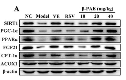
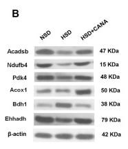
Oil Red O staining observation of liver (×200, scale bars = 100 μm). (B) H&E staining observation of liver (×200, scale bars = 50 μm). (C) The volume density of quantitation of hepatic steatosis (n = 5). (D) The genes expression involved in fatty acid β-oxidation in liver (n = 5). (E) The mRNA expression of genes involved in lipogenesis and FXR in liver (n = 5). (F) Bands of PPARα,PPARγ, ACOX1, FAS, SREBP-1c, SCD1.

9). MiR-103-3p promotes hepatic steatosis to aggravate nonalcoholic fatty liver disease by targeting of ACOX1. MOLECULAR BIOLOGY REPORTS, 2022 (PubMed: 35606603) [IF=2.6]

Application: WB    Species: Mice    Sample: liver tissue

Fig. 3 Antagomir-103-3p alleviated the damage to mice with NAFLD. Mice with NAFLD were fed an HFD for 8 weeks, and Antagomir-NC or Antagomir-103-3p was used for tail vein injection once a week for 2 weeks. A MiR-103-3p expression in mouse liver tissues was examined by qRT-PCR. B Oil Red O staining detected lipid droplet accumulation in mouse liver tissues, and HE staining detected liver tissue lesions in mice. C The TG, ALT, AST and H2O2 contents in mouse serum were examined, while ROS generation and ATP content were examined in mouse tissues. D The protein and mRNA levels of ACOX1, FASN and ACSL1 were examined by western blotting and qRT-PCR, respectively. *P < 0.05 compared with the control group; #P < 0.05 compared with the NAFLD+Antagomir-NC group

10). Ficus hirta Vahl. Ameliorates Nonalcoholic Fatty Liver Disease through Regulating Lipid Metabolism and Gut Microbiota. Oxidative Medicine and Cellular Longevity, 2022 (PubMed: 35592528)

Application: WB    Species: Mouse    Sample: HepG2 cells

Figure 2 Amelioration of lipid accumulation in PA-induced HepG2 cells by FV. (a) Oil red O was used to measure the level of lipid accumulation (magnification 100x, scale bar = 250 μm). (b) The oil red O-positive area was analyzed and quantified. (c–j) The relative mRNA expression levels of FABP1, SCD1, CD36, HMGCR, ACACA, CCL5, IL-1β, and TNF-α were determined by qRT-PCR. (k) The proinflammatory factor protein levels of IL-1β, IL-6, and TNF-α. (l) The protein levels related to lipid metabolism of SREBP-1, ACOX1, CD36, CPT1α, and HMGCR were analyzed by western blotting, and the relative ratios were calculated and expressed as the mean ± SD; n = 3. #P < 0.05 means that the difference between the NC group and the PA group is significant. ∗P < 0.05 means that the difference between the PA group and the FV (30 mg/mL) group or the FV (15 mg/mL) group is significant.

限制条款

产品的规格、报价、验证数据请以官网为准,官网链接:www.affbiotech.com | www.affbiotech.cn(简体中文)| www.affbiotech.jp(日本語)

产品的数据信息为Affinity所有,未经授权不得收集Affinity官网数据或资料用于商业用途,对抄袭产品数据的行为我们将保留诉诸法律的权利。

产品相关数据会因产品批次、产品检测情况随时调整,如您已订购该产品,请以订购时随货说明书为准,否则请以官网内容为准,官网内容有改动时恕不另行通知。

Affinity保证所销售产品均经过严格质量检测。如您购买的商品在规定时间内出现问题需要售后时,请您在Affinity官方渠道提交售后申请。

产品仅供科学研究使用。不用于诊断和治疗。 

产品未经授权不得转售。

Affinity Biosciences将不会对在使用我们的产品时可能发生的专利侵权或其他侵权行为负责。Affinity Biosciences, Affinity Biosciences标志和所有其他商标所有权归Affinity Biosciences LTD.