![Phospho-ULK1 (Ser757)[Ser758] Antibody - Western blot analysis of lysates from 293 cells, using Phospho-ULK1 (Ser757)[Ser758] Antibody.](http://img.affbiotech.cn/images/202210/af4387_62062_phospho_ulk1_ser757_ser758_antibody_1666340908.jpg "Western blot analysis of lysates from 293 cells, using Phospho-ULK1 (Ser757)[Ser758] Antibody. The lane on the left was treated with blocking peptide.")
![Phospho-ULK1 (Ser757)[Ser758] Antibody - Western blot analysis of lysates from RAW264.](http://img.affbiotech.cn/images/202202/af4387_56480_phospho_ulk1_ser757_ser758_antibody_1645777859.jpg "Western blot analysis of lysates from RAW264.7 cells(serum starvation treatment), using Phospho-ULK1 (Ser757)[Ser758] Antibody. The lane on the left was treated with blocking peptide.")
![Phospho-ULK1 (Ser757)[Ser758] Antibody - AF4387 at 1/100 staining Rat heart tissue by IHC-P.](http://img.affbiotech.cn/images/201911/thumb_img/af4387_phospho_ulk1_ser757_ser758_antibody_thumb_P_1574236991419.jpg "AF4387 at 1/100 staining Rat heart tissue by IHC-P. The sample was formaldehyde fixed and a heat mediated antigen retrieval step in citrate buffer was performed. The sample was then blocked and incubated with the primary antibody at 4°C overnight. An HRP conjugated anti-Rabbit antibody was used as the secondary antibody.")
![Phospho-ULK1 (Ser757)[Ser758] Antibody - AF4387 at 1/100 staining Rat skin tissue by IHC-P.](http://img.affbiotech.cn/images/201911/thumb_img/af4387_phospho_ulk1_ser757_ser758_antibody_thumb_P_1574236991407.jpg "AF4387 at 1/100 staining Rat skin tissue by IHC-P. The sample was formaldehyde fixed and a heat mediated antigen retrieval step in citrate buffer was performed. The sample was then blocked and incubated with the primary antibody at 4°C overnight. An HRP conjugated anti-Rabbit antibody was used as the secondary antibody.")
![Phospho-ULK1 (Ser757)[Ser758] Antibody - AF4387 at 1/100 staining Mouse heart tissue by IHC-P.](http://img.affbiotech.cn/images/201911/thumb_img/af4387_phospho_ulk1_ser757_ser758_antibody_thumb_P_1574236990493.jpg "AF4387 at 1/100 staining Mouse heart tissue by IHC-P. The sample was formaldehyde fixed and a heat mediated antigen retrieval step in citrate buffer was performed. The sample was then blocked and incubated with the primary antibody at 4°C overnight. An HRP conjugated anti-Rabbit antibody was used as the secondary antibody.")
![Phospho-ULK1 (Ser757)[Ser758] Antibody - AF4387 at 1/100 staining Mouse liver tissue by IHC-P.](http://img.affbiotech.cn/images/201911/thumb_img/af4387_phospho_ulk1_ser757_ser758_antibody_thumb_P_1574236991535.jpg "AF4387 at 1/100 staining Mouse liver tissue by IHC-P. The sample was formaldehyde fixed and a heat mediated antigen retrieval step in citrate buffer was performed. The sample was then blocked and incubated with the primary antibody at 4°C overnight. An HRP conjugated anti-Rabbit antibody was used as the secondary antibody.")
![Phospho-ULK1 (Ser757)[Ser758] Antibody - AF4387 at 1/100 staining Mouse lung tissue by IHC-P.](http://img.affbiotech.cn/images/201911/thumb_img/af4387_phospho_ulk1_ser757_ser758_antibody_thumb_P_1574236990514.jpg "AF4387 at 1/100 staining Mouse lung tissue by IHC-P. The sample was formaldehyde fixed and a heat mediated antigen retrieval step in citrate buffer was performed. The sample was then blocked and incubated with the primary antibody at 4°C overnight. An HRP conjugated anti-Rabbit antibody was used as the secondary antibody.")
![Phospho-ULK1 (Ser757)[Ser758] Antibody - AF4387 at 1/100 staining Mouse kidney tissue by IHC-P.](http://img.affbiotech.cn/images/201911/thumb_img/af4387_phospho_ulk1_ser757_ser758_antibody_thumb_P_1574236990899.jpg "AF4387 at 1/100 staining Mouse kidney tissue by IHC-P. The sample was formaldehyde fixed and a heat mediated antigen retrieval step in citrate buffer was performed. The sample was then blocked and incubated with the primary antibody at 4°C overnight. An HRP conjugated anti-Rabbit antibody was used as the secondary antibody.")
![Phospho-ULK1 (Ser757)[Ser758] Antibody - peptide-ELISA analysis of AF4387.](http://img.affbiotech.cn/images/pelisa/809/af4387-peptide-elisa.png "peptide-ELISA analysis of AF4387. showing specificity to antigen peptide. Peptides concentration: 1ug/ml.<br>
P-peptide: phospho-peptide. N-peptide: non-phospho-peptide.")
![Phospho-ULK1 (Ser757)[Ser758] Antibody - Figure 5.](http://img.affbiotech.cn/images/cited_image/202112/cite-wx-303-1640949481.jpg "Figure 5.| Autophagy level of the corpus cavernosum (control, n ¼ 6; DMED, n ¼ 6; DMED þ rapamycin, n ¼ 6). A, Representative photomicrographs of LC3B using immunofluorescence (magnification 100). B, Representative Western blot for ULK1, p-ULK1(Ser757),p-ULK1(Ser555), Beclin1, P62, and LC3B (I, II).")
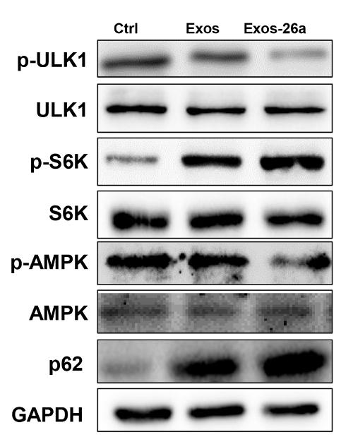
![Phospho-ULK1 (Ser757)[Ser758] Antibody - Fig.](http://img.affbiotech.cn/uploads/202406/e82e5c6c4fb66dcee8636e4f63862cdb.png "Fig. 4. PF, FA, and ATL in combination ameliorated LPS-induced inflammation in BV2 microglia cells by promoting autophagy. (A–B) Quantitative real-time PCR was used to investigate whether the regulation of IL-1β and IL-6 mRNA by PF, FA, and ATL in combination was affected by the autophagy inhibitor Wor. (C) Quantitative real-time PCR was used to detect the expression of Beclin1 mRNA levels. (D–H) Expression of p-AMPK, p-ULK1, Beclin1, LC3-II, and p62 in BV2 cells were examined by Western blotting. (I) IL-6 expression was examined by Western blotting to determine whether Wor affects the anti-inflammatory effect of PF, FA and ATL in combination. (J–K) Immunocytochemistry analysis of LC3 and p62 in cells. LC3 Scale bar = 25 μm, p62 Scale bar = 40 μm. The date was expressed as means ± standard deviation (SD). The analysis was performed using one-way analysis of variance (One-way ANOVA) followed by LSD multiple comparisons.")
产品描述
*The optimal dilutions should be determined by the end user. For optimal experimental results, antibody reuse is not recommended.
*Tips:
WB: 适用于变性蛋白样本的免疫印迹检测. IHC: 适用于组织样本的石蜡(IHC-p)或冰冻(IHC-f)切片样本的免疫组化/荧光检测. IF/ICC: 适用于细胞样本的荧光检测. ELISA(peptide): 适用于抗原肽的ELISA检测.
引用格式: Affinity Biosciences Cat# AF4387, RRID:AB_2844452.
展开/折叠
ATG 1; ATG1; ATG1 autophagy related 1 homolog; ATG1A; Autophagy related protein 1 homolog; Autophagy-related protein 1 homolog; FLJ38455; FLJ46475; hATG1; KIAA0722; Serine/threonine protein kinase ULK1; Serine/threonine protein kinase Unc51.1; Serine/threonine-protein kinase ULK1; ULK 1; ULK1; ULK1_HUMAN; Unc 51 (C. elegans) like kinase 1; UNC 51; Unc 51 like kinase 1; Unc-51 like kinase 1 (C. elegans); Unc-51-like kinase 1; UNC51; UNC51, C. elegans, homolog of; Unc51.1;
抗原和靶标
A synthesized peptide derived from mouse ULK1 around the phosphorylation site of Ser757. This site is equivalent to Ser758 in the human ULK1 sequence.
研究领域
· Cellular Processes > Transport and catabolism > Autophagy - animal. (View pathway)
· Environmental Information Processing > Signal transduction > mTOR signaling pathway. (View pathway)
· Environmental Information Processing > Signal transduction > AMPK signaling pathway. (View pathway)
· Organismal Systems > Aging > Longevity regulating pathway. (View pathway)
文献引用

Application: WB Species: Mouse Sample:
Application: WB Species: rat Sample:

Application: WB Species: Rat Sample:
Application: WB Species: Mice Sample:

Application: WB Species: Human Sample: MDA-MB-468 cells

Application: WB Species: bovine Sample: MAC-T cells
限制条款
产品的规格、报价、验证数据请以官网为准,官网链接:www.affbiotech.com | www.affbiotech.cn(简体中文)| www.affbiotech.jp(日本語)产品的数据信息为Affinity所有,未经授权不得收集Affinity官网数据或资料用于商业用途,对抄袭产品数据的行为我们将保留诉诸法律的权利。
产品相关数据会因产品批次、产品检测情况随时调整,如您已订购该产品,请以订购时随货说明书为准,否则请以官网内容为准,官网内容有改动时恕不另行通知。
Affinity保证所销售产品均经过严格质量检测。如您购买的商品在规定时间内出现问题需要售后时,请您在Affinity官方渠道提交售后申请。产品仅供科学研究使用。不用于诊断和治疗。
产品未经授权不得转售。
Affinity Biosciences将不会对在使用我们的产品时可能发生的专利侵权或其他侵权行为负责。Affinity Biosciences, Affinity Biosciences标志和所有其他商标所有权归Affinity Biosciences LTD.