产品: 磷酸化 TAZ (Ser89) 抗体
货号: AF4315
描述: Rabbit polyclonal antibody to Phospho-TAZ (Ser89)
应用: WB
文献验证: WB
反应: Human, Mouse, Rat
预测: Pig, Zebrafish, Bovine, Horse, Sheep, Rabbit, Dog, Chicken, Xenopus
蛋白号: Q9GZV5
RRID: AB_2844394

浏览相似产品>>

   规格 价格 库存
 100ul RMB¥ 2800 现货
 200ul RMB¥ 3800 现货

货期: 当天发货

联系销售

产品描述

来源:
Rabbit
应用:
WB 1:500-1:2000
*The optimal dilutions should be determined by the end user.
*Tips:

WB: 适用于变性蛋白样本的免疫印迹检测. IHC: 适用于组织样本的石蜡(IHC-p)或冰冻(IHC-f)切片样本的免疫组化/荧光检测. IF/ICC: 适用于细胞样本的荧光检测. ELISA(peptide): 适用于抗原肽的ELISA检测.

反应:
Human, Mouse, Rat
克隆:
Polyclonal
特异性:
Phospho-TAZ (Ser89) Antibody detects endogenous levels of TAZ only when phosphorylated at Ser89.
RRID:
AB_2844394
引用格式: Affinity Biosciences Cat# AF4315, RRID:AB_2844394.
偶联:
Unconjugated.
纯化:
The antibody is from purified rabbit serum by affinity purification via sequential chromatography on phospho-peptide and non-phospho-peptide affinity columns.
保存:
Rabbit IgG in phosphate buffered saline , pH 7.4, 150mM NaCl, 0.02% sodium azide and 50% glycerol. Store at -20 °C. Stable for 12 months from date of receipt.
别名:

展开/折叠

DKFZP586I1419; FLJ27004; FLJ45718; OTTHUMP00000215994; OTTHUMP00000215995; OTTHUMP00000215996; OTTHUMP00000216001; TAZ; Transcriptional co activator with PDZ binding motif; Transcriptional coactivator with PDZ binding motif; Transcriptional coactivator with PDZ-binding motif; WW domain containing transcription regulator 1; WW domain containing transcription regulator protein 1; WW domain-containing transcription regulator protein 1; WWTR 1; WWTR1; WWTR1_HUMAN;

抗原和靶标

免疫原:

A synthesized peptide derived from human TAZ around the phosphorylation site of Ser89.

基因/基因ID:

研究领域

· Environmental Information Processing > Signal transduction > Hippo signaling pathway.   (View pathway)

· Environmental Information Processing > Signal transduction > Hippo signaling pathway - multiple species.   (View pathway)

文献引用

1). An H2 S-BMP6 Dual-Loading System with Regulating Yap/Taz and Jun Pathway for Synergistic Critical Limb Ischemia Salvaging Therapy. Advanced healthcare materials, 2023 (PubMed: 37531238) [IF=10.0]

2). Identification of Gαi3 as a promising molecular oncotarget of pancreatic cancer. Cell death & disease, 2024 (PubMed: 39349432) [IF=8.1]

Application: WB    Species: Mouse    Sample: pancreatic cancer cells

Fig. 7: Gαi3 regulates PKA-Hippo-YAP signaling axis in pancreatic cancer cells. The primary priPC-1 cells stably expressing the Gαi3 shRNA (“sh-Gαi3-s1”), the CRISPR/Cas9-Gαi3-KO construct (“ko-Gαi3-s1”), the Gαi3-expressing lentiviral construct (“OE-Gαi3”), alongside their respective controls (“shC”, “Cas9-C” or “Vec”) were established, and the expression of listed proteins were tested (A–D). Primary priPC-1 cells stably expressing the Gαi3 shRNA (“sh-Gαi3-s1”), as well as the corresponding control (“shC”) underwent cytoplasmic/nuclear protein extraction, and listed proteins in the described cell fraction lysates were measured (E, F), cells were also subjected to immunofluorescence (IF) staining, with representative images presented (G). The priPC-1 cells expressingsh-Gαi3-s1 were treated with H-89 (a PKA inhibitor, 7 μM) or 0.1% DMSO as vehicle control (“Veh”), and expression of listed proteins was shown (H–L). After specified cultivation periods, cell proliferation and migration were examined, with results quantified (I, J). The priPC-1 cells with“sh-Gαi3-s1” were engineered to express either a constitutively active Akt1 mutant (caAkt1, S473D) or a control vector (“Vec”), expression of the described proteins was assessed by Western blotting (K). The designations “Cyt” and “Nuc” refer to cytoplasmic and nuclear proteins, respectively. *P 

3). Pan-cancer analysis predict that FAT1 is a therapeutic target and immunotherapy biomarker for multiple cancer types including non-small cell lung cancer. Frontiers in immunology, 2024 (PubMed: 38855103) [IF=5.7]

Application: WB    Species: Mouse    Sample: lung cancer cells

Figure 8 FAT1 knockdown effects in lung cancer cells. (A) Cell cycle assay showed that FAT1 knockdown induced G0/G1 phase arrest. (B) Western blot results verifying the effect of knockdown FAT1 on the FAK–YAP/TAZ signaling pathway. (C) Immunohistochemical validation of the effect of FAT1 knockdown on the FAK-YAP/TAZ signaling pathway. (D, E) Pictures of dissected mouse tumors and knocking down FAT1 inhibits tumor growth in mice. **P < 0.01, ****P < 0.0001.

4). Dapagliflozin delays renal fibrosis in diabetic kidney disease by inhibiting YAP/TAZ activation. Life sciences, 2023 (PubMed: 37023953) [IF=5.2]

5). Differentiation-inducing factor-1 inhibits EMT by proteasomal degradation of TAZ and YAP in cervical and colon cancer cell lines. European journal of pharmacology, 2025 (PubMed: 39645220) [IF=5.0]

6). Knockdown of HSP110 attenuates hypoxia-induced pulmonary hypertension in mice through suppression of YAP/TAZ-TEAD4 pathway. RESPIRATORY RESEARCH, 2022 (PubMed: 35986277) [IF=4.7]

Application: WB    Species: Mouse    Sample: lung tissues

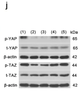
Fig. 3Knockdown of HSP110 inhibits hypoxia-induced autophagy and YAP/TAZ-TEAD4 activity in mice. Relative mRNA level (a) and protein level (b, c) of HSP110 pulmonary arteries in lung tissues in each group (N = 8). d Double immunofluorescence staining of α-SMA (green) and HSP110 (red) in pulmonary arteries (N = 8). White scale bars, 50 μm; Yellow scale bars, 25 μm. White arrows pointed to α-SMA and HSP110 double-positive cells. e Protein levels of LC3II, LC3I, Beclin1, ATG5, ATG7 and p62 in pulmonary arteries (N = 8). f–h Quantitative analysis of relative protein ratio of LC3-II/I and relative protein level of Beclin1, p62, ATG5 and ATG7 (N = 8). i Double immunofluorescence staining of α-SMA (green) and Beclin 1 (red) in pulmonary arteries (N = 8). Scale bars, 50 μm. j Protein levels of p-YAP, YAP, p-TAZ and TAZ in pulmonary arteries (N = 8). k Quantitative analysis of relative protein ratio of p-YAP/t-YAP and p-TAZ/t-TAZ (N = 8). l–n Nuclear protein levels of YAP, TAZ and TEAD4 and quantitative analysis of relative protein level of nuclear YAP, TAZ and TEAD4 (N = 8). o Double immunofluorescence staining of α-SMA (green) and YAP (red) in pulmonary arteries (N = 8). White scale bars, 50 μm; Yellow scale bars, 25 μm. White arrows pointed to α-SMA and YAP double-positive cells. Data are means ± SD from 8 mice per group. *p 

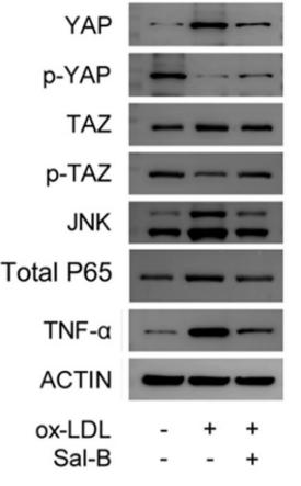
7). Salvianolic acid B ameliorates atherosclerosis via inhibiting YAP/TAZ/JNK signaling pathway in endothelial cells and pericytes. BIOCHIMICA ET BIOPHYSICA ACTA-MOLECULAR AND CELL BIOLOGY OF LIPIDS, 2020 (PubMed: 32739616) [IF=3.9]

Application: WB    Species: mouse    Sample: ECs

Fig. 5.| Influence of Sal-B on inflammatory response during YAP/TAZ/JNK signaling pathway in ECs. (A) Expression levels of YAP, TAZ, JNK, NF-κB and TNF-α were monitored by RT-PCR (n = 3). (B–C, F) Proteins (YAP, p-YAP, TAZ, p-TAZ, JNK, Nuclear NF-κB P65, Total P65 and TNF-α) in the pathway were detected by western blot (n = 3).

8). Endometriosis-derived exosomes encapsulated miR-196a-5p mediate macrophage polarization through regulation of the Hippo pathway. Journal of cell communication and signaling, 2025 (PubMed: 40416727) [IF=3.6]

Application: WB    Species: human    Sample:

FIGURE 6 The effect of the Hippo pathway on macrophage polarization. To investigate whether EMs exosomes@miR-196a-5p affects macrophage polarization through the Hippo pathway, we treated macrophages with EMs exosomes@miR-196a-5p in combination with the Hippo pathway inhibitor XMU-MP-1. (A) RT-qPCR was used to detect macrophage polarization markers (iNOS and CD86 for M1 markers; CD206 and CD163 for M2 markers). (B) Western blot was used to detect the protein expression levels of macrophage polarization markers (iNOS and CD86 for M1 markers; CD206 and CD163 for M2 markers). (C) The proportion of CD86+ (M1) and CD206+ (M2) macrophages was detected using a flow cytometer. (D) Western blot was used to detect Hippo pathway-related proteins (MST1, p-YAP1, YAP1, p-TAZ, and TAZ). Data represent mean ± SD from three independent experiments. XMU-MP-1: Hippo inhibitor. *: P < 0.05, **: P < 0.01, compared with EMs exosomes@miR-196a-5p.

9). Gallic acid from Terminalia chebula inhibited the growth of esophageal carcinoma cells by suppressing the Hippo signal pathway. Iranian Journal of Basic Medical Sciences, 2020 (PubMed: 33235697) [IF=2.1]

限制条款

产品的规格、报价、验证数据请以官网为准,官网链接:www.affbiotech.com | www.affbiotech.cn(简体中文)| www.affbiotech.jp(日本語)

产品的数据信息为Affinity所有,未经授权不得收集Affinity官网数据或资料用于商业用途,对抄袭产品数据的行为我们将保留诉诸法律的权利。

产品相关数据会因产品批次、产品检测情况随时调整,如您已订购该产品,请以订购时随货说明书为准,否则请以官网内容为准,官网内容有改动时恕不另行通知。

Affinity保证所销售产品均经过严格质量检测。如您购买的商品在规定时间内出现问题需要售后时,请您在Affinity官方渠道提交售后申请。

产品仅供科学研究使用。不用于诊断和治疗。 

产品未经授权不得转售。

Affinity Biosciences将不会对在使用我们的产品时可能发生的专利侵权或其他侵权行为负责。Affinity Biosciences, Affinity Biosciences标志和所有其他商标所有权归Affinity Biosciences LTD.