


 By human cancer pathway PCR array, ectopic expression of BCL2L10 up- or down-regulated several genes related to tumor proliferation, apoptosis, metastasis and angiogenesis. (B) Western blot was performed to confirm the downstream gene expression regulated by BCL2L10 in HepG2 cells. GAPDH was used as an internal control. (C) Schematic diagram of the molecular events for BCL2L10 function as a tumor suppressor through regulating cell cycle, proliferation, apoptosis metastasis and angiogenesis effectors.")


 Compound P-3 down-regulated the levels of IL-6 and TNF-α in RAW 264.7. The cell
viability of RAW 264.7 is higher than 80% under the treatment of compound P-3 at indicated
concentrations. (B) Compound P-3 suppressed the expression of STAT3, phosphorylated STAT3, and
Bcl-2 in both HCT-116 and MGC-803 cells. (C) Compound P-3 inhibited the nuclear translocation and
the total amount of STAT3 in both HCT-116 and MGC-803 cells. Data are presented as mean ± SD from
three independent experiments. * P < 0.05, ** P<0.01, *** P<0.001 versus the control group.")
 Representative images of Western blot. (B–I) quantification of the Western blot data by densitometric analysis and normalization to GAPDH (n = 3 independent experiments). **P < 0.01 vs model group.")

 The protein expressions along with quantification of FAK, P-FAK, SRC, P-SRC, STAT3, P-STAT3, Bcl-2, c-Myc, and MMP9 in the FAK-SRC-STAT pathway. (B–D) The relative mRNA expressions of Bcl-2, c-Myc and MMP9. (E) Analysis of MMP9 secretion and its quantification in SCC-9 cells. (F, G) Immunohistochemical staining and analysis of the expression of cyclin G2, p-FAK, p-SRC and p-STAT3. Scale bar = 100 µm. *p < 0.05, **p < 0.01, ***p < 0.001 vs. vector.")

, sgp130 (20 μg/d), and Dexamethasone (20 μg/d).")
 on April 8, 2020.10Gy-irradiation and suppressed secretion after adding 0.8μM JAK1 inhibitor following irradiation. D-G: Representative images and quantitative analysis of ALP activity and mineralized nodule area of osteoblasts co-cultured with different types of CM from irradiated BMSCs (Con CM, Con/JAKi CM, IRIS CM and IRIS/JAKi CM).Magnification, ×100. H-I: Relative mRNA and protein expression levels of ALP and OC of osteoblasts co-cultured with four types of CM from irradiated BMSCs. All data were analyzed from three independent experiments. P values were calculated by Student’s t-test and one-way ANOVA analysis. Results are presented as mean ± SD.*p < 0.01, **p < 0.01, ***p < 0.001. ALP: alkaline phosphatase; OC: osteocalcin; Con CM: CM from control BMSCs; Con/JAKi CM: CM from control BMSCs with JAKi intervention; IRIS CM: CM from irradiation-induced senescent BMSCs; IRIS/JAKi CM: CM from irradiation-induced senescent BMSCs with JAKi intervention. SD:standard deviation. Scale bar: 100μm (D, F).")
 Overexpress USP5 in Panc1 cells activate STAT3 signaling. (B) Knockdown USP5 in AsPC1 cells inhibit STAT3 signaling. Data were quantified by densitometric analysis with ImageJ software. *P < 0.05; **P < 0.01; ***P < 0.001.")
, STAT3 (B). Values are expressed as means ± SD.
*p < 0.05 versus VEH.
#p < 0.05 versus CORT.
CORT, corticosterone group; CORT+EM, CORT + empagliflozin group; STAT3, Signal transducer and activator of transcription 3; T-STAT3, total STAT3; P-STAT3, phosphorylated STAT3; GAPDH, glyceraldehyde 3-phosphate dehydrogenase; SD, standard deviation; TLR4, Toll-like receptor 4; VEH, vehicle group.")
 The protein levels of STAT3, phospho-STAT3 and PARP-1 were measured by western blot analysis. GAPDH was used as an internal control for grayscale analyses. (B and C) The mRNA levels of IL-6 and TGF-β1 were measured by RT-qPCR. The results are presented as the means ± SD (n=10). *P<0.05 vs. control; #P<0.05 vs. DSS group.5-AIQ, 5-aminoisoquinolinone; DSS, dextran sodium sulfate.")


 or hypoxia (H4W, n = 8) for 4 weeks with rmIL-17 (H4W + rmIL-17, n = 6) or not. Scale bars: 50 μm. B Bar chart for cardiomyocytes diameter in the right ventricle of each group. ****p < 0.0001. C Immunofluorescent staining of Bax and caspase-3 of representative histological sections from paraffin-embedded mice right ventricular tissues of each group. Blue for DAPI, red for Bax and caspase-3. Scale bar = 50 μm. D Western blot analysis of p-STAT3 in right ventricular tissue of wide type mice and IL-17 knockout mice (n = 6) and the statistical bar graph. **p = 0.0063")
 inhibitory concentration; p-STAT3, phosopho-STAT3Tyr705")
 The original bands of BDNF, JAK2, p‐JAK2, STAT3, p‐STAT2, TERT and β‐actin. β‐actin was considered as loading controls. (b) The ratio of different proteins to β‐actin was calculated by the band density. *p < .05, **p < .01 vs control group; # p < .05 and ## p < .01 vs D‐gal group")
 miR-214 mRNA levels among colonic were detected by real-time RT-PCR. (B–C) Expressions of STAT3 and p–STAT3 among colonic proteins were examined by Western blot and quantitative data was presented. (D) Immunohistochemistry staining
for p-STAT3 in colon tissue. Arrows denote positive expression. Scale bar: 50 μm. (E) The statistics of positive area expression in p-STAT3. (F) PTEN mRNA levels
among colonic were detected by real-time RT-PCR. (G-H) Expressions and quantitative data of PTEN among colonic proteins were examined by Western blot. *p <
0.05, vs control groups; #p < 0.05, vs DSS groups, n = 6.")
 P53 and (B) P21. Western blotting was applied to evaluate the expression of Stat3 and p-Stat3. *, p<0.05 vs control group, * *, p<0.01 vs control group")

 Western blot and band intensity analysis for the t- and p-AKT, STAT3 and ERK1/2 in B7-H3 KO and mock A549 cells. (C-E) Western blot and band intensity analysis for t-AKT, p-AKT and SIRT1 expression in A549 cells treated with LY294002 (50 µΜ). GAPDH was used as the loading control, and relative phosphorylation level (p-/t-) was determined. Data are representative of three independent experiments. SIRT1, Sirtuin 1; t-, total; p-, phosphorylated; KO, knockout; ns, no significance; NC, negative control.")
 hirudin and 10 μM losartan for 24 h, respectively. (A) The expression of STAT3, MAPK1 and IL-6 were analyzed by qRT-PCR, n = 6. (B) The IL-6 in cell supernatant was assayed by ELISA, n = 5. (C) Western blot for the expression of proteins including STAT3, p-STAT3, MAPK1, p-MAPK1 and IL-6. (D) Quantified by western blot analysis and normalized to control, n = 3. Bars represent the mean ± SD, *p < 0.05, **p < 0.01.")
 IL-17A concentrations in serum samples. (B–C) Expression of IL-17RA protein in colon tissue samples (n = 4 per group). (D) IL-6 concentrations in serum samples (n = 6 per group). (E,F) Nuclear expression of NF-κB-p65 and p-STAT3 proteins in colon tissue samples (n = 4 per group). Results are expressed as mean ± SEM. # p < 0.05, ## p < 0.01, compared with normal mice; * p < 0.05, ** p < 0.01, compared with AOM/DSS-treated mice.")
 Expression of MMP9, p-STAT3, STAT3, Bcl-2, and Bax proteins in colon tissue (n = 4 per group). (C) Immunofluorescence staining of colon tissue, green: p-STAT3, red: MMP9, blue: DAPI. Values are expressed as mean ± SEM. # p < 0.05, ## p < 0.01, compared with normal mice; * p < 0.05, ** p < 0.01, compared with AOM/DSS-treated mice.")
 = 6, ×400). Immunohistochemistry was used to detect (a) NF-κB p65 and (b) p-STAT3 protein levels. The AOD was used as an index to evaluate protein levels. All error bars represent the mean ± standard deviation. ##P < 0.01, compared to the control group; ∗∗P < 0.01, compared to the AR group. SD: Saposhnikovia divaricata, AR: allergic rhinitis, AOD: average optical density, NF-κB: nuclear factor kappa-B, and p-STAT3: phospho-signal transducer and activator of transcription 3.")
, NF-κB (B) was significantly increased in WT and Mer-/- group at day 4 after CLP; however, the expression of phosphorylated STAT1 (p-STAT1) (C), SOCS1 (D), and SOCS3 (E) were significantly decreased at day 4 after CLP. Data are expressed as fold change compared to the Sham + WT group; n = 5 rats per group. *P < 0.05 compared to Sham + WT group,")
 The luciferase activity in the STAT3-SIE-luc-293 cell lysates treated with IL-6 (70 ng/mL) and rhTβ4. (B) The viability of rhTβ4-treated HEK-293 cells. (C) Western blot assay of JAK2, p-JAK2, STAT3 and p-STAT3 expression in rhTβ4-treated A549 cells. (D) Western blot assay of JAK2, p-JAK2, STAT3 and p-STAT3 expression in rhTβ4-treated Mlg cells. (E,F) Immunohistochemical staining analysis of p-JAK2 and p-STAT3 in mice. Scale bar = 50 μm. Data are shown as the mean ± SD.")
 The luciferase activity in the STAT3-SIE-luc-293 cell lysates treated with IL-6 (70 ng/mL) and rhTβ4. (B) The viability of rhTβ4-treated HEK-293 cells. (C) Western blot assay of JAK2, p-JAK2, STAT3 and p-STAT3 expression in rhTβ4-treated A549 cells. (D) Western blot assay of JAK2, p-JAK2, STAT3 and p-STAT3 expression in rhTβ4-treated Mlg cells. (E,F) Immunohistochemical staining analysis of p-JAK2 and p-STAT3 in mice. Scale bar = 50 μm. Data are shown as the mean ± SD.")
 and quantitative analysis (B, C). One-way ANOVA, [pJAK2] F(3, 8) = 6.753, p = 0.0154; [pSTAT3] F(3, 8) = 5.325, p = 0.0245, n = 3. D,E MaR1 inactivated GSK3β pathway in hippocampus shown by Western blotting (D) and quantitative analysis (E). One-way ANOVA, [pS9] F(3, 8) = 7.565, p = 0.0112, n = 3. F Predicted STAT3′ s binding motifs on the promoter of IL-6 were examined by dual luciferase reporter assay. Unpaired t-test, [Motif3] t = 3.546 df = 4, p = 0.0116, n = 3. * p < 0.05; # p < 0.05. Data were presented as mean ± SEM.")
 The colocalization of p-STAT3 (green) and NFAT2 (red) in control and LASP1-overexpressing SW620 cells was assessed by IF staining. The scale bar represents 50 mm. (B) Analysis of the correlation between NFAT2 and p-STAT3 in clinical tissues with high and low expression of NFAT2 by IHC staining. The right panel presents the percentage of patients with high or low expression of CRC tissue. Scale bar represents 50 mm. (C) Endogenous interaction between NFAT2 and p-STAT3 in control and LASP1-overexpressing SW620 cells. (D) Endogenous interaction between NFAT2 and p-STAT3 in SW620 cells after treatment with NIFE or BAY was detected by coIP assays. (E) The transcriptional regulation of the downstream genes COPS5, TWIST1, MMP2, and PD-L1 by NFAT2 and p-STAT3 was detected by ChIP assays. (F) The binding sites between NFAT2 and the downstream genes were confirmed by a dual-luciferase reporter system. (G) Left panel: the expression of NFAT2 and downstream genes in control and NIFE-treated SW620 cells were detected by qPCR. Right panel: the expression alterations in downstream genes in the indicated cells treated with NIFE or BAY were detected by WB. (H) The relationship between NFAT2 and downstream genes was detected from the GEO database. (I) IHC analysis was performed to detect the expression of NFAT2, TWIST1, COPS5, and MMP2 in human CRC tissues. Two representative cases are shown. Percentage of patients with high or low expression of CRC tissue on the right panel. Scale bar represents 50 mm. (G) After treating SW620 cells with CsA (10 mM), VIVIT (10 mM), and FK506 (10 ng/mL) and subsequently separating the nuclear and cytoplasmic proteins, WB analysis was used to detect the level of NFAT2 in the nucleus and p-NFAT2 in the cytoplasm. (H) Representative images of IHC staining analysis of NFAT2 expression in CRC tissues and adjacent nontumor tissues. The bar chart on the right represents the percentage of high and low NFAT2 expression cases in normal and CRC tissues. (I) IHC analysis of NFAT2 and p-NFAT2 expression in nonmetastatic and metastatic CRC tissues. The bar chart on the right represents the percentage of high and low NFAT2 or p-NFAT2 expression cases in nonmetastatic CRC and metastatic CRC tissues.")
 The colocalization of p-STAT3 (green) and NFAT2 (red) in control and LASP1-overexpressing SW620 cells was assessed by IF staining. The scale bar represents 50 mm. (B) Analysis of the correlation between NFAT2 and p-STAT3 in clinical tissues with high and low expression of NFAT2 by IHC staining. The right panel presents the percentage of patients with high or low expression of CRC tissue. Scale bar represents 50 mm. (C) Endogenous interaction between NFAT2 and p-STAT3 in control and LASP1-overexpressing SW620 cells. (D) Endogenous interaction between NFAT2 and p-STAT3 in SW620 cells after treatment with NIFE or BAY was detected by coIP assays. (E) The transcriptional regulation of the downstream genes COPS5, TWIST1, MMP2, and PD-L1 by NFAT2 and p-STAT3 was detected by ChIP assays. (F) The binding sites between NFAT2 and the downstream genes were confirmed by a dual-luciferase reporter system. (G) Left panel: the expression of NFAT2 and downstream genes in control and NIFE-treated SW620 cells were detected by qPCR. Right panel: the expression alterations in downstream genes in the indicated cells treated with NIFE or BAY were detected by WB. (H) The relationship between NFAT2 and downstream genes was detected from the GEO database. (I) IHC analysis was performed to detect the expression of NFAT2, TWIST1, COPS5, and MMP2 in human CRC tissues. Two representative cases are shown. Percentage of patients with high or low expression of CRC tissue on the right panel. Scale bar represents 50 mm. (G) After treating SW620 cells with CsA (10 mM), VIVIT (10 mM), and FK506 (10 ng/mL) and subsequently separating the nuclear and cytoplasmic proteins, WB analysis was used to detect the level of NFAT2 in the nucleus and p-NFAT2 in the cytoplasm. (H) Representative images of IHC staining analysis of NFAT2 expression in CRC tissues and adjacent nontumor tissues. The bar chart on the right represents the percentage of high and low NFAT2 expression cases in normal and CRC tissues. (I) IHC analysis of NFAT2 and p-NFAT2 expression in nonmetastatic and metastatic CRC tissues. The bar chart on the right represents the percentage of high and low NFAT2 or p-NFAT2 expression cases in nonmetastatic CRC and metastatic CRC tissues.")
 The colocalization of p-STAT3 (green) and NFAT2 (red) in control and LASP1-overexpressing SW620 cells was assessed by IF staining. The scale bar represents 50 mm. (B) Analysis of the correlation between NFAT2 and p-STAT3 in clinical tissues with high and low expression of NFAT2 by IHC staining. The right panel presents the percentage of patients with high or low expression of CRC tissue. Scale bar represents 50 mm. (C) Endogenous interaction between NFAT2 and p-STAT3 in control and LASP1-overexpressing SW620 cells. (D) Endogenous interaction between NFAT2 and p-STAT3 in SW620 cells after treatment with NIFE or BAY was detected by coIP assays. (E) The transcriptional regulation of the downstream genes COPS5, TWIST1, MMP2, and PD-L1 by NFAT2 and p-STAT3 was detected by ChIP assays. (F) The binding sites between NFAT2 and the downstream genes were confirmed by a dual-luciferase reporter system. (G) Left panel: the expression of NFAT2 and downstream genes in control and NIFE-treated SW620 cells were detected by qPCR. Right panel: the expression alterations in downstream genes in the indicated cells treated with NIFE or BAY were detected by WB. (H) The relationship between NFAT2 and downstream genes was detected from the GEO database. (I) IHC analysis was performed to detect the expression of NFAT2, TWIST1, COPS5, and MMP2 in human CRC tissues. Two representative cases are shown. Percentage of patients with high or low expression of CRC tissue on the right panel. Scale bar represents 50 mm. (G) After treating SW620 cells with CsA (10 mM), VIVIT (10 mM), and FK506 (10 ng/mL) and subsequently separating the nuclear and cytoplasmic proteins, WB analysis was used to detect the level of NFAT2 in the nucleus and p-NFAT2 in the cytoplasm. (H) Representative images of IHC staining analysis of NFAT2 expression in CRC tissues and adjacent nontumor tissues. The bar chart on the right represents the percentage of high and low NFAT2 expression cases in normal and CRC tissues. (I) IHC analysis of NFAT2 and p-NFAT2 expression in nonmetastatic and metastatic CRC tissues. The bar chart on the right represents the percentage of high and low NFAT2 or p-NFAT2 expression cases in nonmetastatic CRC and metastatic CRC tissues.")
 Protein level and phosphorylation status of STAT3 and Anxa2 were determined by immuno-blotting. (B) The activation of STAT3 was determined via reporter gene assay. (C) The expression of STAT3 target genes was determined by qRT-PCR. (D) The protein level of four VEGFs was determined by immuno-blotting. (E) The concentration of four VEGFs in cell-free supernatant was determined by ELISA. All experiments were performed with triple replicant.")
 Expression levels of PCSK9, LDLR, JAK2, p-STAT3, and SOCS3. β-actin served as a loading control. (b) The bar graph indicates the relative density calculated by Image J software. The results represent experiments performed in triplicate. *p < 0.05 and **p < 0.01.")
 assay. (a) Representative photos are shown (200×). Scale bar = 50 μm. (b) The bar graph indicates IHC staining scores. Data are presented as the mean ± standard error of the mean.")
 staining and immunohistochemical (IHC) staining of transplanted tumor tissue; F: Statistical graph of IHC staining results. All analyses were repeated three times. The data is represented as the mean ± SD. aP < 0.05; bP < 0.01. NC: Negative control; Vector: The blank vector for lentivirus transfection.")
 pathway- related markers in HCT-116 cells was detected by Western blot. We used β-actin as the internal control. All experiments have been performed in triplicate and data were expressed as mean ±SD. ▲p")

 Attenuation in p-JAK2, p-STAT3, p-JAK2/JAK2, p-STAT3/STAT3, CLDN6 and Bcl-2 expression and enhancement of Bax expression and cleaved caspase-3 levels via CLDN6 silencing (*, P")
 Immunofluorescence staining showing that p-STAT3 (red) co-localized with microglia (Iba1, green) and astrocytes (GFAP, green); (C–E) Expression levels of STAT3 and p-STAT3 in BV-2 cells were detected by Western blot assay. All data were derived from the results of three independent experiments. Values are mean ± SEM (n = 3 per group), ***P < 0.001, **P < 0.01, *P < 0.05 (one-way ANOVA followed by Tukey's post hoc test). DAPI: 4′,6-diamidino-2-phenylindole; GFAP: Glial fibrillary acidic protein; Iba1: Ionized calcium binding adapter molecule 1.")
 Immunofluorescence staining showing that p-STAT3 (red) co-localized with microglia (Iba1, green) and astrocytes (GFAP, green); (C–E) Expression levels of STAT3 and p-STAT3 in BV-2 cells were detected by Western blot assay. All data were derived from the results of three independent experiments. Values are mean ± SEM (n = 3 per group), ***P < 0.001, **P < 0.01, *P < 0.05 (one-way ANOVA followed by Tukey's post hoc test). DAPI: 4′,6-diamidino-2-phenylindole; GFAP: Glial fibrillary acidic protein; Iba1: Ionized calcium binding adapter molecule 1.")
 Western blot protein bands; (B-C) Relative protein levels. The data are presented as means ± SD (n = 3).")
 mRNA expression levels of inflammatory factors; (B,D) protein expression levels and phosphorylation of JAK, represented by grayscale values; (C,E) protein expression levels and phosphorylation of STAT3, represented by grayscale values. n = 8. * represents a significant difference, with p < 0.05. ** represents a significant difference, with p < 0.01.")
 The relative gene expression level of the IL6ST in 143B and U2OS cells. (B) The relative gene expression level of the JAK2 in 143B and U2OS cells. (C) The relative gene expression level of the STAT3 in 143B and U2OS cells. (D) The images of protein blots of 143B cells. (E) The images of protein blots of U2OS cells. (F) The relative expression level of diverse proteins (including IL6ST, JAK2, p-JAK2, STAT3, p-STAT3) in 143B cells. (G) The relative expression level of diverse proteins (including IL6ST, JAK2, p-JAK2, STAT3, p-STAT3) in U2OS cells.")
 Multiple immunofluorescence staining of intervertebral discs. (B) Comparison of the expression levels of serum IL-1β, IL-6, TNF-α, PEG2, IL-4, and IL-10. (C, D) The relative expression levels of p-JAK2/JAK2, p-STAT3/STAT3, iNOS/β-actin in the interdisc tissues of rats in each group. The unedited images are referenced in Fig. S2. *: P < 0.05, **: P < 0.01, ***: P < 0.001, ****: P < 0.0001, ns: P>0.05.")
 cells. (a, b) Bavachin induces GSH depletion in a dose-related manner over 24 h. (c, d) Bavachin greatly induces ROS generation in OS cells over 24 h. (e, f) Bavachin increases MDA accumulation in a dose-related manner. (g, h) Western blotting analysis shows the expression of p-STAT3, STAT3, P53, SLC7A11, and GPX4 in OS cells treated with bavachin (10 μM, 20 μM, and 40 μM) for 24 h. &p < 0.01 vs. the control group, #p < 0.001 vs. the control group, ∗p < 0.05, ∗∗p < 0.01, and ∗∗∗p < 0.001.")
, JAK2, PD-L1, and SOCS3 between parental (CT26) and acquired radioresistance (CT26-R) cells. *p")
-induced Janus kinase 2/signal transducer and activator of transcription 3 (JAK2/STAT3) pathway. (a-c) Western blot was used to detect protein expression of JAK2 and STAT3 phosphorylation. Immunohistochemistry was used to detect the expression of JAK2 (d) and STAT3 (e) phosphorylation. ×200, #P < 0.05 (vs. sham group), *P < 0.05 (vs. CLP group). CLP: Cecal ligation and puncture, SFI: Shenfu injection, GAPDH: Glyceraldehyde-3-phosphate dehydrogenase, JAK2/STAT3: Janus kinase 2/signal transducer and activator of transcription 3")
 Western blot analysis on ferroptosis pathway-related protein changes in HCC38 and HCC1937 cells. Graphs are representatives of three independent experiments. The densitometric analysis of SLC7A11 (C,H), GPX4 (D,I), Keap1 (E,J), Nrf2 (G,K) and HO-1 (F,L) in HCC38 cells. For comparing control and CHRNA9 overexpression groups, 25 μM erastin pre-treatment was applied to induce ferroptosis conditions in HCC38 cells. * p < 0.05; ** p < 0.01; *** p < 0.001. (original western blot images see Supplementary Materials)")
 Protein levels of SLC7A11 and GPX4 were detected by Western blot (n = 3). (b) The contents of MDA and GSH were evaluated using commercial kits (n = 3). (c) A tissue iron assay kit was used to determine the iron content (n = 3). (d) Protein levels of p-STAT3 and NLRP3 were detected by Western blot (n = 3).")
 Flow cytometry assay was used for CD4+ T-cell identification, n = 3. (B) Flow cytometry was used for Th17 (CD4+ IL-17+ cells) identification and CD4+ IL-17+ cells proportion analysis, n = 3. (D-F) The contents of (D) IL-6, (E) IL-17, and (F) IL-23 were detected using ELISA. n = 6. (G-I) The relative expressions of (G) STAT3, (H) p-STAT3, and (I) IL-17 were measured using Western blot. n = 3. (J) Representative image of protein bands. n = 3. The “n” means number of experimental replicates. Mean ± standard deviation, one-way analysis of variance was used for statistical analysis. **p")
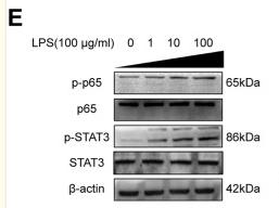
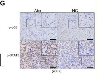
 The protein expressions of p65 and STAT3 (n = 3). (D) The mRNA expressions of FGL1 (n = 6). (E, F) The protein expressions of FGL1 (n = 3). *p < 0.05, **p < 0.01, ***p < 0.001, ****p < 0.0001.")
 Pearson correlation analysis showed the correlation heat map between samples. n = 3. (B) RNA-seq was then performed and differentially expressed genes were subjected to cluster analysis. (C) Differential gene volcano map. (D) GSEA enrichment score curve. (E) Western blot detection of PI3K/Akt and JAK/STAT3 signal pathway-related protein expression. n = 3.")
 Uptake of Dio/siICAM-1@MSN@PDA-CD11b by neutrophils and cardiomyocytes at different time points was analyzed by CLSM (A) and flow cytometry (B). Scale bar: 10 μm. (C) The mRNA expression levels of ICAM-1 in neutrophils under different treatment groups. (D) The protein expression of NF-κB/STAT3/p38-MAPK pathway in neutrophils under different treatment groups. ∗P < 0.05, ∗∗P < 0.01. N = 3 for each experiment.")
 Protein expression levels of hepatic KLF15, total STAT3, and p-STAT3 (n = 3). (B) Hepatic IL-6 levels measured by ELISA (n = 5). (C) Schematic diagram illustrating the proposed mechanism by which YGYZ modulated bile acid metabolism and improved the immune microenvironment through gut microbiota and KLF15 regulation. Data are presented as mean ± SD. Statistical analysis was performed using one-way ANOVA and Tukey’s multiple comparisons test. *p")
 and spleen index (B) of HSF-treated tumor-bearing C57BL/6 mice (n=8). (C) Histological analysis for tumor tissues by H&E staining (n=8). (D) Effect of HSF on the levels of IFN-γ, IL-2, IL-10, and IL-4 cytokine in the serum detected by ELISA (n=8). (E) Effect of HSF on the mRNA expression levels of IFN-γ, IL-2, IL-4, IL-10, and IL-10R detected by qRT-PCR assay (n=8). (F) Effect of HSF on the protein expressions of IL-10R, p-STAT3, STAT3, p-STAT1, and STAT1 in tumor tissues detected by Western blot. Data were showed as each dot for one animal and the exact p shown on plot.")
 The protein expression of α7nAchR, CD68, iNOS, p-JAK2, and p-STAT in M1/M2 cells of each group was detected by Western blot. (A-G) M1 macrophages. (H-N) M2 macrophages. * indicates P < 0.05 compared with M1/M2 group; # indicates P < 0.05 compared with M1/M2+si-NC+FMN group.")

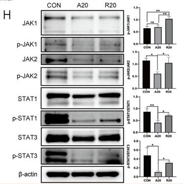
. The Western blot bands of MSGA regulates JAK-STAT signaling pathway, n = 3. (B-E). Grayscale analysis of p-JAK1/JAK1, p-JAK3/JAK3, p-STAT1/STAT1, and p-STAT3/STAT3 expression. Immunofluorescence (IF) images of STAT1 (F) and STAT3 (G) were photoed by laser scanning confocal microscope (Scale bar, 20 μm; Scale bar in representative images, 100 nm), n = 3. MSGA-L (100 μg/mL); MSGA-H (400 μg/mL). One-way ANOVA with Tukey’s post hoc test was used for the statistical analyses. Compared with M0 macrophages, statistically significant differences are indicated.")
. C Expression of TNF-α in different treatment groups by ELISA (n = 5). D The expressions of IL-22, IFN-γ, IL-1β and p-STAT3 proteins in different treatment groups were detected by Western blot. Quantitative data are presented as mean ± standard deviation (n = 5). Statistical significance is indicated as follows: ****P")
; Compared to the sham surgery group:#P < 0.05, ##P < 0.01; Compared with the model group∗ P < 0.05, ∗∗P < 0.01.")
 Western blot experiments confirmed the activation of the CXCR2-mediated JAK–STAT signaling pathway around the haematoma (n = 6). (C, D) Western blot experiments detected the effects of Vinorine on the expression of JAK–STAT signaling pathway-related proteins in the perihaematoma region. (E) Western blot experiments detected the effects of Vinorine on the expression of MMPs in the perihaematoma region. (F) Western blot experiments detected the effects of Vinorine on the expression of apoptosis-related proteins such as BCL-2, Cleaved-Caspase 3, and Cleaved-Caspase 9 in the perihaematoma region. (G) Western blot experiments were conducted to assess the effects of Vinorine on the expression of blood–brain barrier proteins ZO-1 and CLDN5. (H) Western blot experiments were conducted to assess the effects of Vinorine on the expression of neuronal axonal proteins NF200 and PSD95. All data were expressed as mean ± standard deviation (SD). Statistical significance was determined by two-way analysis of variance (ANOVA) and Tukey's multiple comparisons test, *p")
 Western blot images of p-SMAD2, SMAD2, p-STAT3, STAT3, and GAPDH expression in response to vehicle, QBY, S1P, and W146 treatments. (B–G) Densitometric analysis of the corresponding band intensity ratios: (B) p-SMAD2/GAPDH, (C) p-SMAD2/SMAD2, (D) SMAD2/GAPDH, (E) p-STAT3/GAPDH, (F) p-STAT3/STAT3, and (G) STAT3/GAPDH. Data are expressed as mean ± SD (n=3). *P")
 Overexpressing and knocking down tRF-23 respectively led to higher and lower levels of p-JAK2/STAT3. JAK2/STAT3 inhibitor treatment also significantly reduced levels of p-JAK2/STAT3 relative to tRF-23 overexpression in the absence of such inhibition (p-JAK2, normalized to total JAK2; p-STAT3, normalized to total STAT3). Data are presented as the mean ± SD (n = 3 independent experiments with two technical replicates per independent sample). (C) JAK2/STAT3 signaling inhibitor treatment suppressed the osteogenic differentiation of hBMSCs on day 14 relative to those in which tRF-23 was overexpressed without inhibitor treatment. Scale bars: 50 μm. Matrix mineralization and ALP activity were quantified based on absorbance. Data are presented as the mean ± SD (n = 3 independent experiments with two technical replicates per independent sample). (D and E) RUNX2, OCN, and ALP levels on day 14 of hBMSC osteogenesis were detected via qPCR and western blotting. Data are presented as the mean ± SD (n = 3 independent experiments with two technical replicates per independent sample). Data were analyzed using paired two-tailed Student’s t tests. ∗∗p < 0.01 was considered significant. OE, overexpression; shRNA, short hairpin RNA; T-JAK2, total JAK2; T-STAT3, total STAT3.")
 Western blot analysis of key proteins. (G) Immunofluorescence analysis. (H) Multiplex IHC showing the expression levels of target molecules in OS model mice. *p")
Heat map of differentially expressed mRNA. (B) Enrichment entries of DEGs GO analysis. (C) Enrichment entries of DEGs KEGG pathway analysis(top30). (D) qPCR validation of transcriptome sequence data. (E, F) Effects of different concentrations of CMSP(6, 8, 10)μg/mL on expression levels of JAK2/STAT3/c-Myc signal axis related proteins in CAL27 and SCC15 cells treated for 48h. β-Actin to control the load. n=3, compared with the control group")
 Heatmap of the top 30 genes in the DEGs of RELT (C) Clustered module maps of DEGs or WGCNA analysis: (D) Dendrograms of sample traits with scores for WGCNA analysis:(E) Screening of thresholds for WGCNA module analysis .F Correlation and P-value of genes with scores for each module of WGCNA analysis: (G) Scatter plots demonstrating the gene modules with strong correlation: (H) PPL network maps of pivotal genes: W Pivotal genes with GO and KEGG enrichment analyses: O. K Western blot validation of the JAK/STAT pathway. () LASSO coefficients pathway plot and cross-validation curve: . K Wester blot validation of the JAK2/STAT3 pathway in7860 and 769P cell lines (L) ELISA validation of lL-6 secretion in 786O and 769P cell lines (M) Western blot validation of lL-6 recovery experiments in 769P cell lines: N Monoclonal assay validation of cell proliferation: (O) Transwell migration assay validation of cell migration: N Flow cytometry analysis of apoptosis.")

产品描述
*The optimal dilutions should be determined by the end user. For optimal experimental results, antibody reuse is not recommended.
*Tips:
WB: 适用于变性蛋白样本的免疫印迹检测. IHC: 适用于组织样本的石蜡(IHC-p)或冰冻(IHC-f)切片样本的免疫组化/荧光检测. IF/ICC: 适用于细胞样本的荧光检测. ELISA(peptide): 适用于抗原肽的ELISA检测.
引用格式: Affinity Biosciences Cat# AF3293, RRID:AB_2810278.
展开/折叠
1110034C02Rik; Acute Phase Response Factor; Acute-phase response factor; ADMIO; APRF; AW109958; DNA binding protein APRF; FLJ20882; HIES; MGC16063; Signal transducer and activator of transcription 3 (acute phase response factor); Signal transducer and activator of transcription 3; STAT 3; Stat3; STAT3_HUMAN;
抗原和靶标
A synthesized peptide derived from human STAT3 around the phosphorylation site of Tyr705.
研究领域
· Cellular Processes > Cell growth and death > Necroptosis. (View pathway)
· Cellular Processes > Cellular community - eukaryotes > Signaling pathways regulating pluripotency of stem cells. (View pathway)
· Environmental Information Processing > Signal transduction > HIF-1 signaling pathway. (View pathway)
· Environmental Information Processing > Signal transduction > FoxO signaling pathway. (View pathway)
· Environmental Information Processing > Signal transduction > Jak-STAT signaling pathway. (View pathway)
· Human Diseases > Drug resistance: Antineoplastic > EGFR tyrosine kinase inhibitor resistance.
· Human Diseases > Endocrine and metabolic diseases > Insulin resistance.
· Human Diseases > Infectious diseases: Parasitic > Toxoplasmosis.
· Human Diseases > Infectious diseases: Viral > Hepatitis C.
· Human Diseases > Infectious diseases: Viral > Hepatitis B.
· Human Diseases > Infectious diseases: Viral > Measles.
· Human Diseases > Infectious diseases: Viral > Epstein-Barr virus infection.
· Human Diseases > Cancers: Overview > Pathways in cancer. (View pathway)
· Human Diseases > Cancers: Overview > Viral carcinogenesis.
· Human Diseases > Cancers: Overview > Proteoglycans in cancer.
· Human Diseases > Cancers: Overview > MicroRNAs in cancer.
· Human Diseases > Cancers: Specific types > Pancreatic cancer. (View pathway)
· Human Diseases > Cancers: Specific types > Acute myeloid leukemia. (View pathway)
· Human Diseases > Cancers: Specific types > Non-small cell lung cancer. (View pathway)
· Human Diseases > Immune diseases > Inflammatory bowel disease (IBD).
· Organismal Systems > Immune system > Chemokine signaling pathway. (View pathway)
· Organismal Systems > Immune system > Th17 cell differentiation. (View pathway)
· Organismal Systems > Endocrine system > Prolactin signaling pathway. (View pathway)
· Organismal Systems > Endocrine system > Adipocytokine signaling pathway.
文献引用
Application: WB Species: Mouse Sample: N2a cells
Application: IF/ICC Species: Mouse Sample: tumor tissue
Application: WB Species: Mouse Sample: tumor tissue
Application: IHC Species: Mouse Sample: tumor tissue

Application: WB Species: Mouse Sample: Schwann cells

Application: WB Species: human Sample: HCT116 cells

Application: WB Species: human Sample: A549 and H1299 cells

Application: WB Species: Human Sample:
Application: WB Species: Mice Sample:
限制条款
产品的规格、报价、验证数据请以官网为准,官网链接:www.affbiotech.com | www.affbiotech.cn(简体中文)| www.affbiotech.jp(日本語)产品的数据信息为Affinity所有,未经授权不得收集Affinity官网数据或资料用于商业用途,对抄袭产品数据的行为我们将保留诉诸法律的权利。
产品相关数据会因产品批次、产品检测情况随时调整,如您已订购该产品,请以订购时随货说明书为准,否则请以官网内容为准,官网内容有改动时恕不另行通知。
Affinity保证所销售产品均经过严格质量检测。如您购买的商品在规定时间内出现问题需要售后时,请您在Affinity官方渠道提交售后申请。产品仅供科学研究使用。不用于诊断和治疗。
产品未经授权不得转售。
Affinity Biosciences将不会对在使用我们的产品时可能发生的专利侵权或其他侵权行为负责。Affinity Biosciences, Affinity Biosciences标志和所有其他商标所有权归Affinity Biosciences LTD.