产品: PKC epsilon 抗体
货号: AF7845
描述: Rabbit polyclonal antibody to PKC epsilon
应用: WB
文献验证: WB
反应: Human, Mouse, Rat
预测: Pig, Bovine, Sheep, Rabbit, Dog
蛋白号: Q02156
RRID: AB_2844209

浏览相似产品>>

   规格 价格 库存
 50ul RMB¥ 1250 现货
 100ul RMB¥ 2300 现货
 200ul RMB¥ 3000 现货

货期: 当天发货

联系销售

产品描述

来源:
Rabbit
应用:
WB 1:500-1:2000
*The optimal dilutions should be determined by the end user.
*Tips:

WB: 适用于变性蛋白样本的免疫印迹检测. IHC: 适用于组织样本的石蜡(IHC-p)或冰冻(IHC-f)切片样本的免疫组化/荧光检测. IF/ICC: 适用于细胞样本的荧光检测. ELISA(peptide): 适用于抗原肽的ELISA检测.

反应:
Human, Mouse, Rat
克隆:
Polyclonal
特异性:
PKC epsilon Antibody detects endogenous levels of total PKC epsilon.
RRID:
AB_2844209
引用格式: Affinity Biosciences Cat# AF7845, RRID:AB_2844209.
偶联:
Unconjugated.
纯化:
The antiserum was purified by peptide affinity chromatography using SulfoLink™ Coupling Resin (Thermo Fisher Scientific).
保存:
Rabbit IgG in phosphate buffered saline , pH 7.4, 150mM NaCl, 0.02% sodium azide and 50% glycerol. Store at -20 °C. Stable for 12 months from date of receipt.
别名:

展开/折叠

KPCE_HUMAN; MGC125656; MGC125657; nPKC epsilon; nPKC-epsilon; PKCE; Pkcea; PRKCE; Protein kinase C epsilon; Protein kinase C epsilon type;

抗原和靶标

免疫原:

A synthesized peptide derived from human PKC epsilon, corresponding to a region within C-terminal amino acids.

基因/基因ID:

研究领域

· Cellular Processes > Cellular community - eukaryotes > Tight junction.   (View pathway)

· Environmental Information Processing > Signal transduction > cGMP-PKG signaling pathway.   (View pathway)

· Environmental Information Processing > Signal transduction > Sphingolipid signaling pathway.   (View pathway)

· Environmental Information Processing > Signal transduction > Apelin signaling pathway.   (View pathway)

· Human Diseases > Endocrine and metabolic diseases > Type II diabetes mellitus.

· Human Diseases > Endocrine and metabolic diseases > Insulin resistance.

· Human Diseases > Cancers: Overview > MicroRNAs in cancer.

· Organismal Systems > Circulatory system > Vascular smooth muscle contraction.   (View pathway)

· Organismal Systems > Immune system > Fc gamma R-mediated phagocytosis.   (View pathway)

· Organismal Systems > Sensory system > Inflammatory mediator regulation of TRP channels.   (View pathway)

· Organismal Systems > Endocrine system > Aldosterone synthesis and secretion.

文献引用

1). Phosphorylated CPI-17 and MLC2 as Biomarkers of Coronary Artery Spasm-Induced Sudden Cardiac Death. International journal of molecular sciences, 2024 (PubMed: 38474189) [IF=5.6]

Application: WB    Species: Rat    Sample:

Figure 5 PKC was activated during contraction of CAS coronary arteries in vivo (n = 5 per group). Pit-induced PKC translocation in coronary arteries. (A) Representative WB shows PKCα in cytosolic and membrane particulate fractions. Results are expressed as the grayscale ratio of cytoplasmic PKCα to cell membrane PKCα. (B) Representative WB shows PKCδ in cytosolic and membrane particulate fractions. Results are expressed as the grayscale ratio of cytoplasmic PKCδ to cell membrane PKCδ. (C) Representative WB shows PKCε in cytosolic and membrane particulate fractions. Results are expressed as the grayscale ratio of cytoplasmic PKCε to cell membrane PKCε. Data presented as mean ± SEM. ns, no significance; *** p < 0.001 vs. control group. PKC: protein kinase C.

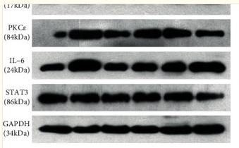
2). Phosphorylation at Ser 727 Increases STAT3 Interaction with PKCε Regulating Neuron–Glia Crosstalk via IL-6-Mediated Hyperalgesia In Vivo and In Vitro. MEDIATORS OF INFLAMMATION, 2022 (PubMed: 35125963) [IF=4.4]

Application: WB    Species: Rat    Sample: spinal cord tissues

Figure 3 Western blot analysis of the proteins related to activated neurocytes during inflammatory pain. Examples (a) and mean values (b–g) of c-Fos, GFAP, Iba-1, PKCε, STAT3, and IL-6 proteins in the spinal cord. Intrathecally injected T-5224 (c-Fos/AP-1 inhibitor, 500 μg/50 μL), minocycline (Mino, 100 μg/50 μL), and L-2-aminoadipic acid (LAA, 1 mg/50 μL) reduced c-Fos, GFAP, Iba-1, PKCε, and IL-6 protein levels in FCA-treated rats, whereas STAT3 expression did not change between groups (P > 0.05). Data were normalized against GAPDH and are expressed as ratios (%) of control. Data are shown as means ± SD (n = 4–5). ∗P < 0.05, ∗∗P < 0.01; ∗∗∗P < 0.001, one-way ANOVA with Bonferroni tests.

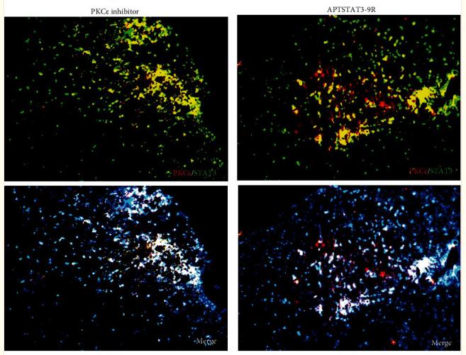
Application: IF/ICC    Species: Rat    Sample: spinal cord tissues

Figure 6 Detection of PKCε and STAT3 coexpression in vivo and their immune complexes in vitro. (a–f) Immunofluorescence staining for PKCε (red) and STAT3 (green) coexpression (yellow) and DAPI (blue in merged image) in spinal cord sections (a–e). Ratios of cells with immunoreactive PKCε-/STAT3 among total cells. (f) Inhibitors of PKCε and STAT3 significantly decreased the coexpression of PKCε/STAT3 after APTSTAT3-9R administration. Bar = 40 μm. Data are shown as means ± SD (n = 6). ∗P < 0.05, ∗∗P < 0.01, ∗∗∗P < 0.001; one-way ANOVA followed by Bonferroni tests.

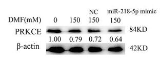
3). Aberrant expression of SNHG12 contributes to N, N-dimethylformamide-induced hepatic apoptosis both in short-term and long-term DMF exposure. Toxicology Research, 2021 (PubMed: 34733487) [IF=2.2]

Application: WB    Species: Human    Sample: HL-7702 cells

Figure 5 Hsa-miR-218-5p promotes HL-7702 cells apoptosis by regulating PRKCE. (A) HL-7702 cells were transfected with hsa-miR-218-5p mimic or NC control for 24 hours. Cell apoptosis was detected by flow cytometry. (B) The apoptotic rate (Q3 + Q2) of HL-7702 cells based on the result of flow cytometry. Expression levels of PRKCE were detected after transfection of hsa-miR-218-5p mimic (C), OE-SNHG12 (E) and sh-SNHG12 (G) by qRT-PCR. GAPDH served as an endogenous control. Expression levels of PRKCE protein in HL-7702 cells were detected after transfection of hsa-miR-218-5p mimic (D), OE-SNHG12 (F) and sh-SNHG12(H) by western blot analysis. Data were shown as mean ± SD. *P < .05 and **P < .01, compared with the control group. DMF, N, N-dimethylformamide; NC (A, B, C, D), mimic negative control; NC (G, H), negative control of sh-SNHG12

限制条款

产品的规格、报价、验证数据请以官网为准,官网链接:www.affbiotech.com | www.affbiotech.cn(简体中文)| www.affbiotech.jp(日本語)

产品的数据信息为Affinity所有,未经授权不得收集Affinity官网数据或资料用于商业用途,对抄袭产品数据的行为我们将保留诉诸法律的权利。

产品相关数据会因产品批次、产品检测情况随时调整,如您已订购该产品,请以订购时随货说明书为准,否则请以官网内容为准,官网内容有改动时恕不另行通知。

Affinity保证所销售产品均经过严格质量检测。如您购买的商品在规定时间内出现问题需要售后时,请您在Affinity官方渠道提交售后申请。

产品仅供科学研究使用。不用于诊断和治疗。 

产品未经授权不得转售。

Affinity Biosciences将不会对在使用我们的产品时可能发生的专利侵权或其他侵权行为负责。Affinity Biosciences, Affinity Biosciences标志和所有其他商标所有权归Affinity Biosciences LTD.