产品: 磷酸化 Annexin A2 (Tyr24) 抗体
货号: AF7096
描述: Rabbit polyclonal antibody to Phospho-Annexin A2 (Tyr24)
应用: WB IHC IF/ICC
文献验证: WB, IF/ICC
反应: Human, Mouse, Rat
预测: Pig, Bovine, Horse, Sheep, Rabbit, Dog, Chicken
蛋白号: P07355
RRID: AB_2843536

浏览相似产品>>

   规格 价格 库存
 100ul RMB¥ 2800 现货
 200ul RMB¥ 3800 现货

货期: 当天发货

联系销售

产品描述

来源:
Rabbit
应用:
WB 1:500-1:2000, IHC 1:50-1:200, IF/ICC 1:100-1:500
*The optimal dilutions should be determined by the end user.
*Tips:

WB: 适用于变性蛋白样本的免疫印迹检测. IHC: 适用于组织样本的石蜡(IHC-p)或冰冻(IHC-f)切片样本的免疫组化/荧光检测. IF/ICC: 适用于细胞样本的荧光检测. ELISA(peptide): 适用于抗原肽的ELISA检测.

反应:
Human, Mouse, Rat
克隆:
Polyclonal
特异性:
Phospho-Annexin A2 (Tyr24) Antibody detects endogenous levels of Annexin A2 only when phosphorylated at Tyr24.
RRID:
AB_2843536
引用格式: Affinity Biosciences Cat# AF7096, RRID:AB_2843536.
偶联:
Unconjugated.
纯化:
The antibody is from purified rabbit serum by affinity purification via sequential chromatography on phospho-peptide and non-phospho-peptide affinity columns.
保存:
Rabbit IgG in phosphate buffered saline , pH 7.4, 150mM NaCl, 0.02% sodium azide and 50% glycerol. Store at -20 °C. Stable for 12 months from date of receipt.
别名:

展开/折叠

Annexin A2; Annexin II; Annexin II, heavy chain; Annexin-2; ANX 2; ANX2; ANX2L4; ANXA2; ANXA2_HUMAN; arylsulfatase B; CAL1H; Calpactin I heavy chain; calpactin I heavy polypeptide (p36); Calpactin I heavy polypeptide; Calpactin-1 heavy chain; chromobindin 8; Chromobindin-8; Epididymis secretory protein Li 270; HEL S 270; LIP2; Lipocortin II; LPC2; LPC2D; p36; P36 protein; PAP-IV; Placental anticoagulant protein IV; Protein I;

抗原和靶标

免疫原:

A synthesized peptide derived from human Annexin A2 around the phosphorylation site of Tyr24.

基因/基因ID:

文献引用

1). (20S) Ginsenoside Rh2 Inhibits STAT3/VEGF Signaling by Targeting Annexin A2. International Journal of Molecular Sciences, 2021 (PubMed: 34502195) [IF=5.6]

Application: WB    Species: Human    Sample: HepG2 cells

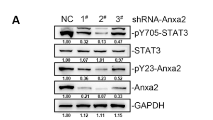
igure 1. Anxa2 regulated the expression of VEGFs. HepG2 cells were treated with three different Anxa2-shRNAs. (A) Protein level and phosphorylation status of STAT3 and Anxa2 were determined by immuno-blotting. (B) The activation of STAT3 was determined via reporter gene assay. (C) The expression of STAT3 target genes was determined by qRT-PCR. (D) The protein level of four VEGFs was determined by immuno-blotting. (E) The concentration of four VEGFs in cell-free supernatant was determined by ELISA. All experiments were performed with triple replicant.

2). Nuclear autoantigenic sperm protein facilitates glioblastoma progression and radioresistance by regulating the ANXA2/STAT3 axis. CNS neuroscience & therapeutics, 2024 (PubMed: 38605477) [IF=4.8]

Application: WB    Species: human    Sample: U87 cells

FIGURE 4 NASP promotes ANXA2-mediated DNA repair. (A) The top 15 proteins potentially associated with NASP were isolated by immunoprecipitation and identified by mass spectrometry in NASP-overexpressing U87 cells. Cell lysates were immunoprecipitated with an antibody against NASP, while an antibody against IgG was used as the negative control. (B) Mass spectrogram of ANXA2. (C, D) Western blot detection of NASP and ANXA2 proteins by reciprocal immunoprecipitation with an antibody against NASP (C) or ANXA2 (D) in U87 and U251 cells. IgG was used as the control. (E) Immunofluorescence detection of NASP, ANXA2, and p-ANXA2 localization in control and NASP-overexpressing U87 cells. Scale bars, 50 μM. (F) Abundance of NASP, ANXA2, and p-ANXA2 in cytoplasmic, nuclear, and total protein fractions from the control and NASP-overexpressing U87 cells estimated by western blotting. (G) Effects of NASP upregulation and ANXA2 downregulation on DNA double-strand breaks in U87 cells after radiotherapy as detected by immunofluorescence. Scale bars, 50 μM. (H) Comet assay showing the effect of NASP upregulation and ANXA2 downregulation on DNA double-strand breaks in U87 cells after RT. Scale bars, 100 μM. Data are presented as mean ± SD from at least three independent experiments. *p 

Application: IF/ICC    Species: human    Sample: U87 cells

FIGURE 4 NASP promotes ANXA2-mediated DNA repair. (A) The top 15 proteins potentially associated with NASP were isolated by immunoprecipitation and identified by mass spectrometry in NASP-overexpressing U87 cells. Cell lysates were immunoprecipitated with an antibody against NASP, while an antibody against IgG was used as the negative control. (B) Mass spectrogram of ANXA2. (C, D) Western blot detection of NASP and ANXA2 proteins by reciprocal immunoprecipitation with an antibody against NASP (C) or ANXA2 (D) in U87 and U251 cells. IgG was used as the control. (E) Immunofluorescence detection of NASP, ANXA2, and p-ANXA2 localization in control and NASP-overexpressing U87 cells. Scale bars, 50 μM. (F) Abundance of NASP, ANXA2, and p-ANXA2 in cytoplasmic, nuclear, and total protein fractions from the control and NASP-overexpressing U87 cells estimated by western blotting. (G) Effects of NASP upregulation and ANXA2 downregulation on DNA double-strand breaks in U87 cells after radiotherapy as detected by immunofluorescence. Scale bars, 50 μM. (H) Comet assay showing the effect of NASP upregulation and ANXA2 downregulation on DNA double-strand breaks in U87 cells after RT. Scale bars, 100 μM. Data are presented as mean ± SD from at least three independent experiments. *p 

3). GNAQ T96S mutation abrogates the ability of wild‐type GNAQ to induce apoptosis by phosphorylating annexin A2 in natural killer/T cell lymphoma. Cancer Science, 2022 (PubMed: 35293080) [IF=4.5]

Application: WB    Species: Mice    Sample: NK cells

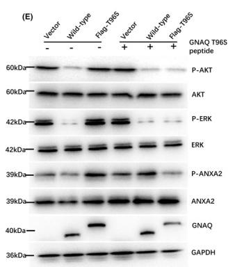
FIGURE 4 GNAQ T96S peptide inhibitor induces apoptosis by competing with annexin A2 (ANXA2) binding to GNAQ T96S in natural killer cells. (A) A schematic diagram showing the different domains of GNAQ. (B) Identification of the interaction between different domains of GNAQ and ANXA2 by co‐immunoprecipitation (IP). M or R refers to the host of antibody. (C) GNAQ T96S peptide blocks the binding of GNAQ to ANXA2 in a dose‐dependent manner. (D) GNAQ T96S peptide significantly induces apoptosis in NK cells. (E) GNAQ peptide affects several signaling pathways, including the AKT, ERK, and ANXA2 signaling pathways. The experiment was independently repeated three times

4). A novel cystathionine γ-lyase inhibitor, I194496, inhibits the growth and metastasis of human TNBC via downregulating multiple signaling pathways. Scientific Reports, 2021 (PubMed: 33903672) [IF=3.8]

Application: WB    Species: Human    Sample: TNBC cells

Figure 6 I194496 inhibits the Anxa2/STAT3 and VEGF/FAK/Paxillin signalling pathways in human TNBC cells. (A, B) The effect of I194496 on Anxa2/STAT3 pathway in MDA-MB-231 cells. *P 

限制条款

产品的规格、报价、验证数据请以官网为准,官网链接:www.affbiotech.com | www.affbiotech.cn(简体中文)| www.affbiotech.jp(日本語)

产品的数据信息为Affinity所有,未经授权不得收集Affinity官网数据或资料用于商业用途,对抄袭产品数据的行为我们将保留诉诸法律的权利。

产品相关数据会因产品批次、产品检测情况随时调整,如您已订购该产品,请以订购时随货说明书为准,否则请以官网内容为准,官网内容有改动时恕不另行通知。

Affinity保证所销售产品均经过严格质量检测。如您购买的商品在规定时间内出现问题需要售后时,请您在Affinity官方渠道提交售后申请。

产品仅供科学研究使用。不用于诊断和治疗。 

产品未经授权不得转售。

Affinity Biosciences将不会对在使用我们的产品时可能发生的专利侵权或其他侵权行为负责。Affinity Biosciences, Affinity Biosciences标志和所有其他商标所有权归Affinity Biosciences LTD.