产品: Cytokeratin 19 抗体
货号: AF0192
描述: Rabbit polyclonal antibody to Cytokeratin 19
应用: WB IHC IF/ICC IHC-F
反应: Human, Mouse, Rat
预测: Bovine, Rabbit, Chicken
分子量: 44kDa; 44kD(Calculated).
蛋白号: P08727
RRID: AB_2833385

浏览相似产品>>

   规格 价格 库存
 50ul RMB¥ 1250 现货
 100ul RMB¥ 2300 现货
 200ul RMB¥ 3000 现货

货期: 当天发货

联系销售

产品描述

来源:
Rabbit
应用:
WB 1:500-1:3000, IHC 1:50-1:200, IF/ICC 1:100-1:500
*The optimal dilutions should be determined by the end user.
*Tips:

WB: 适用于变性蛋白样本的免疫印迹检测. IHC: 适用于组织样本的石蜡(IHC-p)或冰冻(IHC-f)切片样本的免疫组化/荧光检测. IF/ICC: 适用于细胞样本的荧光检测. ELISA(peptide): 适用于抗原肽的ELISA检测.

反应:
Human,Mouse,Rat
预测:
Bovine(100%), Rabbit(100%), Chicken(100%)
克隆:
Polyclonal
特异性:
Cytokeratin 19 Antibody detects endogenous levels of total Cytokeratin 19.
RRID:
AB_2833385
引用格式: Affinity Biosciences Cat# AF0192, RRID:AB_2833385.
偶联:
Unconjugated.
纯化:
The antiserum was purified by peptide affinity chromatography using SulfoLink™ Coupling Resin (Thermo Fisher Scientific).
保存:
Rabbit IgG in phosphate buffered saline , pH 7.4, 150mM NaCl, 0.02% sodium azide and 50% glycerol. Store at -20 °C. Stable for 12 months from date of receipt.
别名:

展开/折叠

40 kDa keratin intermediate filament; CK 19; CK-19; CK19; Cytokeratin 19; Cytokeratin-19; K19; K1C19_HUMAN; K1CS; Keratin 19; Keratin type I 40 kD; Keratin type I 40kD; Keratin type I cytoskeletal 19; Keratin, type I cytoskeletal 19; Keratin, type I, 40 kd; Keratin-19; KRT19; MGC15366;

抗原和靶标

免疫原:
Uniprot:
基因/基因ID:
表达:
P08727 K1C19_HUMAN:

Expressed in a defined zone of basal keratinocytes in the deep outer root sheath of hair follicles. Also observed in sweat gland and mammary gland ductal and secretory cells, bile ducts, gastrointestinal tract, bladder urothelium, oral epithelia, esophagus, ectocervical epithelium (at protein level). Expressed in epidermal basal cells, in nipple epidermis and a defined region of the hair follicle. Also seen in a subset of vascular wall cells in both the veins and artery of human umbilical cord, and in umbilical cord vascular smooth muscle. Observed in muscle fibers accumulating in the costameres of myoplasm at the sarcolemma in structures that contain dystrophin and spectrin.

描述:
K19 a type I cytoskeletal keratin. The keratins are intermediate filament proteins responsible for the structural integrity of epithelial cells and are subdivided into cytokeratins and hair keratins. Keratin 19 differs from all other IF proteins in lacking the C-terminal tail domain.
序列:
MTSYSYRQSSATSSFGGLGGGSVRFGPGVAFRAPSIHGGSGGRGVSVSSARFVSSSSSGAYGGGYGGVLTASDGLLAGNEKLTMQNLNDRLASYLDKVRALEAANGELEVKIRDWYQKQGPGPSRDYSHYYTTIQDLRDKILGATIENSRIVLQIDNARLAADDFRTKFETEQALRMSVEADINGLRRVLDELTLARTDLEMQIEGLKEELAYLKKNHEEEISTLRGQVGGQVSVEVDSAPGTDLAKILSDMRSQYEVMAEQNRKDAEAWFTSRTEELNREVAGHTEQLQMSRSEVTDLRRTLQGLEIELQSQLSMKAALEDTLAETEARFGAQLAHIQALISGIEAQLGDVRADSERQNQEYQRLMDIKSRLEQEIATYRSLLEGQEDHYNNLSASKVL

种属预测

种属预测:

score>80的预测可信度较高,可尝试用于WB检测。*预测模型主要基于免疫原序列比对,结果仅作参考,不作为质保凭据。

Species
Results
Score
Bovine
100
Chicken
100
Rabbit
100
Xenopus
75
Pig
0
Horse
0
Sheep
0
Dog
0
Zebrafish
0
Model Confidence:
High(score>80) Medium(80>score>50) Low(score<50) No confidence

翻译修饰 - P08727 作为底物

Site PTM Type Enzyme
S3 Phosphorylation
Y4 Phosphorylation
R7 Methylation
S9 Phosphorylation
S10 Phosphorylation
T12 Phosphorylation
S13 Phosphorylation
S14 Phosphorylation
S22 Phosphorylation
R24 Methylation
R32 Methylation
S35 Phosphorylation P31749 (AKT1)
S40 Phosphorylation
R43 Methylation
S46 Phosphorylation
S48 Phosphorylation
S49 Phosphorylation
R51 Methylation
S55 Phosphorylation
S58 Phosphorylation
Y61 Phosphorylation
Y65 Phosphorylation
T70 Phosphorylation
S72 Phosphorylation
K81 Ubiquitination
S93 Phosphorylation
Y94 Phosphorylation
K97 Acetylation
K97 Ubiquitination
K111 Ubiquitination
Y116 Phosphorylation
K118 Methylation
K118 Ubiquitination
Y127 Phosphorylation
S128 Phosphorylation
Y130 Phosphorylation
Y131 Phosphorylation
T132 Phosphorylation
T133 Phosphorylation
K140 Ubiquitination
T145 Phosphorylation
K168 Methylation
K168 Sumoylation
K168 Ubiquitination
S178 Phosphorylation
K208 Sumoylation
K208 Ubiquitination
Y213 Phosphorylation
K215 Sumoylation
K215 Ubiquitination
K216 Ubiquitination
S223 Phosphorylation
T224 Phosphorylation
S234 Phosphorylation
S239 Phosphorylation
K247 Ubiquitination
S250 Phosphorylation
S254 Phosphorylation
Y256 Phosphorylation
K265 Ubiquitination
T272 Phosphorylation
S273 Phosphorylation
T275 Phosphorylation
T286 Phosphorylation
S292 Phosphorylation
S294 Phosphorylation
T302 Phosphorylation
S312 Phosphorylation
S315 Phosphorylation
K317 Ubiquitination
T323 Phosphorylation
T327 Phosphorylation
S343 Phosphorylation
K370 Ubiquitination
S371 Phosphorylation
T379 Phosphorylation
Y380 Phosphorylation
S382 Phosphorylation
Y391 Phosphorylation P12931 (SRC)
S395 Phosphorylation
S397 Phosphorylation
K398 Ubiquitination

研究背景

功能:

Involved in the organization of myofibers. Together with KRT8, helps to link the contractile apparatus to dystrophin at the costameres of striated muscle.

组织特异性:

Expressed in a defined zone of basal keratinocytes in the deep outer root sheath of hair follicles. Also observed in sweat gland and mammary gland ductal and secretory cells, bile ducts, gastrointestinal tract, bladder urothelium, oral epithelia, esophagus, ectocervical epithelium (at protein level). Expressed in epidermal basal cells, in nipple epidermis and a defined region of the hair follicle. Also seen in a subset of vascular wall cells in both the veins and artery of human umbilical cord, and in umbilical cord vascular smooth muscle. Observed in muscle fibers accumulating in the costameres of myoplasm at the sarcolemma in structures that contain dystrophin and spectrin.

亚基结构:

Heterotetramer of two type I and two type II keratins. Interacts with PNN and the actin-binding domain of DMD. Interacts with HCV core protein.

(Microbial infection) Interacts with hepatitis C virus/HCV core protein.

蛋白家族:

This keratin differs from all other IF proteins in lacking the C-terminal tail domain.

Belongs to the intermediate filament family.

研究领域

· Organismal Systems > Endocrine system > Estrogen signaling pathway.   (View pathway)

文献引用

1). Enhanced hepatic differentiation of rat bone marrow-derived mesenchymal stem cells in spheroidal aggregate culture on a decellularized liver scaffold. INTERNATIONAL JOURNAL OF MOLECULAR MEDICINE (PubMed: 27314916) [IF=5.4]

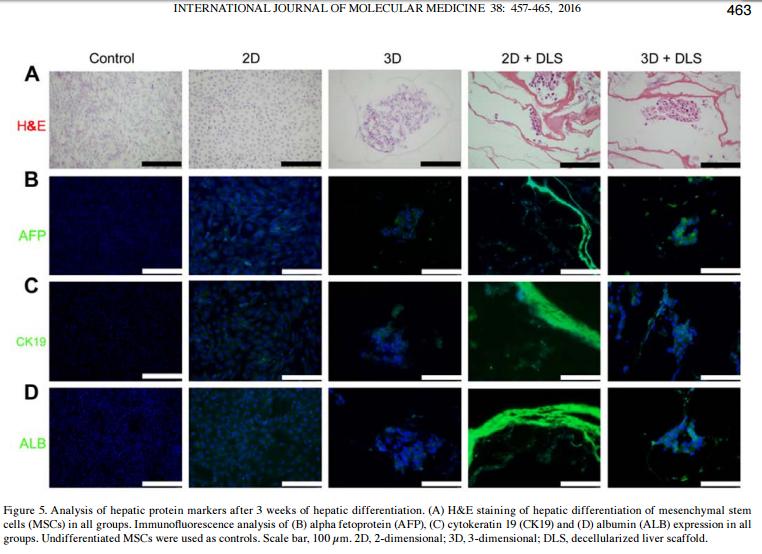
Application: IF/ICC    Species: rat    Sample:

Figure 5. Analysis of hepatic protein markers after 3 weeks of hepatic differentiation. (A) H&E staining of hepatic differentiation of mesenchymal stem cells (MSCs) in all groups. Immunofluorescence analysis of (B) alpha fetoprotein (AFP), (C) cytokeratin 19 (CK19) and (D) albumin (ALB) expression in all groups. Undifferentiated MSCs were used as controls. Scale bar, 100 µm. 2D, 2-dimensional; 3D, 3-dimensional; DLS, decellularized liver scaffold.

2). Human umbilical cord mesenchymal stem cells implantation accelerates cutaneous wound healing in diabetic rats via the Wnt signaling pathway. EUROPEAN JOURNAL OF MEDICAL RESEARCH (PubMed: 30736851) [IF=4.2]

Application: WB    Species: Human    Sample: hUCMSCs

Fig. 5 Activation of the Wnt signaling pathway enhanced hUCMSCs differentiation. a Western blotting was applied to detect the changes in the levels of β-catenin, c-Myc, p63, CK19, and PCNA proteins. b Statistical analysis of a results. c The differentiation degree of hUCMSCs was evaluated by cell morphology under optical microscope (scale: 100 μm). d Statistical analysis on the ratio of cells with round or small polygonal morphology displayed in c;

3). Hepatic differentiation of mouse bone marrow‑derived mesenchymal stem cells using a novel 3D culture system. Molecular Medicine Reports (PubMed: 29152658) [IF=3.4]

Application: IF/ICC    Species: Mouse    Sample:

Figure 4. Analysis of hepatic protein markers in mouse MSCs following 23 days of hepatic differentiation. Green immunofluorescence staining of ALB, AFP and CK19 in undifferentiated MSC control, 2D and 3D groups. Nuclei were stained blue with DAPI. Scale bars, 100 µm. MSCs, mesenchymal stem cells; ALB, albumin; AFP, α-fetoprotein; CK19, cytokeratin-19.

4). Adenomyosis-derived extracellular vesicles endow endometrial epithelial cells with an invasive phenotype through epithelial-mesenchymal transition. Genes & Diseases (PubMed: 33335963)

限制条款

产品的规格、报价、验证数据请以官网为准,官网链接:www.affbiotech.com | www.affbiotech.cn(简体中文)| www.affbiotech.jp(日本語)

产品的数据信息为Affinity所有,未经授权不得收集Affinity官网数据或资料用于商业用途,对抄袭产品数据的行为我们将保留诉诸法律的权利。

产品相关数据会因产品批次、产品检测情况随时调整,如您已订购该产品,请以订购时随货说明书为准,否则请以官网内容为准,官网内容有改动时恕不另行通知。

Affinity保证所销售产品均经过严格质量检测。如您购买的商品在规定时间内出现问题需要售后时,请您在Affinity官方渠道提交售后申请。

产品仅供科学研究使用。不用于诊断和治疗。 

产品未经授权不得转售。

Affinity Biosciences将不会对在使用我们的产品时可能发生的专利侵权或其他侵权行为负责。Affinity Biosciences, Affinity Biosciences标志和所有其他商标所有权归Affinity Biosciences LTD.