产品: GAP43 抗体
货号: DF7766
描述: Rabbit polyclonal antibody to GAP43
应用: WB IHC IF/ICC
文献验证: WB, IHC, IF/ICC
反应: Human, Mouse, Rat
预测: Pig, Bovine, Rabbit, Dog
蛋白号: P17677
RRID: AB_2841232

浏览相似产品>>

   规格 价格 库存
 50ul RMB¥ 1250 现货
 100ul RMB¥ 2300 现货
 200ul RMB¥ 3000 现货

货期: 当天发货

联系销售

产品描述

来源:
Rabbit
应用:
WB 1:1000-3000, IF/ICC 1:100-1:500, IHC 1:50-1:200
*The optimal dilutions should be determined by the end user.
*Tips:

WB: 适用于变性蛋白样本的免疫印迹检测. IHC: 适用于组织样本的石蜡(IHC-p)或冰冻(IHC-f)切片样本的免疫组化/荧光检测. IF/ICC: 适用于细胞样本的荧光检测. ELISA(peptide): 适用于抗原肽的ELISA检测.

反应:
Human, Mouse, Rat
克隆:
Polyclonal
特异性:
GAP43 Antibody detects endogenous levels of total GAP43.
RRID:
AB_2841232
引用格式: Affinity Biosciences Cat# DF7766, RRID:AB_2841232.
偶联:
Unconjugated.
纯化:
The antiserum was purified by peptide affinity chromatography using SulfoLink™ Coupling Resin (Thermo Fisher Scientific).
保存:
Rabbit IgG in phosphate buffered saline , pH 7.4, 150mM NaCl, 0.02% sodium azide and 50% glycerol. Store at -20 °C. Stable for 12 months from date of receipt.
别名:

展开/折叠

Axonal membrane protein GAP 43; Axonal membrane protein GAP-43; B 50; Calmodulin binding protein P 57; F1; GAP 43; GAP43; Growth Associated Protein 43; Growth-associated protein 43; Nerve Growth Related Peptide; Nerve growth related peptide GAP43; NEUM_HUMAN; Neural phosphoprotein B 50; Neural phosphoprotein B-50; Neuromodulin; Neuron growth associated protein 43; PP46; Protein F1; QtrA-11580; QtrA-13071;

抗原和靶标

免疫原:

A synthesized peptide derived from human GAP43, corresponding to a region within C-terminal amino acids.

基因/基因ID:

文献引用

1). SiNiSan ameliorates depression-like behavior in rats by enhancing synaptic plasticity via the CaSR-PKC-ERK signaling pathway. BIOMEDICINE & PHARMACOTHERAPY, 2020 (PubMed: 31958763) [IF=6.9]

Application: WB    Species: rat    Sample: HIP

Fig. 5. |Effects of SNS on synaptic-associated protein in the HIP and PFC of stressed rats. Representative immunoblots for PSD-95, GAP-43, Syn, and Tublin in the HIP(A) and PFC(B) regions. (A) Results of relative protein levels of PSD-95, GAP-43, and Syn in the HIP of rats of each group.

2). Exosomes derived from human induced pluripotent stem cell-derived neural progenitor cells protect neuronal function under ischemic conditions. Neural Regeneration Research, 2021 (PubMed: 33642395) [IF=5.9]

Application: WB    Species: Rat    Sample: neural progenitor cells

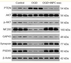
Figure 3 Effect of iPSC-NPC-derived exosomes (OGD+iNPC-exo) on expression of the PTEN/AKT signaling pathway and of synaptic plasticity-related proteins in OGD induced neurons. (A–C) mRNA expression (A) and protein expression (B, C) of the PTEN/AKT signaling pathway and of synaptic plasticity-related proteins (NF200, GAP-43, Synapsin, and PSD95) analyzed by polymerase chain reaction and western blot assay. The mRNA expression is described by the optical density ratio relative to the control group. Protein expression was described by the optical density ratio relative to β-actin. Data are presented as mean ± SD. ***P < 0.001, vs. control group; ###P < 0.001 (one-way analysis of variance with post hoc Bonferroni test). All experiments were repeated three times. GAP43: growth associated protein 43; iPSC-NPCs: induced pluripotent stem cells-derived neural progenitor cells; NF200: neurofilament 200; OGD: oxygen-glucose deprivation; p-Akt: phosphor-Akt; PSD95: postsynaptic density protein 95; PTEN: phosphatase and tensin homolog deleted on chromosome ten.

3). Porcine decellularized nerve matrix hydrogel attenuates neuroinflammation after peripheral nerve injury by inhibiting the TLR4/MyD88/NF-κB axis. Neural regeneration research, 2024 (PubMed: 39589179) [IF=5.9]

4). Early-Life Stress Induces Depression-Like Behavior and Synaptic-Plasticity Changes in a Maternal Separation Rat Model: Gender Difference and Metabolomics Study. Frontiers in Pharmacology, 2020 (PubMed: 32174832) [IF=5.6]

Application: WB    Species: Rat    Sample: hippocampus

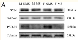
Figure 5 MS reduces the expression of synaptic-plasticity protein. (A) The bands of synaptic-plasticity proteins of SYN, PSD-95, and GAP-43 in the hippocampus by WB. Statistical results indicate the relative protein levels expressed by SYN, GAP-43, and PSD-95. (B) The bands of synaptic-plasticity proteins of SYN, PSD-95, and GAP-43 in cortex by WB. Statistical results indicate the relative protein levels expressed by SYN, GAP-43, and PSD-95. Statistical analyses are performed by two-way ANOVA followed by t-test. Data are presented as mean ± SEM, *p < 0.05, **p < 0.01, n = 3 per group.

5). The role of EndophilinA1 in chronic unpredicted mild stress-induced depression model mice. International immunopharmacology, 2023 (PubMed: 37837716) [IF=4.8]

6). Erythropoietin-Induced Autophagy Protects Against Spinal Cord Injury and Improves Neurological Function via the Extracellular-Regulated Protein Kinase Signaling Pathway. MOLECULAR NEUROBIOLOGY, 2020 (PubMed: 32647973) [IF=4.6]

Application: IF/ICC    Species: rat    Sample:

Fig. 6 |EPO benefit antiinflammation and axonal regeneration can be neutralized by ERK inhibition, and activator can mimic the effect of EPO. a The axonal special protein GAP43 immunofluorescence staining and histogram at 14 days (magnification × 400). b–e The inflammatory marker proteins immunofluorescence staining and histograms at 14 days, including CD86, GFAP, TNF-α, iNOS(magnification × 400). All data presented as mean ± SEM in each group. *p < 0.05 vs. SCI + saline group, **p < 0.01 vs. SCI + saline group. #p < 0.05 comparison between SCI + EPO group and SCI + EPO + PD98059, ##p < 0.01 comparison between SCI + EPO group and SCI + EPO + PD98059

7). GAP-43 targeted indocyanine green-loaded near-infrared fluorescent probe for real-time mapping of perineural invasion lesions in pancreatic cancer in vivo. Nanomedicine : nanotechnology, biology, and medicine, 2023 (PubMed: 37054805) [IF=4.2]

Application: IHC    Species: Mouse    Sample: pancreatic cancer tissues

Fig. 3. Validation of GAP-43 expression in pancreatic cancer samples, data were obtained from the authors' affiliations. (A) Immunohistochemical results showed GAP-43 expression in PNI, normal pancreatic tissues, and pancreatic cancer tissues with non-PNI. Scale bar: 200 μm. (B) The IOD value of GAP-43 staining in PNI, normal tissues, and tumors without PNI. (C) IHC scores differed between tumor and tumor, and between PNI and non-PNI in the tumor, respectively. (D) Survival difference between high and low GAP-43 expression in pancreatic cancer. (D) Immunohistochemical staining of GAP-43(−), GAP-43(+), GAP-43(2+), GAP-43(3+). ****p < 0.0001.

Application: WB    Species: Mouse    Sample: pancreatic cancer tissues

Fig. 5. Validation of probe targeting and toxicity in vitro. (A) Confocal microscopy images of PC12, Miapaca-2 and PC12* after incubation with GAP-43Ra-ICG-PEG or ICG-NP. Magnification:630×. (B) CCK-8 results in probes GAP-43Ra-ICG-PEG and ICG-NP. (C) Fluorescence quantitative differences of GAP-43Ra-ICG-PEG or ICG-NP uptake by PC12, Miapaca-2 and PC12* under confocal microscopy. (D) GAP-43 expression of PC12 and PC12* in western blot. (F) Quantitative analysis of PC12 and PC12*in western blot. PC12*: PC12 cells after co-culture with pancreatic cancer cell line Miapaca-2.****p < 0.0001.

8). TAG-1 Regulates NRP1 in Schwann Cells and Participates in Regulating Nerve Regeneration in Rats with Sciatic Nerve Crush Injury. Neurochemical research, 2024 (PubMed: 39718654) [IF=3.7]

Application: IHC    Species: Rat    Sample: RSC96 cells

Fig. 9 Effects of TAG-1 treatment on the GAP-43 and NCAM expression in the rats with SNI. A The expression of GAP-43 was measured by immunohistochemistry. Scale = 50 μm. Black arrow: GAP-43 expression. The expression of GAP-43 was analyzed using ImageJ software. B The expression of NCAM was measured by immunofluorescence. The mean gray of NCAM was analyzed by ImageJ software. *p 

9). The Effect of Early Maternal Separation Combined With Adolescent Chronic Unpredictable Mild Stress on Behavior and Synaptic Plasticity in Adult Female Rats. Frontiers in Psychiatry, 2021 (PubMed: 33746787) [IF=3.2]

Application: WB    Species: Rat    Sample: Hippocampus

Figure 5 Western blot analysis. The expressions of synaptic plasticity proteins were determined by western blot (A), including PSD-95 (B), GAP-43 (C), and SYN (D). The values represent the mean ± SEM, n = 5. *p < 0.05, **p < 0.01 vs. CON, #p < 0.05, ##p < 0.01 vs. MS. CON, control; MS, maternal separation; CUMS, chronic unpredictable mild stress.

10). Astragaloside IV promotes neuronal axon regeneration by inhibiting the PTEN/AKT pathway. Brain research, 2025 (PubMed: 39793915) [IF=2.7]

加载更多

限制条款

产品的规格、报价、验证数据请以官网为准,官网链接:www.affbiotech.com | www.affbiotech.cn(简体中文)| www.affbiotech.jp(日本語)

产品的数据信息为Affinity所有,未经授权不得收集Affinity官网数据或资料用于商业用途,对抄袭产品数据的行为我们将保留诉诸法律的权利。

产品相关数据会因产品批次、产品检测情况随时调整,如您已订购该产品,请以订购时随货说明书为准,否则请以官网内容为准,官网内容有改动时恕不另行通知。

Affinity保证所销售产品均经过严格质量检测。如您购买的商品在规定时间内出现问题需要售后时,请您在Affinity官方渠道提交售后申请。

产品仅供科学研究使用。不用于诊断和治疗。 

产品未经授权不得转售。

Affinity Biosciences将不会对在使用我们的产品时可能发生的专利侵权或其他侵权行为负责。Affinity Biosciences, Affinity Biosciences标志和所有其他商标所有权归Affinity Biosciences LTD.