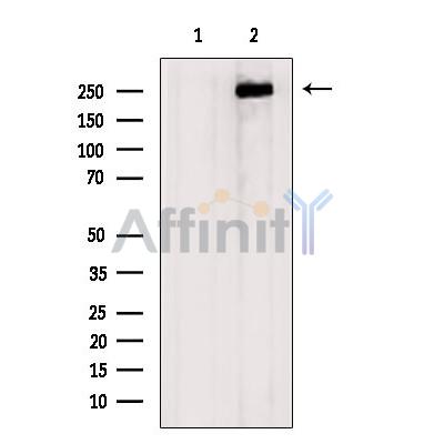
产品: Aggrecan 抗体
货号: DF7561
描述: Rabbit polyclonal antibody to Aggrecan
应用: WB IHC IF/ICC
文献验证: WB, IHC, IF/ICC
反应: Human, Mouse, Rat
预测: Pig, Bovine, Sheep, Rabbit, Dog
蛋白ID: P16112
RRID: AB_2841055

浏览相似产品>>

   规格 价格 库存
 50ul RMB¥ 1250 现货
 100ul RMB¥ 2300 现货
 200ul RMB¥ 3000 现货

货期: 当天发货

联系销售

产品描述

来源:
Rabbit
应用:
WB 1:1000-3000, IF/ICC 1:100-1:500, IHC 1:50-1:200
*The optimal dilutions should be determined by the end user. For optimal experimental results, antibody reuse is not recommended.
*Tips:

WB: 适用于变性蛋白样本的免疫印迹检测. IHC: 适用于组织样本的石蜡(IHC-p)或冰冻(IHC-f)切片样本的免疫组化/荧光检测. IF/ICC: 适用于细胞样本的荧光检测. ELISA(peptide): 适用于抗原肽的ELISA检测.

反应:
Human, Mouse, Rat
克隆:
Polyclonal
特异性:
Aggrecan Antibody detects endogenous levels of total Aggrecan.
RRID:
AB_2841055
引用格式: Affinity Biosciences Cat# DF7561, RRID:AB_2841055.
偶联:
Unconjugated.
纯化:
The antiserum was purified by peptide affinity chromatography using SulfoLink™ Coupling Resin (Thermo Fisher Scientific).
保存:
Rabbit IgG in phosphate buffered saline , pH 7.4, 150mM NaCl, 0.02% sodium azide and 50% glycerol. Store at -20 °C. Stable for 12 months from date of receipt.
别名:

展开/折叠

ACAN; AGC 1; AGC1; AGCAN; Aggrecan 1 (chondroitin sulfate proteoglycan 1, large aggregating proteoglycan, antigen identified by monoclonal antibody A0122); Aggrecan 1; Aggrecan core protein; Aggrecan proteoglycan; Aggrecan structural proteoglycan of cartilage; Aggrecan1; ATEGQV; Cartilage specific proteoglycan core protein; Chondroitin sulfate proteoglycan 1; Chondroitin sulfate proteoglycan 1 large aggregating proteoglycan antigen identified by monoclonal antibody A0122; Chondroitin sulfate proteoglycan core protein 1; CSPG 1; CSPG1; CSPGCP; JSCATE; Large aggregating proteoglycan; mcspg; mgsk16; MSK 16; MSK16; SEDK;

抗原和靶标

免疫原:

A synthesized peptide derived from human Aggrecan, corresponding to a region within N-terminal amino acids.

基因/基因ID:

文献引用

1). Hydrogen Ion Capturing Hydrogel Microspheres for Reversing Inflammaging. Advanced materials (Deerfield Beach, Fla.), 2024 (PubMed: 37699155) [IF=27.4]

2). An anti-inflammatory and neuroprotective biomimetic nanoplatform for repairing spinal cord injury. Bioactive Materials, 2022 (PubMed: 35845318) [IF=18.9]

3). Cartilage-Penetrating Framework Nucleic Acid Nanoparticles Ameliorate Osteoarthritis by Promoting Drug Delivery and Chondrocyte Uptake. Advanced science (Weinheim, Baden-Wurttemberg, Germany), 2025 (PubMed: 40192172) [IF=15.1]

4). CRISPLD2 Attenuates Intervertebral Disc Degeneration by Suppressing Oxidative Stress-Induced Ferroptosis through the miR-548I-IL17A Axis. Advanced science (Weinheim, Baden-Wurttemberg, Germany), 2026 (PubMed: 41514293) [IF=15.1]

5). Glycolysis-Derived Lactate Induces ACSL4 Expression and Lactylation to Activate Ferroptosis during Intervertebral Disc Degeneration. Advanced science (Weinheim, Baden-Wurttemberg, Germany), 2025 (PubMed: 40171826) [IF=15.1]

6). Bioactive Patch for Rotator Cuff Repairing via Enhancing Tendon-to-Bone Healing: A Large Animal Study and Short-Term Outcome of a Clinical Trial. Advanced science (Weinheim, Baden-Wurttemberg, Germany), 2024 (PubMed: 38922803) [IF=15.1]

Application: IF/ICC    Species: human    Sample: BMSCs

Figure 6 Chondrogenic evaluation of UC extracts. A–C) Immunofluorescence staining for Aggrecan, SOX9 and COL-II in BMSCs under different induction conditions. D,E) Positive rate and mean fluorescence intensity of Immunofluorescence. F,G) Western-blot of Aggrecan, SOX9 and COL-II in BMSCs under different induction conditions. H) Chondrogenic pellet evaluation by staining under different induction conditions. I) Positive rate of immunohistochemistry of pellet staining. The results are presented as means ± SD. (*P < 0.05 compared with control.).

Application: WB    Species: human    Sample: BMSCs

Figure 6 Chondrogenic evaluation of UC extracts. A–C) Immunofluorescence staining for Aggrecan, SOX9 and COL-II in BMSCs under different induction conditions. D,E) Positive rate and mean fluorescence intensity of Immunofluorescence. F,G) Western-blot of Aggrecan, SOX9 and COL-II in BMSCs under different induction conditions. H) Chondrogenic pellet evaluation by staining under different induction conditions. I) Positive rate of immunohistochemistry of pellet staining. The results are presented as means ± SD. (*P < 0.05 compared with control.).

7). A high-resolution route map reveals distinct stages of chondrocyte dedifferentiation for cartilage regeneration. Bone research, 2022 (PubMed: 35477573) [IF=14.3]

8). Sustained therapeutic effects of self-assembled hyaluronic acid nanoparticles loaded with α-Ketoglutarate in various osteoarthritis stages. Biomaterials, 2024 (PubMed: 39326362) [IF=12.8]

Application: IHC    Species: Mouse    Sample:

Fig. 9. αKG protects articular cartilage by modulating cartilage matrix homeostasis. Immunohistochemistry staining of Col2a1, Acan, MMP13, and Adamts4 in the knee joint cartilage of advanced OA mice under different treatments.

9). Melatonin attenuates oxidative stress-induced ferroptosis of nucleus pulposus cells and intervertebral disc degeneration via PI3K/AKT-mTOR pathway. International journal of surgery (London, England), 2026 (PubMed: 41729690) [IF=12.5]

10). Cell Shock Absorption via Stress Relaxation Hydrogel Microspheres for Alleviating Endoplasmic Reticulum Stress in Chondrocytes. Research (Washington, D.C.), 2025 (PubMed: 40678150) [IF=11.0]

Application: IF/ICC    Species: Rat    Sample:

Fig. 4. Stressed-relaxed HAMA@Lip exhibits good biocompatibility and alleviates chondrocyte ERS and OA phenotypes. (A) CCK-8 assay results for chondrocytes treated with blank, Lip, stress-relaxed HAMA, and stress-relaxed HAMA@Lip on days 1, 2, and 3 (Blank, Lipo-Wyrgrl@TUDCA; Stress-relaxed HAMA; Stress-relaxed HAMA@Lipo). (B) Cell growth curves under different concentrations of tunicamycin. (C and D) ThT staining results and corresponding fluorescence quantification for different treatment groups (n = 3). (E and F) Col II staining results and corresponding fluorescence quantification for different treatment groups (n = 3). (G and H) Aggrecan staining results and corresponding fluorescence quantification for different treatment groups (n = 3). (I and J) MMP13 staining results and corresponding fluorescence quantification for different treatment groups (n = 3). One-way ANOVA with Tukey’s post hoc test. ns: no significance, *P < 0.05, **P < 0.01.

加载更多

限制条款

产品的规格、报价、验证数据请以官网为准,官网链接:www.affbiotech.com | www.affbiotech.cn(简体中文)| www.affbiotech.jp(日本語)

产品的数据信息为Affinity所有,未经授权不得收集Affinity官网数据或资料用于商业用途,对抄袭产品数据的行为我们将保留诉诸法律的权利。

产品相关数据会因产品批次、产品检测情况随时调整,如您已订购该产品,请以订购时随货说明书为准,否则请以官网内容为准,官网内容有改动时恕不另行通知。

Affinity保证所销售产品均经过严格质量检测。如您购买的商品在规定时间内出现问题需要售后时,请您在Affinity官方渠道提交售后申请。

产品仅供科学研究使用。不用于诊断和治疗。 

产品未经授权不得转售。

Affinity Biosciences将不会对在使用我们的产品时可能发生的专利侵权或其他侵权行为负责。Affinity Biosciences, Affinity Biosciences标志和所有其他商标所有权归Affinity Biosciences LTD.