产品: TRPM7 抗体
货号: DF7513
描述: Rabbit polyclonal antibody to TRPM7
应用: WB IHC IF/ICC
文献验证: WB, IF/ICC
反应: Human, Mouse, Rat
预测: Pig, Bovine, Horse, Sheep, Rabbit, Dog
蛋白号: Q96QT4
RRID: AB_2841012

浏览相似产品>>

   规格 价格 库存
 100ul RMB¥ 2300 现货
 200ul RMB¥ 3000 现货

货期: 当天发货

联系销售

产品描述

来源:
Rabbit
应用:
WB 1:500-3000, IHC 1:50-200, IF/ICC 1:100-1:500
*The optimal dilutions should be determined by the end user.
*Tips:

WB: 适用于变性蛋白样本的免疫印迹检测. IHC: 适用于组织样本的石蜡(IHC-p)或冰冻(IHC-f)切片样本的免疫组化/荧光检测. IF/ICC: 适用于细胞样本的荧光检测. ELISA(peptide): 适用于抗原肽的ELISA检测.

反应:
Human, Mouse, Rat
克隆:
Polyclonal
特异性:
TRPM7 Antibody detects endogenous levels of total TRPM7.
RRID:
AB_2841012
引用格式: Affinity Biosciences Cat# DF7513, RRID:AB_2841012.
偶联:
Unconjugated.
纯化:
The antiserum was purified by peptide affinity chromatography using SulfoLink™ Coupling Resin (Thermo Fisher Scientific).
保存:
Rabbit IgG in phosphate buffered saline , pH 7.4, 150mM NaCl, 0.02% sodium azide and 50% glycerol. Store at -20 °C. Stable for 12 months from date of receipt.
别名:

展开/折叠

ALSPDC; CHAK; CHAK1; Channel kinase 1; Channel-kinase 1; Long transient receptor potential channel 7; LTrpC-7; LTrpC7; Transient receptor potential cation channel subfamily M member 7; TRP PLIK; TRPM7; TRPM7_HUMAN;

抗原和靶标

免疫原:

A synthesized peptide derived from human TRPM7, corresponding to a region within C-terminal amino acids.

基因/基因ID:

研究领域

· Cellular Processes > Cell growth and death > Necroptosis.   (View pathway)

· Cellular Processes > Cell growth and death > Cellular senescence.   (View pathway)

· Organismal Systems > Immune system > NOD-like receptor signaling pathway.   (View pathway)

· Organismal Systems > Digestive system > Mineral absorption.

文献引用

1). Stem Leydig cells support macrophage immunological homeostasis through mitochondrial transfer in mice. Nature communications, 2024 (PubMed: 38459012) [IF=16.6]

Application: WB    Species: Mouse    Sample:

Fig. 5: TRPM7 is needed for mitochondrial transfer via TNT. A Heatmap showing the expression of pattern recognition receptors on BMSCs and SLCs. B Western blot analysis of TRPM7. C Quantitative analysis of TRPM7. Data are presented as the means ± SDs, n = 3 biological replicates for each group, one-way ANOVA. D Flow cytometry analysis of the mitochondrial transfer rate. E Quantitative analysis of the mitochondrial transfer rate. Data are presented as the means ± SDs, n = 3 biological replicates for each group, unpaired two-tailed Student’s t test. F The percentage of mitochondria transferred to macrophages treated with H2O2. G Quantitative analysis of the mitochondrial transfer rate. Data are presented as the means ± SDs. n = 3 biological replicates for each group, unpaired two-tailed Student’s t test. H The percentage of mitochondria transferred to macrophages after treatment with blebbistatin. I Quantitative analysis of the mitochondrial transfer rate. Data are presented as the means ± SDs, n = 3 biological replicates for each group, unpaired two-tailed Student’s t test. J Macrophages cocultured with SLCsMito-DsRed. Scale bar, 10 μm; large scale bar, 5 μm. K Knockdown of TRPM7 reduced mitochondrial transfer rate. L Quantitative analysis of the mitochondrial transfer rate in vivo. Data are presented as the means ± SDs. n = 3 biological replicates for each group, unpaired two-tailed t test. M Bright field diagram of testicular (scale bar, 2 mm) and H&E staining of testis samples (scale bar, 50 μm). N, O Quantification of the testis weight of the whole body and germ cell layers, n = 6 biological replicates for each group. Data are presented as the means ± SDs, one-way ANOVA. P Immunostaining of α-SMA (red) and PNA (green). Scale bar, 50 μm. Q Quantitative analysis of the PNA+ cells. n = 6 biological replicates for each group. Data are presented as the means ± SDs, one-way ANOVA. All box-and-whisker plots denote the maximum (top whisker), 75th (top edge of box), 25th (bottom edge of box), and minimum (bottom whisker) percentiles and the median (line in box). Source data are provided as a Source Data file.

2). Phosphorus magnesium fiber regulates macrophage polarization through TRPM7 to accelerate wound healing. Applied Materials Today, 2023 [IF=7.2]

Application: WB    Species: Mouse    Sample:

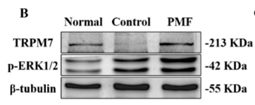
Fig. 4. PMF promoted wound healing through TRPM7-ERK1/2. (A) The release content and schematic diagram of magnesium ions in the wound site on the 3rd, 7th, and 15th days. *P < 0.05, **P < 0.01, ***P < 0.001 vs. control group, n = 3 (B) Western blot analysis of the expression of TRPM7 and phosphorylated ERK1/2 proteins. (C) Quantitative analysis of related protein expression levels in the PMF treatment group compared with the control group. ns: no significance, *P < 0.05, **P < 0.01, ***P < 0.001, n = 3. (D) Immunofluorescent staining images of skin tissue sections on day 7. CD68: pan-macrophage marker (red). TRPM7: (green). Scale bar: 40 μm. (E) Statistical summary of fluorescent expressing of TRPM7/CD68 expressing. ns: no significance

Application: IF/ICC    Species: Mouse    Sample:

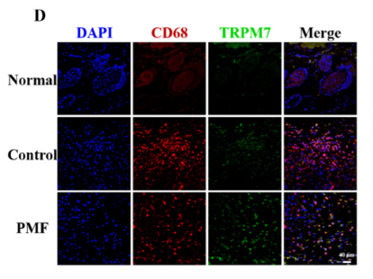
Fig. 4. PMF promoted wound healing through TRPM7-ERK1/2. (A) The release content and schematic diagram of magnesium ions in the wound site on the 3rd, 7th, and 15th days. *P < 0.05, **P < 0.01, ***P < 0.001 vs. control group, n = 3 (B) Western blot analysis of the expression of TRPM7 and phosphorylated ERK1/2 proteins. (C) Quantitative analysis of related protein expression levels in the PMF treatment group compared with the control group. ns: no significance, *P < 0.05, **P < 0.01, ***P < 0.001, n = 3. (D) Immunofluorescent staining images of skin tissue sections on day 7. CD68: pan-macrophage marker (red). TRPM7: (green). Scale bar: 40 μm. (E) Statistical summary of fluorescent expressing of TRPM7/CD68 expressing. ns: no significance

3). Inhibition of the RIP3/MLKL/TRPM7 necroptotic pathway ameliorates diabetes mellitus-induced erectile dysfunction by reducing cell death, fibrosis, and inflammation. Frontiers in pharmacology, 2024 (PubMed: 39329120) [IF=5.6]

4). Gallium ions promote osteoinduction of human and mouse osteoblasts via the TRPM7/Akt signaling pathway. Molecular Medicine Reports, 2020 (PubMed: 32945378) [IF=3.4]

Application: WB    Species: human    Sample: MC3T3 cells

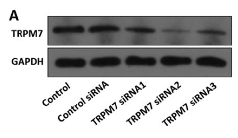
Figure 3. |Effect of TRPM7 siRNAs on TRPM7 protein expression in MC3T3‑E1 and hFOB1.19 cells. (A) Total and (B) membrane TRPM7 protein expression levels in MC3T3 cells following transfection with TRPM7 siRNAs.

5). Novel benzoylurea derivative decreases TRPM7 channel function and inhibits cancer cells migration. Channels (Austin, Tex.), 2024 (PubMed: 39212541) [IF=3.3]

Application: WB    Species: Human    Sample: MCF-7 cells

Figure 4. SUD inhibits TRPM7 expression through PI3K/Akt signaling pathway. (a) Effects of different concentrations SUD on the AKT1 and PI3K mRNA expression in MCF-7 cells examined by qPCR. (b) effects of different concentrations SUD on the AKT1 and PI3K mRNA expression in BGC-823 cells examined by qPCR. ***, **** denote significant difference with control group, p

6). Circ_0021155 can participate in the phenotypic transformation of human vascular smooth muscle cells via the miR-4459/TRPM7 axis. Biochemical and biophysical research communications, 2022 (PubMed: 36155059) [IF=2.5]

限制条款

产品的规格、报价、验证数据请以官网为准,官网链接:www.affbiotech.com | www.affbiotech.cn(简体中文)| www.affbiotech.jp(日本語)

产品的数据信息为Affinity所有,未经授权不得收集Affinity官网数据或资料用于商业用途,对抄袭产品数据的行为我们将保留诉诸法律的权利。

产品相关数据会因产品批次、产品检测情况随时调整,如您已订购该产品,请以订购时随货说明书为准,否则请以官网内容为准,官网内容有改动时恕不另行通知。

Affinity保证所销售产品均经过严格质量检测。如您购买的商品在规定时间内出现问题需要售后时,请您在Affinity官方渠道提交售后申请。

产品仅供科学研究使用。不用于诊断和治疗。 

产品未经授权不得转售。

Affinity Biosciences将不会对在使用我们的产品时可能发生的专利侵权或其他侵权行为负责。Affinity Biosciences, Affinity Biosciences标志和所有其他商标所有权归Affinity Biosciences LTD.