产品: 磷酸化 DRP1 (Ser637) 抗体
货号: DF2980
描述: Rabbit polyclonal antibody to Phospho-DRP1 (Ser637)
应用: WB IHC IF/ICC
文献验证: WB, IF/ICC
反应: Human, Mouse, Rat
预测: Pig, Bovine, Horse, Sheep, Rabbit, Dog, Chicken, Xenopus
蛋白号: O00429
RRID: AB_2840960

浏览相似产品>>

   规格 价格 库存
 50ul RMB¥ 1300 现货
 100ul RMB¥ 2400 现货

货期: 当天发货

联系销售

产品描述

来源:
Rabbit
应用:
WB 1:1000-3000, IHC 1:50-1:200, IF/ICC 1:100-1:500
*The optimal dilutions should be determined by the end user.
*Tips:

WB: 适用于变性蛋白样本的免疫印迹检测. IHC: 适用于组织样本的石蜡(IHC-p)或冰冻(IHC-f)切片样本的免疫组化/荧光检测. IF/ICC: 适用于细胞样本的荧光检测. ELISA(peptide): 适用于抗原肽的ELISA检测.

反应:
Human, Mouse, Rat
克隆:
Polyclonal
特异性:
Phospho-DRP1 (Ser637) Antibody detects endogenous levels of DRP1 only when phosphorylated at Ser637.
RRID:
AB_2840960
引用格式: Affinity Biosciences Cat# DF2980, RRID:AB_2840960.
偶联:
Unconjugated.
纯化:
The antibody is from purified rabbit serum by affinity purification via sequential chromatography on phospho-peptide and non-phospho-peptide affinity columns.
保存:
Rabbit IgG in phosphate buffered saline , pH 7.4, 150mM NaCl, 0.02% sodium azide and 50% glycerol. Store at -20 °C. Stable for 12 months from date of receipt.
别名:

展开/折叠

DLP1; dnm1l; DNM1L_HUMAN; Dnm1p/Vps1p-like protein; dnml1; DRP1; DVLP; Dymple; Dynamin 1 like; Dynamin family member proline-rich carboxyl-terminal domain less; Dynamin like protein; Dynamin related protein 1; Dynamin-1-like protein; Dynamin-like protein 4; Dynamin-like protein; Dynamin-like protein IV; Dynamin-related protein 1; DYNIV 11; EMPF; EMPF1; FLJ41912; HdynIV; VPS1;

抗原和靶标

免疫原:

A synthesized peptide derived from human DRP1 around the phosphorylation site of Ser637.

基因/基因ID:

研究领域

· Cellular Processes > Cell growth and death > Necroptosis.   (View pathway)

· Environmental Information Processing > Signal transduction > TNF signaling pathway.   (View pathway)

· Organismal Systems > Immune system > NOD-like receptor signaling pathway.   (View pathway)

文献引用

1). Nutrient status alters developmental fates via a switch in mitochondrial homeodynamics. Nature communications, 2025 (PubMed: 39893174) [IF=16.6]

Application: IF/ICC    Species: insect    Sample: PG cells

Fig. 2: Pre-NRC starvation promotes mitochondrial tabulation. A The percentage of fragmented mitochondria in PG cells from 72 h AEL to WPP. From left to right, n = 30, 16, 27, 17, 24, and 40, respectively. B Mitochondrial morphology (green) was assessed in PG cells. Magnified images of the highlighted areas (red dotted box) are shown. Right: the percentage of fragmented mitochondria. Nucleus: DAPI (cyan). From left to right, n = 30, 23, 24, and 17, respectively. C–F Representative images of PTTH (C), p-ErK, green, 108 h: n = 24, pre-NRC ST: n = 27), insulin (D, E InR-SPARK and AKT-SPARK, green), and TORC1 signalings (F, p-S6K, green, 108 h: n = 9, pre-NRC ST: n = 8) in PG cells of control and pre-NRC ST larvae. G, H Representative images of mitochondrial morphology (green) in PG cells (168 h AEL) with inhibition of PTTH, insulin, TORC1 signalings G and mitochondria-associated genes H. Magnified images of the highlighted areas (red dotted box) are shown. G From left to right, n = 7, 7, 7, 6, 6, 13, 9, and 7, respectively. H From left to right, n = 32, 19, 19, 20, 12, 16, 17, and 19, respectively. I, J Images showing Marf (I Marf-EGFP, green) and p-Drp1 (J p-Drp1 Ser637 antibody, green) staining in PG cells of control and pre-NRC ST larvae at 108 h AEL. Mitochondria: mito-mCherry (red). Nucleus: DAPI (cyan). From left to right, n = 26, 15, 30, and 22, respectively in (I, J). K, L Images showing ATP-SPARK puncta (green) in PG cells of control and pre-NRC ST or Marf-expressing larvae at 108 h AEL. M, N Representative images of mitochondria morphology (M, green, From left to right, n = 20, 27, 24, and 19, respectively) and ATP-SPARK puncta (N, green) in PG cells (144 h AEL) with PG-specific expression of RasV12, Dp110CA, or Rheb. Magnified images of the highlighted areas (red dotted box) are shown. O, P Images showing p-AMPK levels (p-AMPK, green, 108 h: n = 17, pre-NRC ST: n = 19) O and AMPK-SPARK puncta (green) P in PG cells of controls and pre-NRC ST larvae at 108 h AEL. All Data are presented as mean ± SD. Different lowercase letters represent statistical significance by one-way ANOVA. The genotypes and statistical data are provided in Supplementary Data 1.

2). A novel PGAM5 inhibitor LFHP-1c protects blood–brain barrier integrity in ischemic stroke. Acta Pharmaceutica Sinica B, 2021 (PubMed: 34386325) [IF=14.7]

Application: WB    Species: Rat    Sample: brain microvascular endothelial cells

Figure 4 LFHP-1c targets PGAM5 to facilitate nuclear translocation of NRF2 for endothelial protection in stroke. (A) The expression of PGAM5, p-DRP1 (Ser637), NRF2 and HO-1 were detected at indicated times after LFHP-1c treatment in rBMECs by immunoblotting, n=3 per group. (B) and (C) IP/Western blotting results reveal that LFHP-1c treatment significantly reduced the interaction of PGAM5 with NRF2, n=3 per group. (D) LFHP-1c facilitates nuclear translocation of NRF2 in rBMECs, n=3 per group. (E) Silencing efficiency of siRNA against PGAM5 in rBMECs was verified by immunoblotting, n=3 per group. (F) and (G) LFHP-1c treatment or knockdown of Pgam5 facilitates nuclear translocation of NRF2 in rBMECs, and LFHP-1c treatment after si-PGAM5 did not affect the nuclear translocation of NRF2, n=3 per group. (H) Fluorescence staining of NRF2 shows that LFHP-1c treatment or knockdown of Pgam5 promoted NRF2 nuclear translocation, n=3 per group. The scale bar represents 20 μm. (I) and (J) The protein expressions of PGAM5, p-DRP1 (Ser637), NRF2 and HO-1 in rat brain microvessels were detected at 72 h after tMCAO onsetand representative blots are shown here, n=4 per group. (K) The mRNA expression of Ho-1 and Nqo1 was measured by RT-PCR, n=4–5 per group. Results are expressed as mean±SEM. For Fig. 4C, *** P<0.001 versus IgG plus DMSO group, ## P<0.01 versus PGAM5 plus LFHP-1c (2 μmol/L) group. For Fig. 4D, * P<0.05, *** P<0.001 versus ‘0’ time point group. For Fig. 4E, *** P<0.001 versus scramble group. For Fig. 4G, *** P<0.001 versus scramble plus DMSO group. For Fig. 4J and K, * P<0.05, ** P<0.01, *** P<0.001 versus Sham group; # P<0.05, ## P<0.01, ### P<0.001 versus Vehicle group.

3). The NLRX1-SLC39A7 complex orchestrates mitochondrial dynamics and mitophagy to rejuvenate intervertebral disc by modulating mitochondrial Zn2+ trafficking. Autophagy, 2024 (PubMed: 37876250) [IF=14.6]

4). Mitochondrial Dysfunction by FADDosome Promotes Gastric Mucosal Injury in Portal Hypertensive Gastropathy. International journal of biological sciences, 2024 (PubMed: 38725851) [IF=8.2]

5). Honokiol attenuates mitochondrial fission and cell apoptosis by activating Sirt3 in intracerebral hemorrhage. CHINESE MEDICAL JOURNAL, 2023 (PubMed: 36805606) [IF=7.5]

6). Paeoniflorin Ameliorates Skeletal Muscle Atrophy in Chronic Kidney Disease via AMPK/SIRT1/PGC-1α-Mediated Oxidative Stress and Mitochondrial Dysfunction. Frontiers in Pharmacology, 2022 (PubMed: 35370668) [IF=5.6]

Application: WB    Species: rat    Sample: C2C12 cells

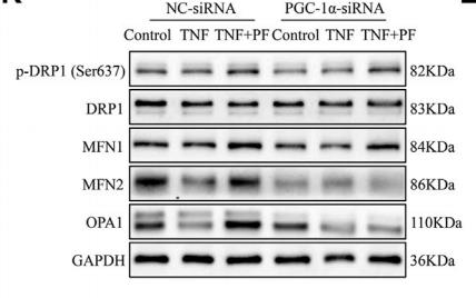
FIGURE 8 | PGC-1α might be important in mediating the effects of PF on TNF-α-treated C2C12 cells.(K) Representative western blots using antibodies against p-DRP1 (Ser637), DRP1, MFN1, MFN2, OPA1, and GAPDH.

7). Inhibition of calpain reduces cell apoptosis by suppressing mitochondrial fission in acute viral myocarditis. CELL BIOLOGY AND TOXICOLOGY, 2022 (PubMed: 34365571) [IF=5.3]

Application: WB    Species: Mice    Sample: NRCMs

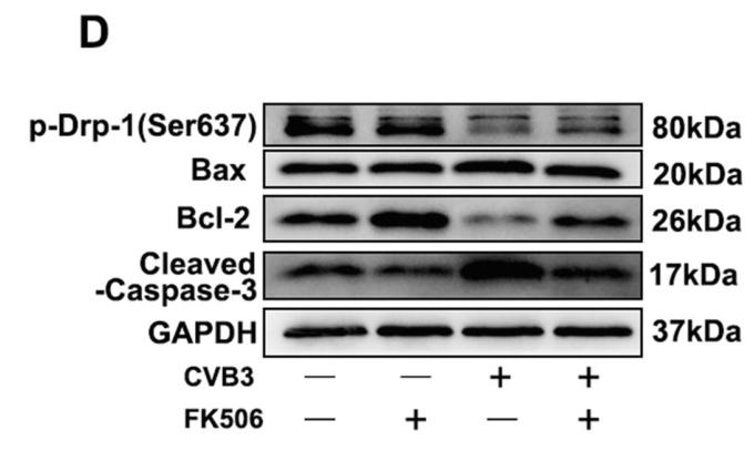
Fig. 8 PD150606 inhibited mitochondrial fragmentation by modulating Drp-1 phosphorylation. a–c The changes of p-Drp- 1(Ser637) and calcineurin A in NRCMs infected with CVB3 with or without PD150606. PD150606 treatment inhibited Drp-1 dephosphorylated at the Ser637 site (b) and calcineu- rin activation (c) after CVB3 infection. d–h The changes of p-Drp-1(Ser637) and apoptosis-related proteins in NRCMs infected with CVB3 with or without FK506 (an inhibitor of calcineurin activation). Dephosphorylation of Drp-1 (e) was activated by calcineurin activation and contributes to apoptosis (f–h) in CVB3-infected NRCMs. *P < 0.05 vs. Con; #P < 0.05 vs. virus group

8). Anemoside B4 alleviates arthritis pain via suppressing ferroptosis-mediated inflammation. Journal of cellular and molecular medicine, 2024 (PubMed: 38334255) [IF=5.3]

Application: WB    Species: Mouse    Sample:

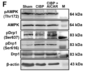
FIGURE 5 (A) The molecular structure of AB4. (B–D) Molecular docking of AB4 with GSK‐3β. The modelled 3D structure of GSK‐3β docked with AB4 (B). The enlarged view of binding site in box (C). The interaction bonds of GSK‐3β with AB4 (D). Bonds showed as yellow dotted lines, and bond lengths were presented as numbers. (E) The titration between AB4 and GSK‐3β. The top panel presents typical calorimetric titration of AB4 with GSK‐3β at 25°C. The bottom panel shows the plots of the heat evolved (kcal) per mol of AB4 added corrected for the heat of with GSK‐3β, against the molar ratio of AB4 to GSK‐3β. Data solid squares were fitted to a single set of the identical sites model, and the solid line represented the best fit. (F) Representative immunofluorescence staining images of GSK‐3β and Drp1 in the spinal dorsal horn of the control, CIA and CIA + AB4 groups. Scale bar = 20 μm. (G) Quantitative analysis of the fluorescence intensity of GSK‐3β and Drp1. (H, I) Western blot analysis and quantitative grey value analysis of pGSK‐3β‐Tyr216, GSK‐3β, pDrp1‐Ser616, pDrp1‐Ser637 and Drp1 level in the spinal cord of the control, CIA and CIA + AB4 groups. Data are presented as mean ± SD (n = 5). *p 

9). Rostellularia procumbens (L) Nees. extract attenuates adriamycin-induced nephropathy by maintaining mitochondrial dynamics balance via SIRT1/PGC-1α signaling pathway activation. Journal of ethnopharmacology, 2025 (PubMed: 39733803) [IF=4.8]

10). Mitochondrial fission enhances IL-6-induced metastatic potential in ovarian cancer via ERK1/2 activation. Cancer science, 2024 (PubMed: 38433313) [IF=4.5]

加载更多

限制条款

产品的规格、报价、验证数据请以官网为准,官网链接:www.affbiotech.com | www.affbiotech.cn(简体中文)| www.affbiotech.jp(日本語)

产品的数据信息为Affinity所有,未经授权不得收集Affinity官网数据或资料用于商业用途,对抄袭产品数据的行为我们将保留诉诸法律的权利。

产品相关数据会因产品批次、产品检测情况随时调整,如您已订购该产品,请以订购时随货说明书为准,否则请以官网内容为准,官网内容有改动时恕不另行通知。

Affinity保证所销售产品均经过严格质量检测。如您购买的商品在规定时间内出现问题需要售后时,请您在Affinity官方渠道提交售后申请。

产品仅供科学研究使用。不用于诊断和治疗。 

产品未经授权不得转售。

Affinity Biosciences将不会对在使用我们的产品时可能发生的专利侵权或其他侵权行为负责。Affinity Biosciences, Affinity Biosciences标志和所有其他商标所有权归Affinity Biosciences LTD.